- Select a language for the TTS:
- UK English Female
- UK English Male
- US English Female
- US English Male
- Australian Female
- Australian Male
- Language selected: (auto detect) - EN
Play all audios:
Access through your institution Buy or subscribe Many vertebrates and invertebrates have cells in the skin which react to light by dispersing or aggregating intracellular pigment granules.
Even in a dish, such cells act like chameleons and change their shading according to the light1. A study by Provencio _et al._, published last month in _Proceedings of the National Academy
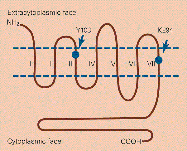
of Sciences_2, now shows that light-sensitive pigment cells in frog skin, called melanophores, express a molecule, melanopsin, which is similar to the rhodopsins used to detect light in the
eye. Melanopsin is also expressed in several other cell types known or thought to be light-sensitive in the frog, including cells in the iris, non-image-forming photoreceptor cells in the
retina, and hypothalamic neurons which may be involved in controlling responses to day/night cycles. All this is hardly unexpected. Rather, the real surprise comes in Provencio and
colleagues' finding that melanopsin is more closely related to the rhodopsins in invertebrate eyes (particularly of scallop, squid and octopus) than to those in vertebrate eyes,
including the frog's own eyes. This is an observation that raises several intriguing issues. This is a preview of subscription content, access via your institution ACCESS OPTIONS Access
through your institution Subscribe to this journal Receive 51 print issues and online access $199.00 per year only $3.90 per issue Learn more Buy this article * Purchase on SpringerLink *
Instant access to full article PDF Buy now Prices may be subject to local taxes which are calculated during checkout ADDITIONAL ACCESS OPTIONS: * Log in * Learn about institutional
subscriptions * Read our FAQs * Contact customer support REFERENCES * Lerner, M. R. _Trends Neurosci_. 17, 142–146 (1994). Google Scholar * Provencio, I., Jiang, G., De Grip, W. J., Hayes,
W. P. & Rollag, M. D. _Proc. Natl Acad. Sci. USA_ 95, 340–345 (1998). Google Scholar * Kojima, D._et al._ _J. Biol. Chem_. 272, 22979–22982 (1997). Google Scholar * Gärtner, W. &
Towner, P. _Photochem. Photobiol_. 62, 1–16 (1995). Google Scholar * Blackshaw, S. & Snyder, S. H. _J. Neurosci_. 17, 8083–8092 (1997). Google Scholar * Soni, B. G. & Foster, R. G.
_FEBS Lett_. 406, 279–283 (1997). Google Scholar * Piatigorsky, J. & Wistow, G. _Science_ 252, 1078–1079 (1991). Google Scholar * Doughtie, D. G. & Rao, K. R. _Cell Tissue Res_.
238, 271–288 (1984). Google Scholar * Callaerts, P., Halder, G. & Gehring, W. J. _Annu. Rev. Neurosci._ 20, 483–532 (1997). Google Scholar * Desplan, C. _Cell_ 91, 861–864 (1997).
Google Scholar * Salvini-Plawen, L. v. & Mayr, E. in _Evolutionary Biology_ Vol. 10 (eds Hecht, M. K., Steere, W. C. & Wallace, B.) 207-263 (Plenum, New York, 1977). * Zuker, C. S.
_Science_ 265, 742–743 (1994). Google Scholar Download references AUTHOR INFORMATION AUTHORS AND AFFILIATIONS * Heinz Arnheiter is in the Laboratory of Developmental Neurogenetics, National
Institute of Neurological Disorders and Stroke, Building 36, Room 5D06, 36 Convent Drive, Bethesda, 20892-4160, Maryland, USA Heinz Arnheiter Authors * Heinz Arnheiter View author
publications You can also search for this author inPubMed Google Scholar RIGHTS AND PERMISSIONS Reprints and permissions ABOUT THIS ARTICLE CITE THIS ARTICLE Arnheiter, H. Eyes viewed from
the skin. _Nature_ 391, 632–633 (1998). https://doi.org/10.1038/35487 Download citation * Issue Date: 12 February 1998 * DOI: https://doi.org/10.1038/35487 SHARE THIS ARTICLE Anyone you
share the following link with will be able to read this content: Get shareable link Sorry, a shareable link is not currently available for this article. Copy to clipboard Provided by the
Springer Nature SharedIt content-sharing initiative