- Select a language for the TTS:
- UK English Female
- UK English Male
- US English Female
- US English Male
- Australian Female
- Australian Male
- Language selected: (auto detect) - EN
Play all audios:
ABSTRACT STUDY DESIGN: A case report. OBJECTIVES: To report a rare case of extension of edema and hemorrhage from initial C4–5 spinal injury to the medulla oblongata. SETTING: Center for
Spinal Disorders and Injuries, Bibai Rosai Hospital, Japan. METHODS: A 68-year-old man with ossification of the posterior longitudinal ligament (OPLL) had sustained tetraplegia after
tumbling over a stone. Initially, the patient was diagnosed with an acute C4–5 spinal cord injury without radiological abnormalities and was treated conservatively. At 7 h after the injury,
the patient had an ascending neurological deficit, which required respiratory assistance. Magnetic resonance imaging revealed a marked swelling of the spinal cord above C4–5 extending to the
medulla oblongata. RESULTS: Retrospective radiological assessment revealed that the spine was unstable at the injury level because of discontinuities in both anterior and posterior
longitudinal ligaments. There was also signal intensity change within the retropharyngeal space at the C4–5 intervertebral disc. This injured segment was highly vulnerable to post-injury
dynamic stenosis and easily sustained secondary neural damage. CONCLUSIONS: This case report emphasizes a careful radiological assessment of latent structural instability in patients with
OPLL in order to detect and prevent deteriorative change in the spinal cord. SIMILAR CONTENT BEING VIEWED BY OTHERS THORACIC LIGAMENTUM FLAVUM OSSIFICATION: A RARE CAUSE OF SPINAL CORD
INJURY WITHOUT TOMOGRAPHIC EVIDENCE OF TRAUMA IN A CAUCASIAN PATIENT. CASE REPORT AND LITERATURE REVIEW Article 09 July 2021 VERY RARE INCIDENCE OF ASCENDING PARALYSIS IN A PATIENT OF
TRAUMATIC SPINAL CORD INJURY: A CASE REPORT Article 26 July 2022 ISOLATED L5 NERVE ROOT INJURY WITHOUT OSSEOUS DISRUPTION IN A CASE OF GUNSHOT INJURY TO THE PAEDIATRIC SPINE—A CASE REPORT
Article 26 April 2022 INTRODUCTION It has been documented that secondary neurologic deterioration after cervical spinal cord injury (CSCI) is one of the most severe management problems that
might affect a patient’s prognosis.1, 2 Although the mechanism causing spinal deterioration is still unclear, a structural instability in the injured segment is one of the most important
factors affecting these detrimental changes.3 Surgical decompression and fixation of the cervical spine is the recommended treatment in a patient with structural damage, such as a spinal
fracture-dislocation.3 Conversely, it is debatable whether tetraplegic patients with no radiological evidence of structural spinal injury should be treated surgically or conservatively.
However, some patients may actually harbor latent structural instability, which may cause secondary neurological deterioration. Therefore, it is important that latent instability should be
promptly detected with a high index of suspicion during the acute management phase of CSCI.4 Here, we document an unusual case, where secondary detrimental change due to latent structural
instability occurred in the medulla oblongata of a patient with ossification of the posterior longitudinal ligament (OPLL) following middle CSCI. CASE REPORT A 68-year-old man tumbled over a
stone and sustained complete tetraplegia. At 2 h later, the patient was found and admitted to a tertiary-care facility. The clinical parameters presented a normal range of the heart rate,
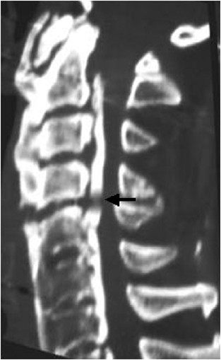
blood pressure, and respiratory rate. Although the patient escaped from the brain, thoracic, and abdominal injuries, his neurological status was complete tetraplegia of C4. Plain X-ray, CT,
and sagittal reformatted CT showed spinal canal stenosis due to OPLL (Figure 1). Although T1-weighted magnetic resonance imaging (MRI) failed to demonstrate any signal changes in the spinal
cord, the T2-weighted image showed a low signal intensity area at the C4–5 level, which indicated intramedullary hemorrhage5 (Figure 2a and b). Initially, the patient was diagnosed with an
acute spinal cord injury at C4–5 without radiological abnormalities and treated conservatively. A Philadelphia collar was used but skull traction was not performed. Intravenous
methylprednisolone was administered according to the recommended National Acute Spinal Cord Injury Study (NASCIS-2) protocol.6 At 7 h after the initial injury, the patient experienced
increased dyspnea and transient loss of consciousness. When the patient regained consciousness, his neurological status changed to complete respiratory tetraplegia with neurological
dysfunction at the C2 level. The patient presented with a normal sensory condition of the face, a sense of taste, and swallowing function. Neurological examination revealed normal function
of the cranial nerves. Despite normal cervical alignment, MRI at the onset of acute respiratory arrest showed a marked swelling of the spinal cord above C2–3 to the medulla oblongata.
T1-weighted MRI showed high signal intensity in the spinal cord from C4–2. T2-weighted MRI demonstrated diffuse heterogeneous high signal intensity within the spinal cord from C4 to the
medulla oblongata. These findings indicated that spinal cord edema and intramedullary hemorrhage had extended cranially from the C4–5 level5 (Figure 3a and b). Retrospective radiological
assessment revealed that the spine had been unstable at the injured level because of a discontinuity in both the anterior and posterior longitudinal ligaments and with a signal intensity
change in the retropharyngeal space between the C4–5 intervertebral discs (Figures 1 and 2b). The patient was referred to our institute 4 weeks after injury. He was managed conservatively
because surgical intervention at such a late stage, after neural damage, would be expected to provide little chance of any neurological improvement. Furthermore, the patient and his family
did not elect for surgery to be performed. MRI obtained 3 months after injury showed that gliosis extended from the medulla oblongata to middle part of the cervical spinal cord, which
exhibited a uniform signal intensity on the T1-weighted image but a high signal intensity on the T2-weighted image (Figure 4a and b). Moreover, hypertrophy of the ligamentum flavum was
observed at C4–5 segment. This finding proved that there was an initial, undetected instability within C4–5 segment, which led to secondary deterioration of the injured spinal cord.
DISCUSSION The incidence of neurological deterioration from spinal injury was reported to be between 1.84, and 5.8%.1, 2 Many reports have documented that hemorrhage, microvascular
infarction, spasm, spinal cord edema, and free radical production are relevant factors affecting this serious condition.7, 8 Lu _et al_9 reported the predictable risk factors for delayed
apnea following middle to lower CSCI in the presence of diffuse and extensive cord lesions, respiratory distress, and bradycardia. In this case, however, the initial spinal cord lesion was
localized to the C4–5 segment and cardiopulmonary functions were normal. The most important point regarding initial management of this case was that the patient was managed as though he had
a stable injury that required no surgical decompression and stabilization. However, in this case, there was mechanical instability in the interrupted portion of OPLL (C4/5 segment). When
mechanical instability exists between the OPLL, spinal cord mechanical stress is usually focused at the same site. Consequently, secondary spinal cord injury often aggravates the
neurological condition. Owing to the fact that CSCI without osteoligamentous damage is quite common in the elderly population with degenerative spinal stenosis including OPLL, CSCI with
spinal stenosis tends to be managed as a stable injury without detailed radiological evaluation. Therefore, it is important to identify any latent mechanical instability using MRI and to
perform adequate management using surgical decompression with or without fusion in the acute phase of injury.4 Both ankylosing spondylitis (AS) and diffuse idiopathic skeletal hyperostosis
are well-known predictive neurological deterioration risk factors.10, 11 Bohlman12 reported that 50% of identified AS patients had a delay in the diagnosis of cervical spine structural
injury and all of them subsequently developed secondary neurological deterioration. Reid _et al_13 documented a high incidence of neurological deterioration due to this delay in diagnosis.
In this case, all cervical spinal segments except C4–5 were fused by ossification of both anterior and posterior longitudinal ligaments. Therefore, any force applied to the cervical spine
would be concentrated on the weak C4–5 segment in a similar situation of a fractured ankylosing spine. Therefore, the C4–5 segment of the spinal cord in our case was vulnerable to postinjury
dynamic stenosis and easily sustained secondary neural damage. The clinical manifestations of patients with medulla oblongata injuries are unclear because such patients rarely survive. This
is the first clinical case report describing the deterioration in the ascending medulla oblongata following middle CSCI. After postmortem neuropathological examination, Ito _et al_14
revealed longitudinal spreading of cord lesions upward into the medulla oblongata in one of eight patients who died after traumatic CSCI. They also documented that there was a completely
necrotic lesion without any cell reaction in several cord segments as well as the initially damaged lesion. They suggested that the spreading of the lesion might be induced by increased
intramedullary pressure that resulted from an intra- and extramedullary circulatory disturbance. In the current case, the critical instability of the C4–5 segment associated with severe
spinal stenosis caused by OPLL could have led to an acutely increased intramedullary pressure, which permitted the extension of the spinal cord lesion. The patient escaped from cranial nerve
damage in spite of medulla oblongata involvement. This may be explained because the involved lesion was located within only the inferior portion of the medulla oblongata that contains no
the cranial nerve nuclei but only the spinal trigeminal nuclei. In addition, there were discrepancies between the patient's neurological status and the MRI findings. Therefore, the
apnea was not necessarily the result of the secondary involvement of the medulla oblongata, but might simply result from the involvement of the C4 segment in which the phrenic nerves are
located. REFERENCES * Famer J et al. Neurologic deterioration after cervical spinal cord injury. _J Spinal Disord_ 1998; 11: 192–196. Google Scholar * Marshall LF, Knowlton S, Garfin SR .
Deterioration following spinal cord injury: a multicenter study. _J Neurosurg_ 1987; 66: 400–404. Article CAS Google Scholar * Mahale YJ, Silver JR . Progressive paralysis after bilateral
facet dislocation of the cervical spine. _J Bone Joint Surg [Britain]_ 1992; 74: 219–223. Article CAS Google Scholar * Benzel EC et al. Magnetic resonance imaging for the evaluation of
patients with occult cervical spine injury. _J Neurosurg_ 1996; 85: 824–829. Article CAS Google Scholar * Flanders AE et al. Acute cervical spine trauma: correlation of MR Imaging
findings with degree of neurologic deficit. _Neuroradiology_ 1990; 177: 25–33. CAS Google Scholar * Bracken MB et al. A randomized controlled trial of methylprednisolone or naloxone in the
treatment of ascute spinal-cord injury. Results of the Second National Acute Spinal Cord Injury Study. _N Engl J Med_ 1990; 322: 1405–1411. Article CAS Google Scholar * Banick N, Hogan
E, Hsu C . The multimolecular cascade of spinal cord injury. _Neurochem Pathol_ 1987; 7: 57–77. Article Google Scholar * Tator CH, Fehlings MG . Review of the secondary injury theory of
acute spinal cord trauma with emphasis on vascular mechanisms. _J Neurosurg_ 1991; 75: 15–26. Article CAS Google Scholar * Lu K et al. Delyed apnea in patients with mid-to lower cervical
spinal cord injury. _Spine_ 2000; 25: 1332–1338. Article CAS Google Scholar * Fox MW, Onofrio BM, Kilgore JE . Neurological complications of ankylosing spondylitis. _J Neurosurg_ 1993;
78: 871–878. Article CAS Google Scholar * Paley D et al. Fractures of the spine in diffuse idiopathic skeletal hyperostosis. _Clin Orthop_ 1991; 267: 22–32. Google Scholar * Bohlman HH .
Acute fractures and dislocations of the cervical spine. _J Bone Joint Surg [America]_ 1979; 61: 1119–1142. Article CAS Google Scholar * Reid DC et al. Etiology and clinical course of
missed spine fractures. _J Trauma_ 1987; 27: 980–986. Article CAS Google Scholar * Ito T et al. Traumatic spinal cord injury: a neuropathological study on the longitudinal spreading of
the lesions. _Acta Neuropathol (Berlin)_ 1997; 93: 13–18. Article CAS Google Scholar Download references AUTHOR INFORMATION AUTHORS AND AFFILIATIONS * Center for Spinal Disorders and
Injuries, Bibai Rosai Hospital, Bibai, Japan H Sudo, H Taneichi & K Kaneda * Department of Orthopaedic Surgery, Hokkaido University Graduate School of Medicine, Sapporo, Japan H Sudo
Authors * H Sudo View author publications You can also search for this author inPubMed Google Scholar * H Taneichi View author publications You can also search for this author inPubMed
Google Scholar * K Kaneda View author publications You can also search for this author inPubMed Google Scholar RIGHTS AND PERMISSIONS Reprints and permissions ABOUT THIS ARTICLE CITE THIS
ARTICLE Sudo, H., Taneichi, H. & Kaneda, K. Secondary medulla oblongata involvement following middle cervical spinal cord injury associated with latent traumatic instability in a patient
with ossification of the posterior longitudinal ligament. _Spinal Cord_ 44, 126–129 (2006). https://doi.org/10.1038/sj.sc.3101803 Download citation * Published: 05 July 2005 * Issue Date:
01 February 2006 * DOI: https://doi.org/10.1038/sj.sc.3101803 SHARE THIS ARTICLE Anyone you share the following link with will be able to read this content: Get shareable link Sorry, a
shareable link is not currently available for this article. Copy to clipboard Provided by the Springer Nature SharedIt content-sharing initiative KEYWORDS * cervical spinal cord injury *
deteriorative change * medulla oblongata * ossification of the posterior longitudinal ligament