- Select a language for the TTS:
- UK English Female
- UK English Male
- US English Female
- US English Male
- Australian Female
- Australian Male
- Language selected: (auto detect) - EN
Play all audios:
ABSTRACT With its antimicrobial and immunomodulating properties, the cathelicidin (LL37) plays an important role in innate immune system. Here, we attempted to alleviate chemically induced
colitis using a lactococci strain that either directly expressed the precursor to LL37, hCAP18 (LL-pSEC:hCAP18), or delivered hCAP18 cDNA to host cells under the control of the
cytomegalovirus promoter (LL-Probi-H1:hCAP18). We also investigated whether the alleviation of symptoms could be explained through modification of the gut microbiota by hCAP18. Mice were
administered daily doses of LL-pSEC:hCAP18 or LL-Probi-H1:hCAP18. On day 7, colitis was induced by DNBS. During autopsy, we assessed macroscopic tissue damage in the colon and collected
tissue samples for the characterization of inflammation markers and histological analysis. Feces were collected at day 7 for 16S DNA sequencing. We also performed a fecal transplant
experiment in which mice underwent colon washing and received feces from _Lactococcus lactis_-treated mice before DNBS-colitis induction. Treatment with LL-Probi-H1:hCAP18 reduced the
severity of colitis symptoms. The protective effects were accompanied by increased levels of IL17A and IL10 in mesenteric lymph node cells. _L. lactis_ administration altered the abundance
of _Lachnospiraceae_ and _Muribaculaceae_. However, fecal transplant from _L. lactis_-treated mice did not improve DNBS-induced symptoms in recipient mice. SIMILAR CONTENT BEING VIEWED BY
OTHERS POTENTIAL OF USING AN ENGINEERED INDOLE LACTIC ACID PRODUCING _ESCHERICHIA COLI_ NISSLE 1917 IN A MURINE MODEL OF COLITIS Article Open access 30 July 2024 ORAL SUPPLEMENTATION WITH
SELECTED _LACTOBACILLUS ACIDOPHILUS_ TRIGGERS IL-17-DEPENDENT INNATE DEFENSE RESPONSE, ACTIVATION OF INNATE LYMPHOID CELLS TYPE 3 AND IMPROVES COLITIS Article Open access 20 October 2022
COLONIZATION WITH UBIQUITOUS PROTIST _BLASTOCYSTIS_ ST1 AMELIORATES DSS-INDUCED COLITIS AND PROMOTES BENEFICIAL MICROBIOTA AND IMMUNE OUTCOMES Article Open access 25 April 2023 INTRODUCTION
Inflammatory bowel diseases (IBD) such as Crohn’s disease (CD) and ulcerative colitis (UC) are multifactorial diseases associated with chronic inflammation of the gastrointestinal tract and
dysbiosis of the patient’s gut microbiota1,2. IBD pathogenesis is characterized by excessive and uncontrolled immune responses against commensal gut bacteria3. While antibiotics and
anti-inflammatory drugs can help to make the condition manageable, IBD can have serious complications and no curative treatment currently exists. The innate immune system plays an important
role in initiating the inflammatory response. Antimicrobial peptides (AMPs)—a broad category of immune molecules that includes defensins and cathelicidins, among others—have various modes of
action4; they may function as wide-spectrum antibiotics that use a variety of methods to kill bacteria and fungi directly5,6 or act as immunomodulators by altering host gene expression,
inducing chemokine production7, or modulating the response of the adaptive immune system8. AMPs have a crucial role in host defense against pathogens and the regulation of inflammatory
responses by immune cells. Many AMPs are typically expressed in response to gut infection and inflammation, and the administration of some of them improves colitis symptoms. HNP1-3, secreted
by neutrophils, are increased in active IBD mucosa9. Low doses of HNP-1 have a protective effect on DSS-induced colitis without affecting cytokines expression levels10. HD5 is also
increased in the colon of IBD patients and has protective effect on DSS-induced colitis9. On the contrary, the expression of cathelicidin is reduced in UC patients and its lowered
circulation level is associated with poor prognosis11. AMPs are currently being investigated as an alternative to antibiotics to prevent the spread of multiple-drug-resistant bacteria12,13.
In the gut, the innate immune response and the production of AMPs play an essential role in maintaining homeostasis. In particular, much attention has focused on the family of AMPs known as
cathelicidins, which are found in both mammals and non-mammals14,15. These peptides have been shown to influence inflammation and wound healing16,17,18, mediate changes in the gut
microbiota, and protect mice against chemically induced colitis. He have seen earlier that LL37 plays an important role in UC patients, who demonstrate a decreased circulating level of this
molecule11,19,20. Human cathelicidin is encoded by the _CAMP_ gene, the translation of which yields hCAP18, a small 18-kDa peptide. Following exocytosis, its C-terminal region is cleaved via
the action of neutrophil proteinase 3 to form the 4.5-kDa peptide LL3721. LL37 is able to disrupt bacterial membranes and thus has direct antibacterial properties22. It has been found in
neutrophil extracellular traps and is also produced at the surface of various epithelia where it helps fight against pathogens23,24,25. Moreover, studies have described the immunomodulating
and -stimulating effects of LL37 in diverse inflammation-related settings such as epithelial cell activation, chemotaxis, angiogenesis, and tissue wound repair. In particular, LL37 has been
shown to use the Fprl1 receptor to mobilize human leucocytes, neutrophils, and T cells26. It is also able to suppress collagen synthesis, as the administration of exogenous cathelicidin in
mice and human colonic fibroblasts has antifibrogenic effects27,28. Multiple studies suggest that the composition of the gut microbiota—which can be modulated by the environment29,30,
diet31,32, or drug treatment33—influences colitis severity. In humans, IBD is clearly associated with gut microbiota alterations although it is still unclear if the effect is causal1. One
study found that mice who were deficient in mCRAMP (the hCAP18 ortholog in mice) were highly sensitive to DSS-induced colitis, which could be prevented by mCRAMP administration. Alterations
were also evident in the gut microbiota of these mice, with increased levels of bacteria from the oral microbiota in their feces34. In the same study, wild-type mice that were co-housed with
mCRAMP-deficient mice became highly sensitive to DSS-induced colitis, suggesting a causal link between gut microbiota and disease severity. In 2013, Divyendu Singh et al. found that
administration of LL37 or mCRAMP could both inhibit LPS-induced IL6 production in human and mouse cell lines. Furthermore, LL37 could up-regulate dsRNA-induced innate response through TLR3
but not mCRAMP35. LL37 can also inhibit LPS-induced pyroptosis of macrophages, modulate the expression of pro-inflammatory cytokines and improve survival in septic mice36. Recently, a
Cecropin-LL37 hybrid peptide was constructed and found to be promising in the treatment of EHEC infections in a mouse model by modulating gut microbiota, inflammation and mucosal barrier37.
Therapies based on the oral administration of AMPs face significant challenges, as these molecules are quickly degraded before reaching the target site. For this reason, effective doses are
typically very high, which raises problems associated with the high production costs and an increased potential for toxic side effects. To more efficiently and cost-effectively produce and
deliver AMPs to their target site, one novel strategy is the use of recombinant probiotics38,39. The concept of probiotics was first introduced by Lilly and Stilwell in 196540; they are live
microorganisms that are considered safe for human consumption, with health benefits provided by their interactions with the gut microbiota or the host intestinal epithelium. Health benefits
aside, probiotics have been proposed as a novel mechanism for drug delivery because of their ability to transit and interact with the intestinal barrier and gut bacteria41. Specifically,
lactic acid bacteria, and _Lactococcus lactis_ in particular, have attracted attention as a result of their health benefits, their lack of toxic metabolites, and their low number of secreted
proteins. To date, these strains have been engineered to serve as vehicles for the production of therapeutic molecules and the delivery of DNA vaccines41,42,43,44. _L. lactis_ that has been
manipulated to contain a eukaryotic expression cassette containing cDNA of a protein of interest can be used as a delivery system for DNA that can then be expressed by the host’s cells
after transfection45,46,47. Here, we administered a recombinant strain of _L. lactis_ that expresses hCAP18 (LL-pSEC:HCAP18) to mice and then induced colitis in these mice through DNBS
intrarectal injection. Additionally, we used _L. lactis_ as a vehicle for the delivery of hCAP18 cDNA (LL-Probi-H1:hCAP18) to the gut epithelium of mice. This second strategy allowed the
direct expression of the peptide by the host’s cells48. We collected feces at day 0 and day 7 after bacterial administration for microbiota sequencing and fecal transplant into other groups
of mice. Our aims were to compare the two strategies of AMP delivery with respect to their effectiveness in alleviating chemically induced colitis in mice, to understand the impact of each
strategy on the host microbiota, and to determine if the mode of action of hCAP18 on the disease is dependent on its modulation of the gut ecosystem of the host. RESULTS DELIVERY OF HCAP18
CDNA ALLEVIATES DNBS-INDUCED SYMPTOMS The functionality of the ProBiH1:hCAP18 vector was verified by Western blot; a specific band at 18 kDa was detected in protein extracts 24 h after
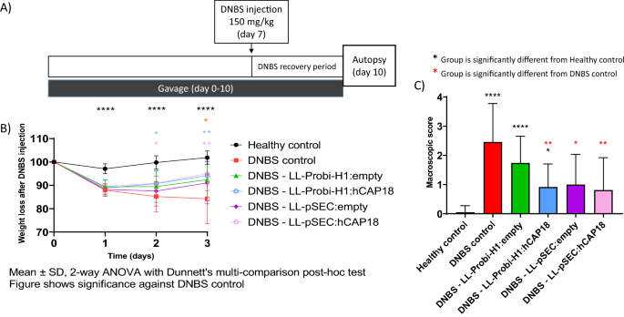
transfection into HEK293 cells (data not shown). We then assessed the effects of the LL-ProBi-H1:empty or LL-Probi-H1:hCAP18 recombinant strains following 7 days of gavage in a mouse model
of acute DNBS-induced colitis (Fig. 1A). One day after DNBS injection, all groups of mice (DNBS control, LL-ProBi-H1:empty, and LL-Probi-H1:hCAP18) had lost 10% of their body weight; three
days after injection, mice in the DNBS control group had lost 15% of their weight and did not show any signs of recovery. Instead, mice that received treatment with either recombinant strain
stabilized their weight after day 1 and began to recover some of the lost weight (Fig. 1B). Macroscopic scores measured 3 days after DNBS injection were significantly better (2.7-fold) in
mice that received LL-Probi-H1:hCAP18 compared to the DNBS control group (Fig. 1C), while mice that were treated with LL-ProBi-H1:empty showed no statistically significant improvement.
Similarly, DNBS injection caused significant thickening of the bowel wall (0.76 mm), which was alleviated by LL-Probi-H1:hCAP18 (0.59 mm) but not LL-ProBi-H1:empty (Fig. 2A). Bowel length
was likewise affected by DNBS injection (7.34 cm in healthy control versus 6.52 cm in DNBS control) and this effect was alleviated by both LL-ProBi-H1:empty and LL-Probi-H1:hCAP18. To
evaluate the effects of LL-Probi-H1:hCAP18 on neutrophil infiltration, we measured the activity of myeloperoxidase (MPO) and the expression of lipocalin-2 in feces (Fig. 2C,D) DNBS injection
induced a significant increase of MPO activity in LL-Probi-H1:empty, LL-pSEC:empty and LL-pSEC:hCAP18 groups. DNBS induced MPO activity of LL-pSEC:empty treated mice was significantly
higher than non-treated control mice. Compared to healthy controls, concentrations of lipocalin-2 in feces were much higher in all groups that received DNBS injections; however, the group
treated with LL-Probi-H1:hCAP18 had significantly lower lipocalin-2 expression than the DNBS control group (Fig. 2D). LL-PSEC:HCAP18 DOES NOT SIGNIFICANTLY IMPROVE DNBS-INDUCED SYMPTOMS
COMPARED TO LL-PSEC:EMPTY The functionality of the pSEC:hCAP18 plasmid was verified by Western blot following nisin induction of the LL-pSEC:hCAP18 strain. A specific band at 18 kDa was
detected (data not shown). We then used this strain to study how the administration of hCAP18 in protein form differed from the effects of cDNA. Weight recovery was not different between
LL-pSEC:empty and LL-pSEC:hCAP18 mice (Fig. 1B). Both LL-pSEC:hCAP18 and LL-pSEC:empty improved macroscopic score and reduced colon thickness compared to DNBS control (Figs. 1C, 2A). There
were no significant differences among groups with respect to lipocalin-2 concentration in feces and MPO activity (Fig. 2C,D). DELIVERY OF HCAP18 DOES NOT ALLEVIATES DSS-INDUCED SYMPTOMS Mice
were given DSS in drinking water for 6 consecutive days (1 day = 24 h cycle) then sacrificed 12 days after the beginning of DSS administration. We found no significant difference in the
weight loss and Disease Activity Index (calculated with the consistency and presence of blood in feces) in our mice (Fig. 3). No further analysis was performed after this experiment.
PROBI-H1:HCAP18 INCREASES LOCAL IL10, IL17A PRODUCTION AND REDUCES IFN-Γ SECRETION We studied the effects of LL-Probi-H1:hCAP18 on the local immune response by measuring cytokine production
in mesenteric lymph node (MLN) cells from our mice following stimulation with PMA ionomycin or CD3+/CD28+. We observed significantly higher levels of the anti-inflammatory cytokine IL-10 in
the MLN cell supernatant of the LL-Probi-H1:hCAP18 group compared to the DNBS and LL-Probi-H1:empty groups following stimulation with PMA ionomycin (Fig. 4A1,A2). Furthermore, following
reactivation with either method, IL17A production was high in the MLN cell supernatants from the LL-Probi-H1:hCAP18 group, suggesting an increased Th17 effector cell response (Fig. 4B1,B2).
Finally, the PMA ionomycin–treated MLN cell supernatants from the LL-Probi-H1:empty group demonstrated high levels of the pro-inflammatory cytokine IFN-γ (Fig. 4C1,C2). We also studied the
expression of TNF-α, IL17A, and IL22 mRNA in colon tissues and found no significant difference between our treatment groups (Fig. S1). COMPARISON OF FECAL MICROBIOTA COMPOSITION AMONG
TREATMENT GROUPS We analyzed fecal microbiota composition by 16S sequencing after 7 days of gavage with PBS glycerol and LL-ProBiH1:empty or LL-ProBiH1:hCAP18. No significant difference in
alpha diversity was found among groups (observed or Shannon index), although mice that received recombinant _L. lactis_ had slightly decreased diversity compared with the PBS glycerol
control group. With respect to the composition of the fecal microbiota, we detected a slight shift in mice that received LL-Probi-H1:empty or LL-Probi-H1:hCAP18 compared to control mice; _L.
lactis_ treatment was found to be responsible for a portion of the variation in the data (PERMANOVA, R2 = 0.114537, F = 1.8079, p = 0.005, see Fig. 5B, Table S1). However, there was no
relationship between patterns of community composition and macroscopic score (PERMANOVA, R2 = 0.16878, F = 1.0152, p = 0.424, see Table S2). We examined a total of 704 OTUs in 31 total
samples; these represented 5 phyla (Fig. 5A), 8 classes, 23 orders, 33 families, and 77 identified genera. Patterns of differential abundance were evaluated using the DESeq2 package in R.
Compared to the control PBS glycerol group, several taxa were differentially abundant in the LL-Probi-H1:hCAP18 group (9 families, 13 OTUs) and LL-Probi-H1:empty group (4 families, 6 OTUs).
In the former comparison, the taxa affected were identified as Muribaculaceae (5 OTUs), Ruminococcaceae (2 OTUs), and Lachnospiraceae (1 OTU) (Fig. 5D, Table S3), while the taxa highlighted
in the latter comparison were assigned to Muribaculaceae (1 OTU), Ruminococcaceae (1 OTU), Clostridia (1 OTU), and Lachnospiraceae (3 OTUs) (Fig. 5C). However, only 1 OTU, from the
Muribaculaceae family (cluster_116), was highlighted by both contrasts (Table S3). Instead, a comparison of the LL-Probi-H1:empty and LL-Probi-H1:hCAP18 groups revealed no patterns of
differential abundance. EFFECTS ON DNBS-INDUCED COLITIS OF FECAL TRANSPLANT FROM MICE TREATED WITH LL-PROBI-H1:EMPTY OR LL-PROBI-H1:HCAP18 In order to evaluate whether, and to what extent,
the protective effects we detected were mediated by shifts in the microbiota, we performed fecal transplants from donor mice who were gavaged with PBS glycerol and either LL-ProBi-H1:empty
or LL-ProBi-H1:hCAP18. Recipient mice were first subjected to colon washes with PEG and then received oral gavages of fecal material from donor mice following the protocol shown in Fig. 6A.
After transplantation, the recipient mice were injected with DNBS in order to induce colitis. There were no significant differences between control and treatment groups with respect to
symptoms such as weight loss (Fig. 6B), colon thickness (Fig. 6C) or macroscopic score (Fig. 6D). There were also no differences in MPO activity (Fig. 6E) among the groups. DISCUSSION In our
study, we investigated the protective effect of human cathelicidin (hCAP18) in DNBS induced colitis, using _L. lactis_ as a vector for the delivery of either the protein or cDNA forms of
this molecule. _L. lactis_ is a food-grade lactic acid bacterium that is considered to be non-invasive and non-colonizing, and has previously demonstrated potential for this type of
treatment. For example, after 7 days of DSS-induced colitis in mice, the administration of 10 log cfu of _L. lactis_ that was engineered to express mCRAMP under the control of nisin was
found to reduce clinical symptoms and tissue damage better than sulfasalazine49 and was associated with lower expression of TNF-α and IL-1β. Another team found similar results with reduction
of pro-inflammatory cytokines IL-6, IL-1β, TNF-α, increased production of IL-10 and upregulation of tight junction proteins ZO-1, ZO-2 and occludin50. Here, we observed that treatment with
the recombinant strain LL-Probi-H1:hCAP18 reduced DNBS-induced colitis symptoms in mice. Interestingly, when we repeated the experiment in a DSS-induced model of colitis we found no
significant effect on symptoms induced by DSS (Fig. 3). The discrepancy between the results of our DSS experiment and this previous work may be explained by the different protocols used:
unlike the earlier study, we included a recovery period of 5 days after DSS treatment and used a cathelicidin of human origin. However, DSS acts as a toxin against the gut epithelium and
induces injury51 whereas ethanol-induced permeability of the gut epithelium will allow DNBS to penetrate the colonic tissues and trigger the host’s innate and adaptive immune responses52.
This difference in mechanism could partly explain why hCAP18 is inefficient in DSS-induced colitis. Further investigation is needed to fully address this issue. One of our main goals was to
compare how the effects of hCAP18 differ depending on its mode of delivery: exogenous delivery (LL-pSEC:hCAP18) or endogenous production after delivery via DNA vaccine (LL-Probi-H1:hCAP18).
We first verified that the LL-pSEC:hCAP18 strain was able to produce hCAP18 and that Probi-H1:hCAP18 could induce hCAP18 production in mammal cells using Western blot analysis (Fig. S2). We
then characterized the effects of both strains in mice with DNBS-induced colitis. Treatment with LL-Probi-H1:hCAP18 improved disease symptoms compared to DNBS control (see Fig. 1 and 2;
weight recovery, bowel length, bowel thickness, and macroscopic tissue damage). The administration of LL-Probi-H1:empty also had a positive effect on weight recovery and bowel length;
however, compared to healthy controls, expression of the pro-inflammatory cytokine IFN-γ was significantly higher in the MLN cells of mice in this group. Treatment with LL-Probi-H1:hCAP18
restored IFN-γ to normal levels (Fig. 4C1). Symptoms in the mice treated with LL-pSEC:empty or LL-pSEC:hCAP18 were also significantly ameliorated compared to the DNBS control group, but
there was no difference between these two treatment groups (Figs. 1 and 2). It thus appeared that _L. lactis_ by itself had protective properties toward DNBS-induced symptoms that could mask
the effects of the hCAP18 it produced. This was unexpected as our previous experiments with this _L. lactis_ strain found this bacterium could exacerbate DNBS-induced colitis weight loss
and had no positive effects on the macroscopic score53. However, a multitude of _L. lactis_ strains have protective effects in colitis models54,55,56 and it is possible that yet unidentified
variables played a role in the exacerbation of DNBS-induced colitis previously observed. We note that despite the decrease in weight loss and tissue damage, _L. lactis_ still increased the
production of the pro-inflammatory cytokine IFN-γ, which is consistent with our previous findings57. Additionally, we observed that endogenous production of hCAP18, as induced by DNA vaccine
delivery, strengthened the protective effects of this bacterium; mice treated with LL-Probi-H1:hCAP18 were more protected from developing symptoms of colitis than those who were treated
with LL-pSEC:hCAP18. This result may be due to differences in the final processing step of hCAP18. As noted earlier, in order to produce the antimicrobial peptide LL37, hCAP18 must be
processed by proteinase 3, which is mainly produced and secreted by neutrophils13. We can hypothesize that hCAP18 delivered into the lumen by LL-pSEC:hCAP18 is less accessible to proteinase
3 than hCAP18 produced by epithelial cells after delivery by LL-Probi-H1:hCAP18. Although this point remains to be clarified, our results confirm the potential of _L. lactis_ as a vehicle
for the production of therapeutic molecules via DNA vaccine delivery. Treatment with LL-Probi-H1:hCAP18 was also associated with elevated levels of the anti-inflammatory cytokine IL10. IL10
is an important regulator of intestinal homeostasis and prevents pro-inflammatory responses by T-cells58 and macrophages59. Surprisingly, we also found increased levels of putatively
proinflammatory IL17A in MLNs of this group. High levels of this cytokine have been found in patients with an active form of ulcerative colitis60, but the beneficial or detrimental nature of
its effects are still up for debate. IL17 KO mice subjected to DSS-induced colitis show increased survival and reduced tissue damage compared to wild-type controls58. Anti-IL17 treatments
such as Ixekizumab, Brodalumab, or Secukinumab are FDA-approved and widely used in the treatment of psoriasis61 but, paradoxically, the use of IL17 blockers to treat IBD has been shown to
exacerbate symptoms62,63,64. Similarly, the use of anti-IL17 monoclonal antibodies was found to exacerbate DSS-induced colitis in mice and increase the expression of pro-inflammatory markers
such as TNF-α, IFN-γ, and IL-665. Other research has reported an association in colitis between IL17 and preservation of epithelial barrier integrity through stimulation of cell
proliferation and AMP production66. A 2020 study suggested that IL17A deficiency in chronic colitis upregulates IL6 expression and leads to the recruitment of RORγt + innate lymphoid cells
in a negative feedback loop; the authors proposed this mechanism to explain the discrepancies between the insensitivity of IL17A-KO mice to acute colitis and the otherwise negative
correlation between IL17A and worsening symptoms67. However, increased IL17A levels are not necessarily associated with worse symptoms in all models of acute colitis; its effects are heavily
dependent on the surrounding circumstances. For example, IL10−/− mice spontaneously develop colitis that is greatly aggravated by IL17A deficiency68, while Minns et al. found that mCRAMP is
required for Th17 differentiation and reported increased production of IL17A in the context of inflammation69. In order to investigate the importance of the anti-microbial activity of
hCAP18 in the protective effects noted here, we studied how an increase in host production of hCAP18, and potentially LL37, affected the composition of the gut microbiota. Previous findings
suggested that mCRAMP deficiency modified the microbiota in such a way as to make wild-type cage mates of mCRAMP−/− mice more susceptible to DSS-induced colitis34. We characterized the
microbiota of mice using 16S sequencing of fecal samples and observed a slight but significant shift in community composition in LL-Probi-H1:hCAP18-treated mice compared to the control
group. Multiple OTUs assigned to family _Muribaculaceae_ increased in abundance, whereas certain members of _Lachnospiraceae_ declined (Fig. 4). However, we must keep in mind that the
sequencing method we used only gives us a global view of the microbiota composition, and it is difficult to determine if such minor changes are relevant. For this reason, we also tested if
the microbiota from treated mice could protect untreated recipient mice from inflammation. We found that fecal transplantation from LL-Probi-H1:hCAP18–treated mice to PEG-washed recipient
mice did not significantly reduce symptoms of DNBS-induced colitis (Fig. 5). One potential explanation of this result is that these wild-type mice had normal expression of mCRAMP during
inflammation, and additional cathelicidin might not have changed the microbiota in a helpful way. Although, it is possible that the antimicrobial effects of hCAP18 are specific to
human-associated microbiota and therefore would not have much impact on mice-associated species. To fully understand the effects of LL37 on the microbiota, further investigation should be
done on a local scale by analyzing the composition of the mucosal microbiota. MATERIALS AND METHODS PLASMID CLONING AND _L. LACTIS_ TRANSFORMATION hCAP18 synthetic cDNA optimized for _L.
lactis_ expression was cloned into a pSEC plasmid as described in70 to create pSEC:hCAP18; this was then transformed in _L. lactis_ NZ9000 to create the recombinant strain LL-pSEC:hCAP18, in
which the induction of hCAP18 was nisin-dependent. hCAP18 synthetic cDNA optimized for expression in mice was cloned into the proBi-H1 plasmid to create proBi-H1:hCAP18; this plasmid vector
contained the repA and repC origin of replication, the chloramphenicol resistance gene, and the expression cassette from pcDNA3 (Invitrogen) as published before71. The resulting vector was
3.7 kb long. The proBi-H1:hCAP18 plasmid was transformed in _Lactococcus lactis_ MG1363 to create the recombinant strain LL-ProBi-H1:hCAP18. BACTERIAL STRAIN PREPARATION LL-pSEC bacteria
were preincubated at 30 °C overnight in M17 broth medium with 0.5% glucose and chloramphenicol (10 µg/mL). The optical density (OD) of the initial culture was 0.2; nisin (10 ng/mL) was added
1 h after the start of incubation and bacteria were incubated for another 1.5 h (OD = 1.2). Bacteria were harvested by centrifugation at 8000×_g_ for 5 min at 4 °C and washed 3 times with
PBS. They were concentrated 5 times in PBS with 16% glycerol and frozen until gavage. LL-Probi-H1 bacteria were preincubated in the same conditions; initial culture OD was 0.2 and culturing
ended at OD 1.7–1.8. Bacteria were harvested by centrifugation at 8000×_g_ for 5 min at 4 °C and washed 3 times with PBS. They were concentrated 10 times in PBS with 16% glycerol and 109 cfu
of bacteria were administered orally to mice every day. Cell counts were quantified on M17 with 0.5% glucose (10 µg/mL of chloramphenicol) agar plates to assess viable cfu concentrations.
HEK293 CULTURE CELL FOR PROTEIN EXPRESSION OF PLASMID Probi-H1:hCAP18 functionality was checked by transfection in HEK293 cells with Probi-H1:empty as control. The HEK293 cell line came from
the American Type Culture Collection (ATCC). Cells were grown in Dulbecco’s modified Eagle’s medium + DMEM with GlutaMAX (Gibco) supplemented with 10% heat-inactivated fetal bovine serum
(Eurobio Scientific), 1% sodium pyruvate (Gibco), and 1% streptomycin/penicillin (Sigma-Aldrich), and incubated at 37 °C in a humidified atmosphere with 10% CO2. Cells were passed at 80%
confluence and seeded in 24-well plates at 8*10^4 cells/well, then incubated for 24 h. Transfection of 50 ng of plasmids was performed with Lipofectamine LTX with Plus Reagent (Invitrogen)
in opti-MEM medium (Gibco, Life Technologies). HEK293 cells were co-incubated with their plasmid mix for 4 h after which the medium was replaced with complete medium. After 24 h of
incubation, the supernatant was removed and cells were lysed in Passive Lysis Buffer 1X (Promega). Protein expression was then checked by western blot. PROTEIN EXTRACTION FROM _LACTOCOCCUS
LACTIS_ To control the expression of hCAP18 by LL-pSEC:hCAP18, bacteria were incubated overnight in 10 mL of broth medium and harvested by centrifugation at 8000×_g_ for 5 min at 4 °C.
Bacteria were physically lysed with Precellys in cOmplete Protease Inhibitor Cocktail (Roche) and cell debris was removed by centrifugation at 8000×_g_ for 5 min. Protein expression was then
checked by western blot. WESTERN BLOT Protein extracts were boiled at 95 °C for 5 min in Laemmli buffer. Gel electrophoresis was performed with 4–20% Mini-PROTEAN TGX Precast Protein Gels
(Bio-Rad) and PageRuler Prestained Protein Ladder (Thermoscientific). Proteins were then transferred on membrane using Trans-Blot Turbo Transfer Pack (Bio-Rad). Nitrocellulose membrane were
saturated in tris-buffered saline, 0.1% tween, 5% dry milk. Primary antibody was the monoclonal LL-37/CAP-18, Human, mAb 3D11 (Hycult Biotech) and secondary antibody was goat-produced
Anti‑Mouse IgM (µ) Affinity purified antibody HRP. The substrate mixture used was the Clarity Western ECL Substrate (Bio-Rad). Digital imaging was performed on the ChemiDoc MP System.
EXPERIMENTAL CONDITIONS FOR MICE Male C57BL/6JRj mice (6 weeks old) were purchased from Janvier Labs (France) and kept at the INRAE animal facility in accordance with institutional ethical
guidelines. The study was approved by the COMETHEA ethics committee (“Comité d’Ethique en Expérimentation Animale”) of the Centre INRAE of Jouy-en-Josas and AgroParisTech
(n°16744-201807061805486). It was reported in accordance with the ARRIVE guidelines. They were fed ad libitum, given autoclaved tap water, and housed in groups of four mice per cage in an
air-conditioned room with controlled temperature and circadian cycle (12 h light, 12 h dark). _LACTOCOCCUS LACTIS_ ADMINISTRATION AND INDUCTION OF COLITIS Colitis was induced by a single
intrarectal injection of DNBS (150 mg/kg of body weight) dissolved in 30% ethanol. Mice were anesthetized prior to injection. Control mice were injected with 30% ethanol. Mice were monitored
daily for weight loss after intrarectal injection. For the DNBS experiments, mice were divided into six different groups: healthy control, DNBS control, DNBS + 109 cfu LL-pSEC:empty (nisin
induction), DNBS + 109 cfu LL-ProBiH1:empty (DNA delivery system), DNBS + 109 cfu LL-pSEC:hCAP18, or DNBS + 109 cfu LL-ProBiH1:hCAP18. LL-pSEC strains were tested 2 × 8 mices replicates and
LL-ProBiH1 strains in 3 × 8 replicates. Bacteria were administered daily by intragastric gavage for 7 days prior to intrarectal injection of DNBS. Gavages continued until the end of the
experiment, 3 days after DNBS injection. After DNBS injections, mice were monitored daily for weight loss. Mice were sacrificed by cervical dislocation at day 3 after injection, the abdomen
was opened by midline incision, the colon and Mesenteric Lymphatic Node (MLN) were removed. MLN were stocked in DMEM medium on ice. They were later mashed and counted before stimulation with
antiCD3 and anti-CD28 antibodies for 48 h at 37 °C and 10% CO2. Colons were opened longitudinally, cleaned and measured for scoring. The _L. lactis_ treatments on DSS-induced colitis were
done in two separate experiments of 8 mice per group. We added 2% dextran sulfate sodium salt (MPBio) in the drinking water for 6 consecutive days (1 day = 24 h hours). Mice were sacrificed
by cervical dislocation at day 13 after DSS induction. Mice were monitored daily for weight loss and Disease Activity Index (DAI) which was evaluated following the protocol by Cooper et
al.72. PEG WASH AND FECAL TRANSPLANTATION In the fecal transplantation group, mice were fasted and the cage litter was changed 1 h before colon washing, in which mice were fed 200 µL
polyethylene glycol (PEG) four consecutive times, with a 20-min interval between feedings, as described in73. They were then fed 200 µL fecal content by intragastric gavage 3 h after the
final PEG dose. Fecal content was given to mice twice a week for 3 weeks after the initial PEG wash. MACROSCOPIC SCORING AND HISTOLOGIC SAMPLING Macroscopic tissue damage was assessed the
day of autopsy using the Wallace score74. Macroscopic criteria for scoring include the presence of adhesions between the colon and other organs, colon wall thickening, gastrointestinal
transit issues, ulcers and hyperemia. Colon thickness was measured at the anal extremity and colon length was measured between the anal extremity and caecal junction. CHEMOKINES SECRETION BY
STIMULATED LYMPHOCYTES Mesenteric Lymph Nodes (MLN) were taken from mice during autopsy. They were crushed and filtered (70 µm, BD biosciences) in DMEM (Gibco). Lymphocytes were counted by
flow cytometry (Accuri C6, Dutscher) and suspended in DMEM with 100 Unit of Streptomycin, Penicillin (Sigma) and 10% Fetal Calf Serum (FCS) (Eurobio) at a concentration of 2.5 × 10⁵ cells/mL
either in 24 wells plate (Costar) pre-incubated with anti-CD3 and anti-CD28 antibodies, 4 µg/mL of each antibody (eBioscience) in PBS or stimulated with PMA ionomycin (eBioscience). Plates
were incubated 48 h at 37 °C, 10% of CO2 and cytokine level was assessed by ELISA MAX Standard Set Mouse (BioLegend). The cytokines tested were Th1-related cytokine (IFNγ); Th17-related
cytokine (IL17) and Treg– related cytokine (IL10). MYELOPEROXIDASE ACTIVITY IN COLON Colon samples were homogenized using a Precellys Evolution (Bertin Instruments) in a
hexadecyltrimethylammonium bromide buffer (Sigma-Aldrich). MPO was detected with O-dianisidine solution (Sigma-Aldrich) with 1% hydrogen peroxide solution (Sigma-Adrich) added just before
measurement. OD was measured using Infinite M200 PRO (TECAN) and normalized to colon mass. MRNA EXTRACTION AND QPCR RNA extraction was performed with the RNeasy Mini Kit (Qiagen) according
to the manufacturer’s recommendations, with the use of the RNase-Free DNase Set (Qiagen). Reverse transcription was performed with an High-Capacity cDNA Reverse Transcription Kit (Applied
Biosystems). Real-Time PCR was performed with SYBR MasterMix dTTP Blue (Takyon Rox) with the primers and their source described in Table 1. Primers were synthetized by Eurofins Scientific.
DNA EXTRACTION AND 16S DNA SEQUENCING DNA was extracted using the protocol of Godon et al.75. DNA concentration and purity were determined using a NanoDrop instrument. We used the KAPA HiFi
HotStart ReadyMix with 10 ng of extracted DNA for PCR amplification of the V3–V4 region of 16S rDNA. The amplification was performed with the primers MSQ-16SV3F
(CTTTCCCTACACGACGCTCTTCCGATCTACGGRAGGCWGCAG) and MSQ-16SV4R (GGAGTTCAGACGTGTGCTCTTCCGATCTTACCAGGGTATCTAATCCT). PCR conditions were: 95 °C for 3 min; 30 cycles at 98 °C for 20 s, 65 °C for 30
s, and 72 °C for 1 min; and a final extension of 72 °C for 10 min. Amplification products were checked via electrophoresis on 2% agarose gel. Band size was approximately 500 bp. Sequencing
was performed at the @BRIDGe facility at INRA Jouy-en-Josas using the Illumina Miseq (250 × 2 bp) system. STATISTICAL ANALYSIS OF SEQUENCING DATA 16S sequencing data were uploaded to the
Galaxy platform76,77 at https://galaxy.migale.inra.fr/. We used the FROGS pipeline to produce abundance tables of operational taxonomic units (OTUs). Denoising and clustering were performed
with SWARM, chimera removal with VSEARCH, and affiliation with RDP Classifier using the 16S_SILVA_Pintail100_138 database. OTUs with a minimum presence of 0.005% in all sequences were kept.
OTUs were filtered for a minimum identity of 97% and a minimum coverage of 95%. Measures were calculated of α- (observed and Shannon index) and β-diversity (Bray–Curtis dissimilarity score).
Analysis of differential expression was performed using RStudio version 3.6.1 with the DESeq2 package. STATISTICAL ANALYSIS OF MACROSCOPIC SYMPTOMS AND INFLAMMATORY MARKERS Graphics were
created in Prism-GraphPad. Macroscopic results are represented as mean ± standard deviation (SD). Data was checked for normal distribution with the Shapiro–Wilk test. Outliers were removed
from normally distributed data using the ROUT method (Q = 1%). All histograms were analyzed using non-parametric one-way ANOVA (Kruskal–Wallis) with Dunn’s post-hoc test. ELISA and qPCR
results were represented in Tukey boxplots. Two-way analysis of variance (ANOVA) with Turkey’s post-hoc test was performed on weight or DAI progression data if no values were missing due to
protocol interruption. If there were missing values, two-way ANOVA with Dunnett's multi-comparison post-hoc test was performed. In all figures, *P < 0.05, **P < 0.01, and ***P
< 0.001. ETHICS APPROVAL This study was carried out in accordance with the guidelines of the local ethics committee. DATA AVAILABILITY
https://data.inrae.fr/dataset.xhtml?persistentId=doi:10.15454/CJ26OO. REFERENCES * Khan, I. _et al._ Alteration of gut microbiota in inflammatory bowel disease (IBD): Cause or consequence?
IBD treatment targeting the gut microbiome. _Pathogens_ 8, 126 (2019). Article CAS PubMed Central Google Scholar * Nishida, A. _et al._ Gut microbiota in the pathogenesis of inflammatory
bowel disease. _Clin. J. Gastroenterol._ 11, 1–10 (2018). Article PubMed Google Scholar * Zhang, M. _et al._ Interactions between intestinal microbiota and host immune response in
inflammatory bowel disease. _Front. Immunol._ 8, 1 (2017). Article Google Scholar * Kahlenberg, J. M. & Kaplan, M. J. Little peptide, big effects: The role of LL-37 in inflammation and
autoimmune disease. _J. Immunol._ 191, 4895–4901 (2013). Article CAS PubMed Google Scholar * Hancock, R. E. W. & Falla, T. J. Antimicrobial peptides: Broad-spectrum antibiotics from
nature the cationic peptides of nature. _Clin. Microbiol. Infect._ 1, 226–229 (1996). Article CAS PubMed Google Scholar * Diamond, G., Beckloff, N., Weinberg, A. & Kisich, K. The
roles of antimicrobial peptides in innate host defense. _Curr. Pharm. Des._ 15, 2377–2392 (2009). Article CAS PubMed PubMed Central Google Scholar * Niyonsaba, F. _et al._ Antimicrobial
peptides human β-defensins stimulate epidermal keratinocyte migration, proliferation and production of proinflammatory cytokines and chemokines. _J. Invest. Dermatol._ 127, 594–604 (2007).
Article CAS PubMed Google Scholar * Oppenheim, J. J., Biragyn, A., Kwak, L. W. & Yang, D. Roles of antimicrobial peptides such as defensins in innate and adaptive immunity. _Ann.
Rheum. Dis._ 62, 17–21 (2003). Article Google Scholar * Ho, S., Pothoulakis, C. & Wai Koon, H. Antimicrobial peptides and colitis. _Curr. Pharm. Des._ 19, 40–47 (2012). Google Scholar
* Maeda, T. _et al._ Low concentrations of human neutrophil peptide ameliorate experimental murine colitis. _Int. J. Mol. Med._ 38, 1777–1785 (2016). Article CAS PubMed PubMed Central
Google Scholar * Tran, D.H.-N. _et al._ Circulating cathelicidin levels correlate with mucosal disease activity in ulcerative colitis, risk of intestinal stricture in Crohn’s disease, and
clinical prognosis in inflammatory bowel disease. _BMC Gastroenterol._ 17, 63 (2017). Article PubMed PubMed Central CAS Google Scholar * Mwangi, J., Hao, X., Lai, R. & Zhang, Z. Y.
Antimicrobial peptides: New hope in the war against multidrug resistance. _Zool. Res._ 40, 488–505 (2019). Article PubMed PubMed Central Google Scholar * Wang, S., Zeng, X., Yang, Q.
& Qiao, S. Antimicrobial peptides as potential alternatives to antibiotics in food animal industry. _Int. J. Mol. Sci._ 17, 226–229 (2016). ADS CAS Google Scholar * Alford, M. A.,
Baquir, B., Santana, F. L., Haney, E. F. & Hancock, R. E. W. Cathelicidin host defense peptides and inflammatory signaling: Striking a balance. _Front. Microbiol._ 11, 1902 (2020).
Article PubMed PubMed Central Google Scholar * Cheng, Y. _et al._ Evolution of the avian β-defensin and cathelicidin genes. _BMC Evol. Biol._ 15, 1–17 (2015). Article CAS Google
Scholar * Huynh, E., Penney, J., Caswell, J. & Li, J. Protective effects of protegrin in dextran sodium sulfate-induced murine colitis. _Front. Pharmacol._ 10, 156 (2019). Article CAS
PubMed PubMed Central Google Scholar * Wu, J. _et al._ A frog cathelicidin peptide effectively promotes cutaneous wound healing in mice. _Biochem. J._ 475, 2785–2799 (2018). Article
CAS PubMed Google Scholar * Grönberg, A., Mahlapuu, M., Ståhle, M., Whately-Smith, C. & Rollman, O. Treatment with LL-37 is safe and effective in enhancing healing of hard-to-heal
venous leg ulcers: A randomized, placebo-controlled clinical trial. _Wound Repair Regen._ 22, 613–621 (2014). Article PubMed Google Scholar * Sharifi, A., Hosseinzadeh-Attar, M. J.,
Vahedi, H. & Nedjat, S. A randomized controlled trial on the effect of vitamin D3 on inflammation and cathelicidin gene expression in ulcerative colitis patients. _Saudi J.
Gastroenterol._ 22, 316–323 (2016). Article PubMed PubMed Central Google Scholar * Sun, L., Wang, W., Xiao, W. & Yang, H. The roles of cathelicidin LL-37 in inflammatory bowel
disease. _Inflamm. Bowel Dis._ 22, 1986–1991 (2016). Article PubMed Google Scholar * Sørensen, O. E. _et al._ Human cathelicidin, hCAP-18, is processed to the antimicrobial peptide LL-37
by extracellular cleavage with proteinase 3. _Blood_ 97, 3951–3959 (2001). Article PubMed Google Scholar * Thennarasu, S. _et al._ Antimicrobial and membrane disrupting activities of a
peptide derived from the human cathelicidin antimicrobial peptide ll37. _Biophys. J._ 98, 248–257 (2010). Article ADS CAS PubMed PubMed Central Google Scholar * Braff, M. H., Zaiou,
M., Fierer, J., Nizet, V. & Gallo, R. L. Keratinocyte production of cathelicidin provides direct activity against bacterial skin pathogens. _Infect. Immun._ 73, 6771–6781 (2005). Article
CAS PubMed PubMed Central Google Scholar * Hiemstra, P. S., McCray, P. B. & Bals, R. The innate immune function of airway epithelial cells in inflammatory lung disease. _Eur.
Respir. J._ 45, 1150–1162 (2015). Article CAS PubMed PubMed Central Google Scholar * Gordon, Y. J. _et al._ Human cathelicidin (LL-37), a multifunctional peptide, is expressed by ocular
surface epithelia and has potent antibacterial and antiviral activity. _Curr. Eye Res._ 30, 385–394 (2005). Article CAS PubMed PubMed Central Google Scholar * De Yang, B. _et al._
LL-37, the neutrophil granule- and epithelial cell-derived cathelicidin, utilizes formyl peptide receptor-like 1 (FPRL1) as a receptor to chemoattract human peripheral blood neutrophils,
monocytes, and T cells. _J. Exp. Med._ 192, 1069–1074 (2000). Article CAS PubMed PubMed Central Google Scholar * Yoo, J. H. _et al._ Antifibrogenic effects of the antimicrobial peptide
cathelicidin in murine colitis-associated fibrosis. _Cell. Mol. Gastroenterol. Hepatol._ 1, 55–74 (2015). Article PubMed Google Scholar * Park, H. J. _et al._ Collagen synthesis is
suppressed in dermal fibroblasts by the human antimicrobial peptide LL-37. _J. Invest. Dermatol._ 129, 843–850 (2009). Article CAS PubMed Google Scholar * Fan, T. J. _et al._
Environmental factors modify the severity of acute DSS colitis in caspase-11-deficient mice. _Inflamm. Bowel Dis._ 24, 2394–2403 (2018). Article PubMed PubMed Central Google Scholar *
Bilski, J. _et al._ Can exercise affect the course of inflammatory bowel disease? Experimental and clinical evidence. _Pharmacol. Rep._ 68, 827–836 (2016). Article PubMed Google Scholar *
Sideri, A. _et al._ Effects of obesity on severity of colitis and cytokine expression in mouse mesenteric fat: Potential role of adiponectin receptor 1. _Am. J. Physiol. Liver Physiol._
308, G591–G604 (2015). CAS Google Scholar * Llewellyn, S. R. _et al._ Interactions between diet and the intestinal microbiota alter intestinal permeability and colitis severity in mice.
_Gastroenterology_ 154, 1037-1046.e2 (2018). Article PubMed Google Scholar * Munyaka, P. M., Eissa, N., Bernstein, C. N., Khafipour, E. & Ghia, J. E. Antepartum antibiotic treatment
increases offspring susceptibility to experimental colitis: A role of the gut microbiota. _PLoS ONE_ 10, e0142536 (2015). Article PubMed PubMed Central CAS Google Scholar * Yoshimura,
T. _et al._ The antimicrobial peptide CRAMP is essential for colon homeostasis by maintaining microbiota balance. _J. Immunol._ 200, 2174–2185 (2018). Article CAS PubMed Google Scholar *
Singh, D., Qi, R., Jordan, J. L., Mateo, L. S. & Kao, C. C. The human antimicrobial peptide LL-37, but not the mouse ortholog, mCRAMP, can stimulate signaling by poly(I:C) through a
FPRL1-dependent pathway. _J. Biol. Chem._ 288, 8258–8268 (2013). Article CAS PubMed PubMed Central Google Scholar * Hu, Z. _et al._ Antimicrobial cathelicidin peptide LL-37 inhibits the
pyroptosis of macrophages and improves the survival of polybacterial septic mice. _Int. Immunol._ 28, 245–253 (2016). Article CAS PubMed PubMed Central Google Scholar * Wei, X. _et
al._ A novel cecropin-LL37 hybrid peptide protects mice against EHEC infection-mediated changes in gut microbiota, intestinal inflammation, and impairment of mucosal barrier functions.
_Front. Immunol._ 11, 1361 (2020). Article CAS PubMed PubMed Central Google Scholar * Mandal, S. M., Silva, O. N. & Franco, O. L. Recombinant probiotics with antimicrobial peptides:
A dual strategy to improve immune response in immunocompromised patients. _Drug Discov. Today_ 19, 1045–1050 (2014). Article CAS PubMed Google Scholar * De Azevedo, M. _et al._ In vitro
and in vivo characterization of DNA delivery using recombinant _Lactococcus lactis_ expressing a mutated form of _L. monocytogenes_ internalin A. _BMC Microbiol._ 12, 299 (2012). Article
PubMed PubMed Central CAS Google Scholar * Lilly, D. M. & Stillwell, R. H. Probiotics: Growth-promoting factors produced by microorganisms. _Science_ 147, 747–748 (1965). Article
ADS CAS PubMed Google Scholar * Carvalho, R. D. D. O. _et al._ Use of wild type or recombinant lactic acid bacteria as an alternative treatment for gastrointestinal inflammatory
diseases: A focus on inflammatory bowel diseases and mucositis. _Front. Microbiol._ 8, 800 (2017). Article PubMed PubMed Central Google Scholar * Tavares, L. M. _et al._ Novel strategies
for efficient production and delivery of live biotherapeutics and biotechnological uses of _Lactococcus lactis_: The lactic acid bacterium model. _Front. Bioeng. Biotechnol._ 8, 517166
(2020). Article PubMed PubMed Central Google Scholar * Bermúdez-Humarán, L. G. _et al._ Effects of intranasal administration of a leptin-secreting _Lactococcus lactis_ recombinant on
food intake, body weight, and immune response of mice. _Appl. Environ. Microbiol._ 73, 5300–5307 (2007). Article ADS PubMed PubMed Central CAS Google Scholar * Bermúdez-Humarán, L. G.
_et al._ Engineering lactococci and lactobacilli for human health. _Curr. Opin. Microbiol._ 16, 278–283 (2013). Article PubMed CAS Google Scholar * Guimarâes, V. D. _et al._ Use of
native lactococci as vehicles for delivery of DNA into mammalian epithelial cells. _Appl. Environ. Microbiol._ 72, 7091–7097 (2006). Article ADS PubMed PubMed Central CAS Google Scholar
* de Azevedo, M. _et al._ Recombinant invasive _Lactococcus lactis_ can transfer DNA vaccines either directly to dendritic cells or across an epithelial cell monolayer. _Vaccine_ 33,
4807–4812 (2015). Article PubMed CAS Google Scholar * Mancha-Agresti, P. _et al._ Recombinant invasive _Lactococcus lactis_ carrying a DNA vaccine coding the Ag85A antigen increases
INF-γ, IL-6, and TNF-α cytokines after intranasal immunization. _Front. Microbiol._ 8, 1263 (2017). Article PubMed PubMed Central Google Scholar * Chatel, J. M. _et al._ In vivo transfer
of plasmid from food-grade transiting lactococci to murine epithelial cells. _Gene Ther._ 15, 1184–1190 (2008). Article CAS PubMed Google Scholar * Wong, C. C. M. _et al._ Protective
effects of cathelicidin-encoding _Lactococcus lactis_ in murine ulcerative colitis. _J. Gastroenterol. Hepatol._ 27, 1205–1212 (2012). Article CAS PubMed Google Scholar * Li, J. _et al._
Recombinant CRAMP-producing _Lactococcus lactis_ attenuates dextran sulfate sodium-induced colitis by colonic colonization and inhibiting p38/NF-κB signaling. _Food Nutr. Res._ 65, 1–11
(2021). Article Google Scholar * Eichele, D. D. & Kharbanda, K. K. Dextran sodium sulfate colitis murine model: An indispensable tool for advancing our understanding of inflammatory
bowel diseases pathogenesis. _World J. Gastroenterol._ 23, 6016–6029 (2017). Article CAS PubMed PubMed Central Google Scholar * Morampudi, V. _et al._ DNBS/TNBS colitis models:
providing insights into inflammatory bowel disease and effects of dietary fat. _J. Vis. Exp._ 84, e51297. https://doi.org/10.3791/51297 (2014). Article CAS Google Scholar * Breyner, N. M.
_et al._ Oral delivery of pancreatitis-associated protein by _Lactococcus lactis_ displays protective effects in dinitro-benzenesulfonic-acid-induced colitis model and is able to modulate
the composition of the microbiota. _Environ. Microbiol._ 21, 4020 (2019). Article CAS PubMed PubMed Central Google Scholar * Liu, M. _et al._ Protective effects of a novel probiotic
strain, _Lactococcus lactis_ ML2018, in colitis: in vivo and in vitro evidence. _Food Funct._ 10, 1132–1145 (2019). Article PubMed Google Scholar * Luerce, T. D. _et al._
Anti-inflammatory effects of _Lactococcus lactis_ NCDO 2118 during the remission period of chemically induced colitis. _Gut Pathog._ 6, 33 (2014). Article PubMed PubMed Central Google
Scholar * Nishitani, Y. _et al._ _Lactococcus lactis_ subsp. cremoris FC alleviates symptoms of colitis induced by dextran sulfate sodium in mice. _Int. Immunopharmacol._ 9, 1444–1451
(2009). Article CAS PubMed Google Scholar * Carvalho, R. D. _et al._ Secretion of biologically active pancreatitis-associated protein I (PAP) by genetically modified dairy _Lactococcus
lactis_ NZ9000 in the prevention of intestinal mucositis. _Microb. Cell Fact._ 16, 27 (2017). Article PubMed PubMed Central CAS Google Scholar * Rodrigues, V. F. _et al._ Acute
infection with _Strongyloides venezuelensis_ increases intestine production IL-10, reduces Th1/Th2/Th17 induction in colon and attenuates dextran sulfate sodium-induced colitis in BALB/c
mice. _Cytokine_ 111, 72–83 (2018). Article CAS PubMed Google Scholar * Ip, W. K. E., Hoshi, N., Shouval, D. S., Snapper, S. & Medzhitov, R. Anti-inflammatory effect of IL-10
mediated by metabolic reprogramming of macrophages. _Science_ 356, 513–519 (2017). Article ADS CAS PubMed PubMed Central Google Scholar * Krawiec, P. & Pac-Kożuchowska, E. Serum
interleukin 17A and interleukin 17F in children with inflammatory bowel disease. _Sci. Rep._ 10, 12617 (2020). Article ADS PubMed PubMed Central CAS Google Scholar * Canavan, T. N.,
Elmets, C. A., Cantrell, W. L., Evans, J. M. & Elewski, B. E. Anti-IL-17 medications used in the treatment of plaque psoriasis and psoriatic arthritis: A comprehensive review. _Am. J.
Clin. Dermatol._ 17, 33–47 (2016). Article PubMed Google Scholar * Wang, J. _et al._ Rapid onset of inflammatory bowel disease after receiving secukinumab infusion. _ACG Case Rep. J._ 5,
e56 (2018). Article PubMed PubMed Central Google Scholar * Targan, S. R. _et al._ A randomized, double-blind, placebo-controlled phase 2 study of brodalumab in patients with
moderate-to-severe Crohn’s disease. _Am. J. Gastroenterol._ 111, 1599–1607 (2016). Article ADS CAS PubMed Google Scholar * Hueber, W. _et al._ Secukinumab, a human anti-IL-17A
monoclonal antibody, for moderate to severe Crohn’s disease: Unexpected results of a randomised, double-blindplacebo- controlled trial. _Gut_ 61, 1693–1700 (2012). Article CAS PubMed
Google Scholar * Ogawa, A., Andoh, A., Araki, Y., Bamba, T. & Fujiyama, Y. Neutralization of interleukin-17 aggravates dextran sulfate sodium-induced colitis in mice. _Clin. Immunol._
110, 55–62 (2004). Article CAS PubMed Google Scholar * Whibley, N. & Gaffen, S. L. Gut-busters: IL-17 ain’t afraid of no IL-23. _Immunity_ 43, 620–622 (2015). Article CAS PubMed
PubMed Central Google Scholar * Park, C. H., Lee, A., Ahn, S. B., Eun, C. S. & Han, D. S. Role of innate lymphoid cells in chronic colitis during anti-IL-17A therapy. _Sci. Rep._ 10,
1–11 (2020). CAS Google Scholar * Tachibana, M. _et al._ Ablation of IL-17A leads to severe colitis in IL-10-deficient mice: Implications of myeloid-derived suppressor cells and NO
production. _Int. Immunol._ 32, 187–201 (2019). Article PubMed Central CAS Google Scholar * Minns, D. _et al._ The neutrophil antimicrobial peptide cathelicidin promotes Th17
differentiation. _Nat. Commun._ 12, 1–16 (2021). Article ADS CAS Google Scholar * Miyoshi, A. _et al._ Controlled production of stable heterologous proteins in _Lactococcus lactis_.
_Appl. Environ. Microbiol._ 68, 3141–3146 (2002). Article ADS CAS PubMed PubMed Central Google Scholar * Meynier, M. _et al._ AhR/IL-22 pathway as new target for the treatment of
post-infectious irritable bowel syndrome symptoms. _Gut Microbes_ 14, 2022997 (2022). Article PubMed Central CAS Google Scholar * Cooper, H. S., Murthy, S. N. S., Shah, R. S. &
Sedergran, D. J. Clinicopathologic study of dextran sulfate sodium experimental murine colitis. _Lab. Investig._ 69, 238–250 (1993). CAS PubMed Google Scholar * Wrzosek, L. _et al._
Transplantation of human microbiota into conventional mice durably reshapes the gut microbiota. _OPEN_ 8, 6854 (2018). Google Scholar * Jl, W., Wk, M., Gp, M. & Pl, B. Inhibition of
leukotriene synthesis markedly accelerates healing in a rat model of inflammatory bowel disease. _Gastroenterology_ 96, 29–36 (1989). Article Google Scholar * Godon, J. J., Zumstein, E.,
Dabert, P., Habouzit, F. & Moletta, R. Molecular microbial diversity of an anaerobic digestor as determined by small-subunit rDNA sequence analysis. _Appl. Environ. Microbiol._ 63,
2802–2813 (1997). Article ADS CAS PubMed PubMed Central Google Scholar * Afgan, E. _et al._ The Galaxy platform for accessible, reproducible and collaborative biomedical analyses: 2018
update. _Nucleic Acids Res._ 46, W537–W544 (2018). Article CAS PubMed PubMed Central Google Scholar * Escudié, F. _et al._ FROGS: Find, rapidly, OTUs with galaxy solution.
_Bioinformatics_ 34, 1287–1294 (2018). Article PubMed CAS Google Scholar Download references ACKNOWLEDGEMENTS Colon tissues were sampled and fixed in paraformaldehyde solution and
processed for paraffin embedding. Hematoxylin-eosin staining, paraffin embedding, sample section, and microscopy were performed at the histology facility of UMR 1313 GABI, 78350,
Jouy-en-Josas, France. Sequencing was performed at the @BRIDGe facility at INRA Jouy-en-Josas using the Illumina Miseq (250x2 bp) system. FUNDING Coordenação de Aperfeiçoamento de Pessoal de
Nível Superior (CAPES)—CAPES-COFECUB 720/11. AUTHOR INFORMATION AUTHORS AND AFFILIATIONS * Université Paris Saclay, INRAE, AgroParisTech, UMR1319, MICALIS, 78352, Jouy en Josas, France
Esther Borras Noguès, Camille Kropp, Laureline Bétemps, Cassiana de Sousa, Florian Chain, Sandrine Auger, Philippe Langella & Jean-Marc Chatel * Institute of Biological Sciences, Federal
University of Minas Gerais, Belo-Horizonte, MG, Brazil Cassiana de Sousa & Vasco Azevedo Authors * Esther Borras Noguès View author publications You can also search for this author
inPubMed Google Scholar * Camille Kropp View author publications You can also search for this author inPubMed Google Scholar * Laureline Bétemps View author publications You can also search
for this author inPubMed Google Scholar * Cassiana de Sousa View author publications You can also search for this author inPubMed Google Scholar * Florian Chain View author publications You
can also search for this author inPubMed Google Scholar * Sandrine Auger View author publications You can also search for this author inPubMed Google Scholar * Vasco Azevedo View author
publications You can also search for this author inPubMed Google Scholar * Philippe Langella View author publications You can also search for this author inPubMed Google Scholar * Jean-Marc
Chatel View author publications You can also search for this author inPubMed Google Scholar CONTRIBUTIONS E.B.N. performed the experiments, analyzed the data and drafted the manuscript,
C.K., L.B., C.d.S. and F.C. participated in the experiments, S.A. participated in the analysis, J.-M.C. corrected the manuscript and supervised the experiments, J.-M.C. and P.L. conceived
the study. CORRESPONDING AUTHOR Correspondence to Jean-Marc Chatel. ETHICS DECLARATIONS COMPETING INTERESTS The authors declare no competing interests. ADDITIONAL INFORMATION
PUBLISHER'S NOTE Springer Nature remains neutral with regard to jurisdictional claims in published maps and institutional affiliations. SUPPLEMENTARY INFORMATION SUPPLEMENTARY FIGURES.
SUPPLEMENTARY TABLES. RIGHTS AND PERMISSIONS OPEN ACCESS This article is licensed under a Creative Commons Attribution 4.0 International License, which permits use, sharing, adaptation,
distribution and reproduction in any medium or format, as long as you give appropriate credit to the original author(s) and the source, provide a link to the Creative Commons licence, and
indicate if changes were made. The images or other third party material in this article are included in the article's Creative Commons licence, unless indicated otherwise in a credit
line to the material. If material is not included in the article's Creative Commons licence and your intended use is not permitted by statutory regulation or exceeds the permitted use,
you will need to obtain permission directly from the copyright holder. To view a copy of this licence, visit http://creativecommons.org/licenses/by/4.0/. Reprints and permissions ABOUT THIS
ARTICLE CITE THIS ARTICLE Noguès, E.B., Kropp, C., Bétemps, L. _et al._ _Lactococcus lactis_ engineered to deliver hCAP18 cDNA alleviates DNBS-induced colitis in C57BL/6 mice by promoting
IL17A and IL10 cytokine expression. _Sci Rep_ 12, 15641 (2022). https://doi.org/10.1038/s41598-022-19455-3 Download citation * Received: 17 March 2022 * Accepted: 30 August 2022 * Published:
19 September 2022 * DOI: https://doi.org/10.1038/s41598-022-19455-3 SHARE THIS ARTICLE Anyone you share the following link with will be able to read this content: Get shareable link Sorry,
a shareable link is not currently available for this article. Copy to clipboard Provided by the Springer Nature SharedIt content-sharing initiative