- Select a language for the TTS:
- UK English Female
- UK English Male
- US English Female
- US English Male
- Australian Female
- Australian Male
- Language selected: (auto detect) - EN
Play all audios:
ABSTRACT Prostate-specific membrane antigen (PSMA) is a promising target for the treatment of advanced prostate cancer (PC) and various solid tumors. Although PSMA-targeted
radiopharmaceutical therapy (RPT) has enabled significant imaging and prostate-specific antigen (PSA) responses, accumulating clinical data are beginning to reveal certain limitations,
including a subgroup of non-responders, relapse, radiation-induced toxicity, and the need for specialized facilities for its administration. To date non-radioactive attempts to leverage PSMA
to treat PC with antibodies, nanomedicines or cell-based therapies have met with modest success. We developed a non-radioactive prodrug, SBPD-1, composed of a small-molecule PSMA-targeting
moiety, a cancer-selective cleavable linker, and the microtubule inhibitor monomethyl auristatin E (MMAE). SBPD-1 demonstrated high binding affinity to PSMA (_K_i = 8.84 nM) and selective
cytotoxicity to PSMA-expressing PC cell lines (IC50 = 3.90 nM). SBPD-1 demonstrated a significant survival benefit in two murine models of human PC relative to controls. The highest dose
tested did not induce toxicity in immunocompetent mice. The high specific targeting ability of SBPD-1 to PSMA-expressing tumors and its favorable toxicity profile warrant its further
development. SIMILAR CONTENT BEING VIEWED BY OTHERS DEVELOPMENT OF 177LU-SCFVD2B AS A POTENTIAL IMMUNOTHERANOSTIC AGENT FOR TUMORS OVEREXPRESSING THE PROSTATE SPECIFIC MEMBRANE ANTIGEN
Article Open access 09 June 2020 IN VITRO AND IN VIVO COMPARATIVE STUDY OF A NOVEL 68GA-LABELED PSMA-TARGETED INHIBITOR AND 68GA-PSMA-11 Article Open access 27 September 2021 ADVANCES IN
PSMA-TARGETED THERAPY FOR PROSTATE CANCER Article 28 May 2021 INTRODUCTION Prostate-specific membrane antigen (PSMA) is over-expressed on the membrane of aggressive forms of prostate cancer
(PC)1,2, other human cancers3, and endothelial cells of tumor neovasculature1. PSMA can also be engineered into T cells as a reporter for imaging or targeted killing4,5. Those attributes
have made PSMA a highly leveraged marker for imaging and targeted therapy of PSMA-expressing tumors6,7,8,9 or cell-based therapies equipped with PSMA as a reporter10. Radiopharmaceutical
therapy (RPT) targeting advanced PC has been tested in clinical trials to good effect for patients who are refractory to currently approved therapies11,12,13. Despite those promising
results, PSMA-targeted RPT still has limitations. RPT using beta-particle emitters, e.g., 177Lu, have enabled substantial imaging and prostate-specific antigen (PSA) responses with minimal
side effects, but patients tend to relapse14,15. Clinical trials with alpha-particle emitters, e.g., 225Ac, have shown even better tumor responses, but also more severe toxicities including
lethal renal failure in preclinical models, xerostomia, and alacrima16,17,18. Furthermore, administration of RPT requires specialized facilities for management of radioactivity. In part
because of those shortcomings, PSMA-targeted therapies other than RPT are actively sought19,20,21. The prodrug concept has been developed to avoid unwanted side effects of potent drugs with
a narrow therapeutic window22. The prodrug itself is inactive and becomes the active pharmaceutical ingredient only through a specific interaction at the target site, such as through
enzymatic cleavage of an ester or peptide bond. Although PSMA-targeted RPT has shown a degree of clinical success as noted above, an additional specificity-conferring mechanism beyond the
over-expression of PSMA in malignant tissues may provide an even greater measure of safety, as PSMA is expressed in some normal tissues, notably kidney23,24. A similar concept has been
tested in the form of an antibody–drug conjugate (ADC) using a humanized anti-PSMA monoclonal antibody conjugated to monomethyl auristatin E (MMAE) through a valine-citrulline linker25,26.
MMAE is a very potent microtubule inhibitor used for an early ADC approved by the US FDA, Brentuximab vedotin27. Brentuximab vedotin used a valine-citrulline linker28 between the drug and
the antibody, which is a dipeptide designed to be enzymatically cleaved by cathepsin B, a lysosomal protease over-expressed in malignant cells29. That PSMA ADC demonstrated a high
therapeutic index in preclinical models of prostate tumors refractory to docetaxel25. A recent phase I trial, however, revealed that despite the prodrug approach the minimal effective dose
(1.8 mg/kg) was too close to the maximum tolerated dose (2.5 mg/kg) and patients suffered from neutropenia, peripheral neuropathy, and an increase in liver transaminases30. The toxicity may
have been due to an unfavorable pharmacokinetic profile of the administered antibody, such as prolonged circulation, resulting in accumulation of free drug, as has been observed in clinical
studies with other ADCs31. We synthesized
(6_S_,9_S_,24_S_,28_S_)-1-amino-6-((4-((5S,8S,11S)-11-((_S_)-sec-butyl)-12-(2-((_S_)-2-((1_R_,2_R_)-3-(((1_S_,2_R_)-1-hydroxy-1-phenylpropan-2-yl)amino)-1-methoxy-2-methyl-3-oxopropyl)pyrrolidin-1-yl)-2-oxoethyl)-5,8-diisopropyl-4,10-dimethyl-3,6,9-trioxo-2,13-dioxa-4,7,10-triazatetradecyl)phenyl)carbamoyl)-9-isopropyl-1,8,11,18,26-pentaoxo-2,7,10,19,25,27-hexaazatriacontane-24,28,30-tricarboxylic
acid (SBPD-1), a PSMA-targeted prodrug using a low-molecular-weight, urea-based PSMA targeting moiety conjugated to monomethyl auristatin E (MMAE) through a valine-citrulline linker. We
evaluated its target specificity, serum stability, cytotoxicity against PSMA-expressing tumors, and in vivo toxicity. RESULTS SBPD-1 BINDS WITH HIGH AFFINITY TO PSMA AND CONTAINS A CATHEPSIN
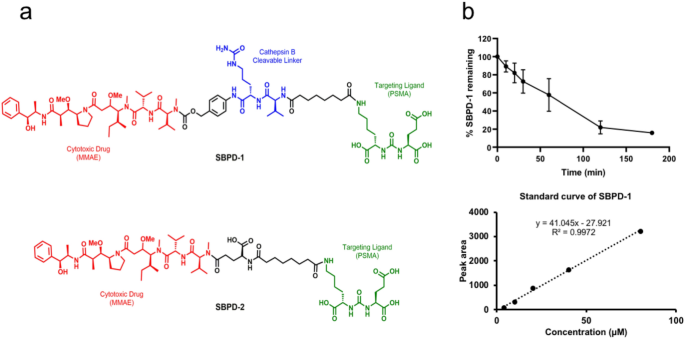
B CLEAVABLE LINKER. To achieve a specific, high-affinity interaction with PSMA we used the low-molecular-weight (LMW) scaffold Lys-Glu-Urea-DSS originally developed in our laboratory32. The
synthetic tubulin inhibitor MMAE was conjugated to the Lys-Glu-Urea-DSS via a cathepsin B cleavable valine-citrulline linker (SBPD-1) or non-cleavable linker (SBPD-2), as a control to
determine the utility of the linker (Fig. 1a). Synthesis of SBPD-1 began with known amine 133, which on treatment with previously reported Lys-Glu-Urea-DSS32 in the presence of
diisopropylethylamine afforded 2 in 85% yield (Supplementary Fig. S1). Compound 2 was further converted into activated carbonate 3 in 45% yield by treating with bis(4-nitrophenyl) carbonate
and subsequent reaction with MMAE, followed by deprotection to realize target conjugate SBPD-1 in 20% combined yield (Supplementary Fig. S1). Synthesis of SBPD-2 began with previously
reported Lys-Glu-Urea-DSS32, which on treatment with L-glutamic acid α-tert-butyl ester in the presence of diisopropylethylamine in DMF, afforded 4 in 70% yield (Supplementary Fig. S2).
Compound 4 was subsequently reacted with MMAE followed by deprotection to provide target conjugate SBPD-2 in 20% combined yield (Supplementary Fig. S2). PSMA inhibitory capacity, a surrogate
for affinity, was measured according to a previously described assay34. Both conjugates, SBPD-1 and SBPD-2, demonstrated high affinity to PSMA with _K_i values of 8.84 nM (95% CI
5.00–15.63) and 3.0 nM (95% CI 1.94–4.67), respectively. We tested if SBPD-1 could release MMAE when incubated with recombinant cathepsin B in vitro and found that MMAE was efficiently
released (80%) within 3 h of incubation (Fig. 1b). SBPD-1 SELECTIVELY KILLS PSMA-EXPRESSING PC CELLS IN VITRO We evaluated the cytotoxicity of SBPD-1 and SBPD-2 in PSMA-expressing PC3 PIP
and PSMA-negative PC3 flu cells in vitro35,36. SBPD-1 demonstrated IC50 values of 3.9 nM (95% CI 2.8–5.5 nM) and 151.1 nM (95% CI 104.1–219.3 nM) for PSMA + PC3 PIP and PSMA − PC3 flu cells,
respectively, indicating selectivity for PSMA-expressing cells. The IC50 value of 151.1 nM for PSMA − PC3 flu cells suggests release of some MMAE to enable non-selective cell kill in vitro.
SBPD-2 demonstrated IC50 values of 4.8 μM (95% CI 0.8–28.5 μM) and 5.8 μM (95% CI 0.7–47.2 μM) for PSMA + PC3 PIP and PSMA− PC3 flu cells, respectively, indicating a lack of potency
regardless of PSMA expression and the need for cleavage of MMAE from the targeting moiety. MMAE alone proved exquisitely potent in both cell lines, demonstrating an IC50 value of 39.2 pM
(95% CI 19.5–78.7 pM) and 40.0 pM (95% CI 21.2–75.4 pM) at 48 h for PSMA+ PC3 PIP and PSMA− PC3 flu cells, respectively (Fig. 2). SBPD-1 SELECTIVELY KILLS PSMA-EXPRESSING PC XENOGRAFTS IN
VIVO Prior to in vivo potency we evaluated the stability of SBPD-1 and SBPD-2 in human and murine serum. SBPD-1 remained intact in human serum out to 48 h of incubation (Fig. 3). While 90%
of SBPD-2 remained intact for 48 h in murine serum (data not shown), SBPD-1 was metabolized more quickly (Fig. 3). While more than 80% of SBPD-1 was intact in serum at 8 h of incubation,
less than half represented parent compound at 24 h, and the majority of the prodrug was fully degraded by 48 h of incubation. It has been reported that the valine-citrulline linker is stable
in human and monkey serum but that it can be hydrolyzed in mouse plasma via extracellular carboxylesterase 1c37,38. Based on those stability results, we applied small, fractionated doses
for the murine efficacy study to avoid systemic toxicity that could affect the overall survival of the test animals. To evaluate efficacy in preclinical models of human PC, we initially
employed xenograft tumor models derived from PSMA+ PC3 PIP and PSMA− PC3 flu cells in NOD/SCID/IL2Rγnull (NSG) mice. Three weeks after injection of the cells, the average tumor volume
reached 62.4 (± 11.6) mm3, and mice were treated with 20, 40 and 80 μg/kg of SBPD-1 via daily intraperitoneal (IP) injection for 30 days, n = 5. We monitored tumor growth and overall animal
welfare (Fig. 4a). Animals were scored ‘dead’ when the tumor reached 4-times its original volume (Fig. 4b). Tumors in non-treated, control mice for both tumor types, in PSMA+ PC3 PIP mice
treated with 20 μg/kg and in PSMA− PC3 flu mice with all three doses, grew rapidly and all animals so treated were euthanized on day 20 post-initiation of treatment (Fig. 4b). The median
survival time for non-treated groups of animals harboring either PSMA+ PC3 PIP or PSMA − PC3 flu tumors was 15 days. For animals harboring PSMA+ PC3 PIP tumors, the median survival time of
the group treated with 20 μg/kg was 17 days. The median survival times for group harboring PSMA − PC3 flu tumors treated with 20, 40, and 80 μg/kg were 15, 15, and 20 days, respectively.
Doses of 40 and 80 μg/kg delivered to animals harboring PSMA + PC3 PIP tumors cleared the tumors such that they were undetectable by the completion of treatment (Fig. 4a). Approximately 1
week was required to be able to re-measure previously undetectable tumors in the group treated at 40 μg/kg. Two weeks were required for re-appearance of tumors in animals treated with the 80
μg/kg dose. In animals harboring PSMA + PC3 PIP tumors, both the 40 and 80 μg/kg doses provided significant survival benefits as the median survival times were 54 days [_P_ = 0.003,
Log-rank (Mantel-Cox) test] and 69 days (_P_ = 0.003), respectively (Fig. 4b). Urine protein level and specific gravity measured for all test animals on Days 9 and 20 were normal, indicating
that no acute renal toxicity occurred at any dose tested (Supplementary Table S1). SBPD-1 IS EFFECTIVE IN AN EXPERIMENTAL METASTATIC MODEL OF PSMA-EXPRESSING PC To evaluate efficacy of
SBPD-1 on established metastatic tumors, we used a PSMA-expressing experimental metastatic model of human PC18. The model has 100% penetrance, and consistently develops lesions in the liver
(100%), kidney (100%), and bone (40%). PSMA + PC3/ML/PSMA cells were administered to NSG mice intravenously (IV) and tumors were allowed to establish for 4 weeks. PC3/ML/PSMA cells express
firefly luciferase as an imaging reporter to allow us to monitor tumor development via weekly bioluminescence imaging (BLI). Mice were treated with 40, 80 and 160 μg/kg of SBPD-1 via daily
IP injection for 30 days, n = 5. We increased the doses to compensate for the lower expression of PSMA on PC3/ML/PSMA cells compared with that of PSMA + PC3 PIP tumors (Supplementary Figure
S3). The 40 μg/kg dose did not show survival benefit to non-treated control mice, with median survival times of 47 days for each group. Mice treated at the 80 and 160 μg/kg dose levels,
however, exhibited significant survival benefits, with median survival of 56 days [_P_ = 0.003, Log-rank (Mantel-Cox) test] and 58 days (_P_ = 0.003), respectively (Fig. 5, Supplementary
Figure S4). SBPD-1 IS NON-TOXIC TO C57BL/6 MICE We evaluated potential toxicity of SBPD-1 in immunocompetent animals. We administered MMAE (80 μg/kg), SBPD-1 (160 μg/kg), and 5% DMSO to
healthy C57BL/6 mice (n = 5). We monitored animals for 80 days after initiation of administration. As previously reported39, MMAE demonstrated severe toxicity as all treated mice required
euthanasia during treatment due to weight loss (Fig. 6b). Mice injected with vehicle or SBPD-1 did not show any signs of toxicity and steadily gained weight (Fig. 6a). We removed lung,
liver, kidneys, salivary and lacrimal glands from all tested animals at Day 80 after initiation of SBPD-1 treatment. Histopathological examination revealed no tissue damage (Fig. 6c). We
also obtained peripheral blood from mice injected with vehicle, SBPD-1, and healthy untreated animals, and prepared serum for chemistry studies (n = 5). Blood urea nitrogen (BUN),
creatinine, glucose, alkaline phosphatase (ALP), total protein (T-Pro), and alanine aminotransferase (ALT) analyses showed that animals injected with either vehicle or SBPD-1 did not show
differences in these values compared with those from untreated mice (Supplementary Table S2). Complete blood counts from the mice also showed no abnormalities except for lower white blood
cell count for mice injected with SBPD-1, which may have resulted from the relative instability of the cathepsin B linker in murine serum and subsequent bone marrow toxicity of MMAE37,40.
DISCUSSION Prostate-specific membrane antigen (PSMA) was first identified as a marker for PC through cloning of a monoclonal antibody raised against the patient-derived PC cell line,
LNCaP41. Since PSMA was discovered to be the same as the _N_-acetyl-l-aspartyl-l-glutamate peptidase I (NAALADase I)42, PSMA has been pursed as a target for diagnostic imaging of advanced PC
with various low-molecular-weight agents35,43,44,45,46. Anti-PSMA antibodies have also been tested as PSMA-targeting entities for both molecular imaging and therapy of PC6,7,8,47,48. Other
therapeutic approaches such as PSMA targeted-nanoparticles loaded with an anti-cancer drug49,50 or photodynamic therapy51,52,53 have been tested in preclinical and clinical settings.
PSMA-targeted RPT has provided a new alternative to managing patients with advanced PC refractory to other therapies54,55. Recent prospective trials of 177Lu-based therapies have
demonstrated substantial imaging and PSA responses56,57. Fewer side effects than other systemic therapies, such as hormonal or chemotherapy, have repeatedly been shown58. Nevertheless,
approximately 50% of patients were non-responders, and the majority of responders relapsed, requiring further cycles or other options55. Questions about long-term toxicity of this method
remain, particularly for α-particle emitting versions of RPT17,18,59,60. Although PSMA-targeted RPT is promising and fraught with fewer adverse events compared to the conventional cytotoxic
therapies, radiation exposure to normal organs can result in xerostomia or other off-target effects16,17,18,59,60. A PSMA-targeted prodrug equipped with additional specificity to malignant
cells may provide an enhanced therapeutic index. Several PSMA-targeted prodrugs were tested in both preclinical and clinical settings. Kularatne et al_._ tested various cytotoxic drugs as a
form of prodrug by conjugating them to the PSMA-targeted agent, 2-[3-(1,3-dicarboxy propyl)ureido] pentanedioic acid61. Those prodrugs utilized a disulfide linker to enable drug release in
the reducing environment of the cytoplasm. Some of the tested drugs exhibited cytotoxicity to PSMA-expressing LNCaP cells at single- or double-digit nanomolar concentration levels. However,
in vivo safety and efficacy of those drugs have not been tested. Mipsagargin (G-202) is a prodrug consisting of an analog of thapsigargin conjugated to a PSMA-cleavable peptide62.
Thapsigargin is a potent inhibitor of the sarcoplasmic/endoplasmic reticulum calcium adenosine triphosphatase (SERCA) pump essential for cell viability. Mipsagargin was used to target the
PSMA-expressing tumor neovasculature of various solid cancers. Despite promising preclinical and phase I results62, phase II trials showed no clinical benefit for advanced hepatocellular
carcinoma63. A PSMA-targeted antibody-MMAE conjugate (ADC) has been tested and showed favorable preclinical efficacy25,26. However, in a phase I trial with that conjugate, the therapeutic
window proved narrow, necessitating modification of dose selection if the compound were to advance further30. The authors of that trial hypothesized that the toxicity may have been due to
the systemic concentration of free MMAE released from the antibody30. The results from the corresponding phase II trial were recently published64. Toxicity was noted shortly after the
initiation of the trial—particularly neutropenia and neuropathy—such that a dose reduction was necessary for it to continue. A partial radiologic response was obtained in only 2 of 119
participants, with none reporting a complete response64. Please note that we used PSMA + PC3 PIP cells to generate subcutaneous tumors that may not precisely reflect the case as it may occur
in patients. Although we did not measure the number of PSMA molecules per PSMA + PC3 PIP cell in the current study, we have previously shown there to be an order of magnitude higher PSMA
expression in these cells than in LNCaP cells, which are patient-derived18. However, PSMA + PC3/ML/PSMA cells used for the metastatic model have comparable PSMA expression to that of LNCaP
cells. Nevertheless, we used the PSMA + PC3 PIP/PSMA − PC3 flu cells to generate subcutaneous tumors in order to minimize the number of variables between cells used, as these lines are
otherwise isogenic, and to see if any signal could be obtained in this proof-of-principal study. Future studies will explore tumor models that have a variety of levels of PSMA expression,
including those that are more in line with what is seen in human specimens. SBPD-1 was designed for safe delivery of the potent toxin MMAE to maximize its therapeutic index. There are three
layers of specificity of this agent for malignant cells. First, there is high-affinity, specific PSMA targeting followed by internalization of drug-bound PSMA. Notably PSMA tends to localize
to the centrosome upon internalization65 enabling it to deliver a drug that interrupts microtubule formation to the compartment in which it can be most effective. Second, MMAE is released
only upon enzymatic cleavage by cathepsin B, which is upregulated in the lysosomes of cancer cells29. The same drug with non-cleavable linker (SBPD-2) showed about 7,100-fold less potency in
PSMA + cancer cells (Fig. 2). Third, MMAE inhibits microtubule polymerization, an essential process for cell division of cancer cells. A further advantage of the small-molecule approach is
that drug conjugates tend to have superior tumor penetration and more rapid clearance from non-target sites than do ADCs66. Since prior reports37,38 as well as our results (Fig. 3a) have
suggested that the valine-citrulline linker is unstable in murine serum, we modified the dosing plan to consist of several fractionated doses. Our in vivo safety results with an
immunocompetent murine model showed no toxicity with the highest doses tested in the efficacy study (Fig. 6, Supplementary Table S2). It is likely that a clinical dosing plan could consist
of less frequent administration as the valine-citrulline linker has been reported to be stable in human plasma38. In summary, we have generated and tested in vivo a low-molecular-weight,
PSMA-targeted prodrug that demonstrated tumor penetration and specificity sufficient to provide survival differences between PSMA + tumor-bearing animals and animals bearing isogenic tumors
devoid of PSMA, including in a metastatic model. Furthermore, despite carrying the potent anti-tumor agent MMAE, the conjugate was non-toxic. We believe that lower toxicity was due to the
controlled environment to which MMAE was delivered, by virtue of the presence of a cathepsin B cleavable linker in the molecule. Compounds of this class or those employing similar strategies
may enable safe and effective targeting of PSMA-expressing lesions in patients. METHODS GENERAL METHODS AND MATERIALS FOR SYNTHESES OF PRODRUGS Experiments were carried out in compliance
with ARRIVE guidelines. Detailed methods for the syntheses of prodrugs are described in the Supplementary Information. Commercially available reagents and solvents for syntheses were
analytical grade and used without further purification. Diisopropylethylamine (DIPEA), triflouroacetic acid (TFA), 4-(Dimethyl amino) pyridine (DMAP), pyridine (Py) and
_N_-(3-dimethylaminopropyl)-_N_-ethylcarbodiimide (EDC) were purchased from Sigma-Aldrich (Allentown, PA, USA). l-Glutamic acid 5-tert-butyl ester, bis(4-nitrophenyl) carbonate and
1-hydroxybenzotriazole hydrate (HOBt) were purchased from Chem-Impex International (Wood Dale, IL, USA), disuccinimidyl suberate was purchased from TCI America (Pittsburgh, PA, USA) and
monomethyl auristatin E (MMAE) was purchased from BroadPharm (San Diego, CA, USA). High performance liquid chromatographic (HPLC) purification of final compounds (SBPD-1 and SBPD-2) was
performed using a C18 Luna 10 mm × 250 mm column (Phenomenex, Torrance, CA, USA) on an Agilent 1260 infinity LC system (Santa Clara, CA, USA) and eluted with water (0.1% TFA) (A) and CH3CN
(0.1% TFA) (B). 1H NMR spectra were recorded on a Bruker Ultrashield 500 MHz spectrometer. Chemical shifts (δ) are reported in parts per million (ppm) downfield by reference to proton
resonances resulting from incomplete deuteration of the NMR solvent and the coupling constants (J) was reported in Hertz (Hz). High resolution mass spectra were obtained by the University of
Notre Dame Mass Spectrometry and Proteomics Facility, Notre Dame, IN using ESI by direct infusion on a Bruker micrOTOF-II. CATHEPSIN B CLEAVAGE Release of MMAE from prodrugs by a
recombinant cathepsin B was analyzed using a modified method from previously published work33. Prodrug stock solutions (80 µL, 10 mM) were added to the 1.92 mL cathepsin B (MilliporeSigma,
Cat# C8571, Burlington, MA, USA) containing buffer (25 mM acetate, 1 mM EDTA, pH 5, pre-warmed at 37 °C) at the final concentration of 30 nM (cathepsin B)and 40 µM (prodrug). Aliquots (200
µL) were periodically removed and enzymatic activity was stopped by the addition of thioprotease inhibitor E-64 (30 nM in the final solution, MilliporeSigma, Cat# E3132). The samples were
centrifuged and the supernatants were analyzed by HPLC (Waters 600 E coupled with Varian prostar detector, Milford, MA, USA). Samples were prepared at 0, 10, 20, 30, 60, and 120 min.
Experiments were performed in triplicate. PSMA AFFINITY AND IN VITRO CYTOTOXICITY PSMA affinities of SBPD-1and SBPD-2 were measured using the modified Amplex Red glutamic acid/glutamate
oxidase assay as previously described34. PSMA-expressing PC3-PIP, PSMA-negative PC3-flu, PSMA-positive PC3/ML/PSMA and PSMA-negative PC3/ML were maintained as previously described18. One
thousand cells (PC3-PIP or PC3-flu) were seeded in 96 well plates 24 h prior to drug treatment. Drug was added to each well in serial dilution and incubated for 24, 48 or 72 h. Cell
viability was measured using TACS XTT Cell Proliferation Assay (Trevigen, Cat# 4891-25-K, Gaithersburg, MD) at each time point according to the manufacturer’s protocol. IC50 values were
calculated using GraphPad Prism 7 software. SERUM STABILITY Serum stability of prodrugs was analyzed using a modified method from previously published work67. Prodrug stock solution (80 µL,
1 mM) was mixed with human serum (320 µL) purchased from Millipore Sigma (Cat# H4522, Saint Louis, MO, USA). Five 50 µL fractions corresponding to five-time points (2, 4, 6, 24, and 48 h)
were removed in separate vials from the above mixture and incubated at 37 °C. Aliquots of 25 µL were removed at 2, 4, 6, 24, and 48 h from the respective vial and diluted with cold ice CH3OH
(125 µL) to precipitate proteins. The samples were centrifuged, and the supernatants were analyzed by HPLC [λ 220 nm, 250 mm × 4.6 mm Phenomenex Luna C18 column, solvent gradient: 61% H2O
(0.1% TFA) and 39% ACN (0.1% TFA) isocratic for 30 min at a flow rate of 1 mL/min. SBPD-1 eluted at 12.1 min]. Murine prodrug stock solution (32 µL, 10 mM) was incubated with 100% mouse
serum (final concentration of the serum was 80% after the mixing with prodrug solution) at 37 °C. Proteins were precipitated as above. Aliquots of 25 µL were evaluated at the same time
points as above. The samples were centrifuged, and the supernatants were analyzed by HPLC as above. Stability was calculated based on the peak area of the prodrug at each time point.
Experiments were performed in triplicate. PRECLINICAL EVALUATION OF SBPD-1 Animal studies were performed under the guidance of a protocol approved by the Johns Hopkins Animal Care and Use
Committee and performed in compliance with the Animal Welfare Act regulations and Public Health Service (PHS) Policy. Johns Hopkins University has an approved PHS assurance. NSG
(NOD/SCID/IL2Rγnull) mice were purchased from the Johns Hopkins University Sydney Kimmel Comprehensive Cancer Center Animal Resources Core. C57BL/6 mice were purchased from Jackson
Laboratory (Bar Harbor, ME, USA). NSG mice were injected with 1.5 million PC3/PIP or 1 million PC3/flu cells at the lower left flank. Two weeks after the injection of cells, mice were
treated with 20, 40, 80 μg/kg of SBPD-1 formulated in 100 μL of sterile saline via daily intraperitoneal (IP) injection for 30 days. Tumor volumes were measure twice per week. Urinalysis was
performed using URS-10 Urine Reagent Strips (LW Scientific Inc. Lawrenceville, GA). For the metastatic model, NSG mice were injected with 0.75 million PC3/ML/PSMA cells via the tail vein.
Four weeks after the injection mice were treated with 40, 80, 160 μg/kg of SBPD-1 formulated in 100 μL of sterile water via daily intraperitoneal injection for 30 days. BLI was performed
weekly using the IVIS Spectrum in vivo imaging system (Perkin Elmer, Waltham, MA). IN VIVO TOXICITY Male C57BL/6 mice were purchased from Jackson Laboratory. Ten-week-old mice were injected
with the indicated doses of MMAE (formulated in 5% DMSO), SBPD-1 (formulated in saline) or 5% DMSO intraperitoneally (daily for 30 days, n = 5). Animals were monitored daily for weight
changes and other abnormalities for 80 days. Animals were euthanized in a CO2 chamber at day 80, and blood, lung, liver, kidney, salivary gland, and lacrimal gland were collected for
complete blood counts, blood chemistry, and histopathological analyses. Complete blood counts including white blood cells (WBC), red blood cells (RBC), hemoglobin (HGB), hematocrit (HCT),
mean corpuscular volume (MCV), mean corpuscular hemoglobin (MCH), mean corpuscular hemoglobin concentration (MCHC), and platelet (PLT) were measured using scil Vet ABC Hematology Analyzer
(scil animal care company, Gurnee, IL). Blood chemistry parameters including blood urea nitrogen (BUN), glucose (GLU), Alkaline Phosphatase (ALP), total protein (T-Pro), Alanine
aminotransferase (ALT) and Creatinine (Cre) were measured with Spotchem EZ chemistry analyzer (Arkray USA, Edina, MN). Hematoxylin and eosin slides were generated for five organs and
examined by certified veterinary pathologist. DATA AVAILABILITY All data used in this submission are included in the body of manuscript or in the Supplementary Information. Additional data
related to the paper are available upon request. REFERENCES * Foss, C. A., Mease, R. C., Cho, S. Y., Kim, H. J. & Pomper, M. G. GCPII imaging and cancer. _Curr. Med. Chem._ 19, 1346–1359
(2012). Article CAS PubMed PubMed Central Google Scholar * Kiess, A. P. _et al._ Prostate-specific membrane antigen as a target for cancer imaging and therapy. _Q. J. Nucl. Med. Mol.
Imaging_ 59, 241–268 (2015). CAS PubMed PubMed Central Google Scholar * Nimmagadda, S. _et al._ Low-level endogenous PSMA expression in nonprostatic tumor xenografts is sufficient for in
vivo tumor targeting and imaging. _J. Nucl. Med._ 59, 486–493. https://doi.org/10.2967/jnumed.117.191221 (2018). Article CAS PubMed PubMed Central Google Scholar * Castanares, M. A.
_et al._ Evaluation of prostate-specific membrane antigen as an imaging reporter. _J. Nucl. Med._ 55, 805–811. https://doi.org/10.2967/jnumed.113.134031 (2014). Article CAS PubMed Google
Scholar * Minn, I. _et al._ Imaging CAR T cell therapy with PSMA-targeted positron emission tomography. _Sci. Adv._ 5, eaaw5096. https://doi.org/10.1126/sciadv.aaw5096 (2019). Article ADS
CAS PubMed PubMed Central Google Scholar * Huang, C. T. _et al._ Development of 5D3-DM1: A novel anti-prostate-specific membrane antigen antibody-drug conjugate for PSMA-positive
prostate cancer therapy. _Mol. Pharm._ https://doi.org/10.1021/acs.molpharmaceut.0c00457 (2020). Article PubMed PubMed Central Google Scholar * Rosenfeld, L. _et al._ Nanobodies
targeting prostate-specific membrane antigen for the imaging and therapy of prostate cancer. _J. Med. Chem._ 63, 7601–7615. https://doi.org/10.1021/acs.jmedchem.0c00418 (2020). Article CAS
PubMed PubMed Central Google Scholar * Petrylak, D. P. _et al._ PSMA ADC monotherapy in patients with progressive metastatic castration-resistant prostate cancer following abiraterone
and/or enzalutamide: Efficacy and safety in open-label single-arm phase 2 study. _Prostate_ 80, 99–108. https://doi.org/10.1002/pros.23922 (2020). Article CAS PubMed Google Scholar *
Machulkin, A. E. _et al._ Synthesis and biological evaluation of PSMA-targeting paclitaxel conjugates. _Bioorg. Med. Chem. Lett._ 29, 2229–2235. https://doi.org/10.1016/j.bmcl.2019.06.035
(2019). Article CAS PubMed Google Scholar * Minn, I., Rowe, S. P. & Pomper, M. G. Enhancing CAR T-cell therapy through cellular imaging and radiotherapy. _Lancet Oncol._ 20,
e443–e451. https://doi.org/10.1016/S1470-2045(19)30461-9 (2019). Article CAS PubMed Google Scholar * Rowe, S. P. _et al._ Prostate-specific membrane antigen-targeted radiohalogenated PET
and therapeutic agents for prostate cancer. _J. Nucl. Med._ 57, 90S-96S. https://doi.org/10.2967/jnumed.115.170175 (2016). Article CAS PubMed PubMed Central Google Scholar * Miyahira,
A. K. _et al._ Meeting report from the prostate cancer foundation PSMA theranostics state of the science meeting. _Prostate_ https://doi.org/10.1002/pros.24056 (2020). Article PubMed
PubMed Central Google Scholar * Violet, J. _et al._ Long-term follow-up and outcomes of retreatment in an expanded 50-patient single-center phase II prospective trial of (177)Lu-PSMA-617
theranostics in metastatic castration-resistant prostate cancer. _J. Nucl. Med._ 61, 857–865. https://doi.org/10.2967/jnumed.119.236414 (2020). Article PubMed PubMed Central Google
Scholar * Miyahira, A. K. _et al._ Meeting report from the Prostate Cancer Foundation PSMA-directed radionuclide scientific working group. _Prostate_ 78, 775–789.
https://doi.org/10.1002/pros.23642 (2018). Article PubMed Google Scholar * Yordanova, A. _et al._ Outcome and safety of rechallenge [(177)Lu]Lu-PSMA-617 in patients with metastatic
prostate cancer. _Eur. J. Nucl. Med. Mol. Imaging_ 46, 1073–1080. https://doi.org/10.1007/s00259-018-4222-x (2019). Article PubMed Google Scholar * Kratochwil, C. _et al._ Targeted
alpha-therapy of metastatic castration-resistant prostate cancer with (225)Ac-PSMA-617: Dosimetry estimate and empiric dose finding. _J. Nucl. Med._ 58, 1624–1631.
https://doi.org/10.2967/jnumed.117.191395 (2017). Article CAS PubMed Google Scholar * Kratochwil, C. _et al._ 225Ac-PSMA-617 for PSMA-targeted alpha-radiation therapy of metastatic
castration-resistant prostate cancer. _J. Nucl. Med._ 57, 1941–1944. https://doi.org/10.2967/jnumed.116.178673 (2016). Article CAS PubMed Google Scholar * Kiess, A. P. _et al._
(2S)-2-(3-(1-Carboxy-5-(4–211At-astatobenzamido)pentyl)ureido)-pentanedioic acid for PSMA-targeted alpha-particle radiopharmaceutical therapy. _J. Nucl. Med._ 57, 1569–1575.
https://doi.org/10.2967/jnumed.116.174300 (2016). Article CAS PubMed PubMed Central Google Scholar * Zhang, A. X. _et al._ A remote arene-binding site on prostate specific membrane
antigen revealed by antibody-recruiting small molecules. _J. Am. Chem. Soc._ 132, 12711–12716. https://doi.org/10.1021/ja104591m (2010). Article CAS PubMed PubMed Central Google Scholar
* Kasten, B. B., Liu, T., Nedrow-Byers, J. R., Benny, P. D. & Berkman, C. E. Targeting prostate cancer cells with PSMA inhibitor-guided gold nanoparticles. _Bioorg. Med. Chem. Lett._
23, 565–568. https://doi.org/10.1016/j.bmcl.2012.11.015 (2013). Article CAS PubMed Google Scholar * Leconet, W. _et al._ Anti-PSMA/CD3 bispecific antibody delivery and antitumor activity
using a polymeric depot formulation. _Mol. Cancer Ther._ 17, 1927–1940. https://doi.org/10.1158/1535-7163.MCT-17-1138 (2018). Article CAS PubMed Google Scholar * Rautio, J., Meanwell,
N. A., Di, L. & Hageman, M. J. The expanding role of prodrugs in contemporary drug design and development. _Nat. Rev. Drug Discov._ 17, 559–587. https://doi.org/10.1038/nrd.2018.46
(2018). Article CAS PubMed Google Scholar * Kinoshita, Y. _et al._ Expression of prostate-specific membrane antigen in normal and malignant human tissues. _World J. Surg._ 30, 628–636.
https://doi.org/10.1007/s00268-005-0544-5 (2006). Article PubMed Google Scholar * Silver, D. A., Pellicer, I., Fair, W. R., Heston, W. D. & Cordon-Cardo, C. Prostate-specific membrane
antigen expression in normal and malignant human tissues. _Clin. Cancer Res._ 3, 81–85 (1997). CAS PubMed Google Scholar * Wang, X., Ma, D., Olson, W. C. & Heston, W. D. In vitro and
in vivo responses of advanced prostate tumors to PSMA ADC, an auristatin-conjugated antibody to prostate-specific membrane antigen. _Mol. Cancer Ther._ 10, 1728–1739.
https://doi.org/10.1158/1535-7163.MCT-11-0191 (2011). Article CAS PubMed Google Scholar * Ma, D. _et al._ Potent antitumor activity of an auristatin-conjugated, fully human monoclonal
antibody to prostate-specific membrane antigen. _Clin. Cancer Res._ 12, 2591–2596. https://doi.org/10.1158/1078-0432.CCR-05-2107 (2006). Article CAS PubMed Google Scholar * Bartlett, N.
L. _et al._ Retreatment with brentuximab vedotin in patients with CD30-positive hematologic malignancies. _J. Hematol. Oncol._ 7, 24. https://doi.org/10.1186/1756-8722-7-24 (2014). Article
CAS PubMed PubMed Central Google Scholar * Lu, J., Jiang, F., Lu, A. & Zhang, G. Linkers having a crucial role in antibody-drug conjugates. _Int. J. Mol. Sci._ 17, 561.
https://doi.org/10.3390/ijms17040561 (2016). Article CAS PubMed PubMed Central Google Scholar * Vigneswaran, N. _et al._ Variable expression of cathepsin B and D correlates with highly
invasive and metastatic phenotype of oral cancer. _Hum. Pathol._ 31, 931–937. https://doi.org/10.1053/hupa.2000.9035 (2000). Article CAS PubMed Google Scholar * Petrylak, D. P. _et al._
Phase 1 study of PSMA ADC, an antibody-drug conjugate targeting prostate-specific membrane antigen, in chemotherapy-refractory prostate cancer. _Prostate_ 79, 604–613.
https://doi.org/10.1002/pros.23765 (2019). Article CAS PubMed Google Scholar * Masters, J. C., Nickens, D. J., Xuan, D., Shazer, R. L. & Amantea, M. Clinical toxicity of antibody
drug conjugates: A meta-analysis of payloads. _Invest. New Drugs_ 36, 121–135. https://doi.org/10.1007/s10637-017-0520-6 (2018). Article CAS PubMed Google Scholar * Banerjee, S. R. _et
al._ Sequential SPECT and optical imaging of experimental models of prostate cancer with a dual modality inhibitor of the prostate-specific membrane antigen. _Angew. Chem. Int. Ed. Engl._
50, 9167–9170. https://doi.org/10.1002/anie.201102872 (2011). Article CAS PubMed PubMed Central Google Scholar * Dubowchik, G. M. _et al._ Cathepsin B-labile dipeptide linkers for
lysosomal release of doxorubicin from internalizing immunoconjugates: Model studies of enzymatic drug release and antigen-specific in vitro anticancer activity. _Bioconjug. Chem._ 13,
855–869 (2002). Article CAS PubMed Google Scholar * Chen, Y. _et al._ Radiohalogenated prostate-specific membrane antigen (PSMA)-based ureas as imaging agents for prostate cancer. _J.
Med. Chem._ 51, 7933–7943. https://doi.org/10.1021/jm801055h (2008). Article CAS PubMed PubMed Central Google Scholar * Mease, R. C. _et al._
N-[N-[(S)-1,3-Dicarboxypropyl]carbamoyl]-4-[18F]fluorobenzyl-l-cysteine, [18F]DCFBC: A new imaging probe for prostate cancer. _Clin. Cancer Res._ 14, 3036–3043.
https://doi.org/10.1158/1078-0432.CCR-07-1517 (2008). Article CAS PubMed PubMed Central Google Scholar * Chang, S. S. _et al._ Five different anti-prostate-specific membrane antigen
(PSMA) antibodies confirm PSMA expression in tumor-associated neovasculature. _Cancer Res._ 59, 3192–3198 (1999). CAS PubMed Google Scholar * Dorywalska, M. _et al._ Molecular basis of
valine-citrulline-PABC linker instability in site-specific ADCs and its mitigation by linker design. _Mol. Cancer Ther._ 15, 958–970. https://doi.org/10.1158/1535-7163.MCT-15-1004 (2016).
Article CAS PubMed Google Scholar * Anami, Y. _et al._ Glutamic acid-valine-citrulline linkers ensure stability and efficacy of antibody-drug conjugates in mice. _Nat. Commun._ 9, 2512.
https://doi.org/10.1038/s41467-018-04982-3 (2018). Article ADS CAS PubMed PubMed Central Google Scholar * Qi, R. _et al._ Nanoparticle conjugates of a highly potent toxin enhance
safety and circumvent platinum resistance in ovarian cancer. _Nat. Commun._ 8, 2166. https://doi.org/10.1038/s41467-017-02390-7 (2017). Article ADS CAS PubMed PubMed Central Google
Scholar * Donaghy, H. Effects of antibody, drug and linker on the preclinical and clinical toxicities of antibody-drug conjugates. _MAbs_ 8, 659–671.
https://doi.org/10.1080/19420862.2016.1156829 (2016). Article CAS PubMed PubMed Central Google Scholar * Horoszewicz, J. S., Kawinski, E. & Murphy, G. P. Monoclonal antibodies to a
new antigenic marker in epithelial prostatic cells and serum of prostatic cancer patients. _Anticancer Res._ 7, 927–935 (1987). CAS PubMed Google Scholar * Carter, R. E., Feldman, A. R.
& Coyle, J. T. Prostate-specific membrane antigen is a hydrolase with substrate and pharmacologic characteristics of a neuropeptidase. _Proc. Natl. Acad. Sci. USA_ 93, 749–753.
https://doi.org/10.1073/pnas.93.2.749 (1996). Article ADS CAS PubMed PubMed Central Google Scholar * Pomper, M. G. _et al._ 11C-MCG: Synthesis, uptake selectivity, and primate PET of a
probe for glutamate carboxypeptidase II (NAALADase). _Mol. Imaging_ 1, 96–101. https://doi.org/10.1162/153535002320162750 (2002). Article CAS PubMed Google Scholar * Foss, C. A. _et
al._ Radiolabeled small-molecule ligands for prostate-specific membrane antigen: In vivo imaging in experimental models of prostate cancer. _Clin. Cancer Res._ 11, 4022–4028.
https://doi.org/10.1158/1078-0432.CCR-04-2690 (2005). Article CAS PubMed Google Scholar * Wone, D. W., Rowley, J. A., Garofalo, A. W. & Berkman, C. E. Optimizing
phenylethylphosphonamidates for the inhibition of prostate-specific membrane antigen. _Bioorg. Med. Chem._ 14, 67–76. https://doi.org/10.1016/j.bmc.2005.07.056 (2006). Article CAS PubMed
Google Scholar * Kularatne, S. A., Wang, K., Santhapuram, H. K. & Low, P. S. Prostate-specific membrane antigen targeted imaging and therapy of prostate cancer using a PSMA inhibitor as
a homing ligand. _Mol. Pharm._ 6, 780–789. https://doi.org/10.1021/mp900069d (2009). Article CAS PubMed Google Scholar * Yao, D. _et al._ The utility of monoclonal antibodies in the
imaging of prostate cancer. _Semin. Urol. Oncol._ 20, 211–218. https://doi.org/10.1053/suro.2002.36250 (2002). Article CAS PubMed Google Scholar * Psimadas, D., Valotassiou, V., Alexiou,
S., Tsougos, I. & Georgoulias, P. Radiolabeled mAbs as molecular imaging and/or therapy agents targeting PSMA. _Cancer Invest._ 36, 118–128.
https://doi.org/10.1080/07357907.2018.1430816 (2018). Article CAS PubMed Google Scholar * Von Hoff, D. D. _et al._ Phase I STUDY of PSMA-targeted docetaxel-containing nanoparticle
BIND-014 in patients with advanced solid tumors. _Clin. Cancer Res._ 22, 3157–3163. https://doi.org/10.1158/1078-0432.CCR-15-2548 (2016). Article CAS Google Scholar * Autio, K. A. _et
al._ Safety and efficacy of BIND-014, a docetaxel nanoparticle targeting prostate-specific membrane antigen for patients with metastatic castration-resistant prostate cancer: A phase 2
clinical trial. _JAMA Oncol._ 4, 1344–1351. https://doi.org/10.1001/jamaoncol.2018.2168 (2018). Article PubMed PubMed Central Google Scholar * Chen, Y. _et al._ A PSMA-targeted
theranostic agent for photodynamic therapy. _J. Photochem. Photobiol. B_ 167, 111–116. https://doi.org/10.1016/j.jphotobiol.2016.12.018 (2017). Article CAS PubMed Google Scholar * Lutje,
S. _et al._ Development and characterization of a theranostic multimodal anti-PSMA targeting agent for imaging, surgical guidance, and targeted photodynamic therapy of PSMA-expressing
tumors. _Theranostics_ 9, 2924–2938. https://doi.org/10.7150/thno.35274 (2019). Article CAS PubMed PubMed Central Google Scholar * Wang, X. _et al._ Theranostic agents for photodynamic
therapy of prostate cancer by targeting prostate-specific membrane antigen. _Mol. Cancer Ther._ 15, 1834–1844. https://doi.org/10.1158/1535-7163.MCT-15-0722 (2016). Article CAS PubMed
Google Scholar * Rahbar, K. _et al._ German multicenter study investigating 177Lu-PSMA-617 radioligand therapy in advanced prostate cancer patients. _J. Nucl. Med._ 58, 85–90.
https://doi.org/10.2967/jnumed.116.183194 (2017). Article CAS PubMed Google Scholar * Yadav, M. P., Ballal, S., Sahoo, R. K., Dwivedi, S. N. & Bal, C. Radioligand therapy with
(177)Lu-PSMA for metastatic castration-resistant prostate cancer: A systematic review and meta-analysis. _Am. J. Roentgenol._ 213, 275–285. https://doi.org/10.2214/AJR.18.20845 (2019).
Article Google Scholar * Hofman, M. S. _et al._ [(177)Lu]-PSMA-617 radionuclide treatment in patients with metastatic castration-resistant prostate cancer (LuPSMA trial): A single-centre,
single-arm, phase 2 study. _Lancet Oncol._ 19, 825–833. https://doi.org/10.1016/S1470-2045(18)30198-0 (2018). Article CAS PubMed Google Scholar * Aghdam, R. A. _et al._ Efficacy and
safety of (177)Lutetium-prostate-specific membrane antigen therapy in metastatic castration-resistant prostate cancer patients: First experience in West Asia—a prospective study. _World J.
Nucl. Med._ 18, 258–265. https://doi.org/10.4103/wjnm.WJNM_66_18 (2019). Article PubMed PubMed Central Google Scholar * Fendler, W. P., Rahbar, K., Herrmann, K., Kratochwil, C. &
Eiber, M. (177)Lu-PSMA radioligand therapy for prostate cancer. _J. Nucl. Med._ 58, 1196–1200. https://doi.org/10.2967/jnumed.117.191023 (2017). Article CAS PubMed Google Scholar *
Kratochwil, C. _et al._ Targeted alpha-therapy of metastatic castration-resistant prostate cancer with (225)Ac-PSMA-617: Swimmer-plot analysis suggests efficacy regarding duration of tumor
control. _J. Nucl. Med._ 59, 795–802. https://doi.org/10.2967/jnumed.117.203539 (2018). Article CAS PubMed Google Scholar * Kratochwil, C. _et al._ Targeted alpha therapy of mCRPC:
Dosimetry estimate of (213)Bismuth-PSMA-617. _Eur. J. Nucl. Med. Mol. Imaging_ 45, 31–37. https://doi.org/10.1007/s00259-017-3817-y (2018). Article CAS PubMed Google Scholar * Kularatne,
S. A. _et al._ Synthesis and biological analysis of prostate-specific membrane antigen-targeted anticancer prodrugs. _J. Med. Chem._ 53, 7767–7777. https://doi.org/10.1021/jm100729b (2010).
Article CAS PubMed Google Scholar * Denmeade, S. R. _et al._ Engineering a prostate-specific membrane antigen-activated tumor endothelial cell prodrug for cancer therapy. _Sci. Transl.
Med._ 4, 140ra186. https://doi.org/10.1126/scitranslmed.3003886 (2012). Article CAS Google Scholar * Mahalingam, D. _et al._ A phase II, Multicenter, Single-Arm Study of Mipsagargin
(G-202) as a second-line therapy following sorafenib for adult patients with progressive advanced hepatocellular carcinoma. _Cancers (Basel)_ https://doi.org/10.3390/cancers11060833 (2019).
Article Google Scholar * Petrylak, D. P. _et al._ PSMA ADC monotherapy in patients with progressive metastatic castration-resistant prostate cancer following abiraterone and/or
enzalutamide: Efficacy and safety in open-label single-arm phase 2 study. _Prostate_ https://doi.org/10.1002/pros.23922 (2019). Article PubMed Google Scholar * Kiess, A. P. _et al._ Auger
radiopharmaceutical therapy targeting prostate-specific membrane antigen. _J. Nucl. Med._ 56, 1401–1407. https://doi.org/10.2967/jnumed.115.155929 (2015). Article CAS PubMed Google
Scholar * Ovacik, M. & Lin, K. Tutorial on monoclonal antibody pharmacokinetics and its considerations in early development. _Clin. Transl. Sci._ 11, 540–552.
https://doi.org/10.1111/cts.12567 (2018). Article PubMed PubMed Central Google Scholar * Chu, D. S., Johnson, R. N. & Pun, S. H. Cathepsin B-sensitive polymers for
compartment-specific degradation and nucleic acid release. _J. Control Release_ 157, 445–454. https://doi.org/10.1016/j.jconrel.2011.10.016 (2012). Article CAS PubMed Google Scholar
Download references ACKNOWLEDGEMENTS This work was supported by NIH CA058236, EB024495, CA13475, CA184228. AUTHOR INFORMATION Author notes * These authors contributed equally: Srikanth
Boinapally and Hye-Hyun Ahn. AUTHORS AND AFFILIATIONS * Russell H. Morgan Department of Radiology and Radiological Science, Johns Hopkins Medical Institutions, Baltimore, MD, USA Srikanth
Boinapally, Hye-Hyun Ahn, Bei Cheng, Mary Brummet, Hwanhee Nam, Sangeeta R. Banerjee, Il Minn & Martin G. Pomper * Department of Molecular and Comparative Pathobiology, Johns Hopkins
Medical Institutions, Baltimore, MD, USA Kathleen L. Gabrielson Authors * Srikanth Boinapally View author publications You can also search for this author inPubMed Google Scholar * Hye-Hyun
Ahn View author publications You can also search for this author inPubMed Google Scholar * Bei Cheng View author publications You can also search for this author inPubMed Google Scholar *
Mary Brummet View author publications You can also search for this author inPubMed Google Scholar * Hwanhee Nam View author publications You can also search for this author inPubMed Google
Scholar * Kathleen L. Gabrielson View author publications You can also search for this author inPubMed Google Scholar * Sangeeta R. Banerjee View author publications You can also search for
this author inPubMed Google Scholar * Il Minn View author publications You can also search for this author inPubMed Google Scholar * Martin G. Pomper View author publications You can also
search for this author inPubMed Google Scholar CONTRIBUTIONS M.G.P. conceived the study. S.B., I.M., H.-H.A., S.R.B. and M.G.P. designed the experiments. S.B., I.M., H.-H.A., B.C., M.B.,
H.N. and K.L.G. performed the experiments and analyzed the data, wrote and revised the manuscript. M.G.P. and I.M. supervised the study, wrote and revised the manuscript. CORRESPONDING
AUTHOR Correspondence to Martin G. Pomper. ETHICS DECLARATIONS COMPETING INTERESTS The authors declare no competing interests. ADDITIONAL INFORMATION PUBLISHER'S NOTE Springer Nature
remains neutral with regard to jurisdictional claims in published maps and institutional affiliations. SUPPLEMENTARY INFORMATION SUPPLEMENTARY INFORMATION RIGHTS AND PERMISSIONS OPEN ACCESS
This article is licensed under a Creative Commons Attribution 4.0 International License, which permits use, sharing, adaptation, distribution and reproduction in any medium or format, as
long as you give appropriate credit to the original author(s) and the source, provide a link to the Creative Commons licence, and indicate if changes were made. The images or other third
party material in this article are included in the article's Creative Commons licence, unless indicated otherwise in a credit line to the material. If material is not included in the
article's Creative Commons licence and your intended use is not permitted by statutory regulation or exceeds the permitted use, you will need to obtain permission directly from the
copyright holder. To view a copy of this licence, visit http://creativecommons.org/licenses/by/4.0/. Reprints and permissions ABOUT THIS ARTICLE CITE THIS ARTICLE Boinapally, S., Ahn, HH.,
Cheng, B. _et al._ A prostate-specific membrane antigen (PSMA)-targeted prodrug with a favorable in vivo toxicity profile. _Sci Rep_ 11, 7114 (2021).
https://doi.org/10.1038/s41598-021-86551-1 Download citation * Received: 30 November 2020 * Accepted: 09 March 2021 * Published: 29 March 2021 * DOI:
https://doi.org/10.1038/s41598-021-86551-1 SHARE THIS ARTICLE Anyone you share the following link with will be able to read this content: Get shareable link Sorry, a shareable link is not
currently available for this article. Copy to clipboard Provided by the Springer Nature SharedIt content-sharing initiative