- Select a language for the TTS:
- UK English Female
- UK English Male
- US English Female
- US English Male
- Australian Female
- Australian Male
- Language selected: (auto detect) - EN
Play all audios:
ABSTRACT Living cells can rapidly adjust their metabolic activities in response to external stimuli, leading to fluctuations in intracellular temperature and reactive oxygen species (ROS)
levels. Monitoring these parameters is essential for understanding cellular metabolism, particularly during dynamic biological processes. In this study, we present a bifunctional nanoprobe
capable of simultaneous measurement of ROS levels and temperature within single cells. The nanoprobe features two individually addressable nanoelectrodes, with platinum (Pt) and nickel (Ni)
coatings on both sides. At the tip, these two metal layers form a nano-thermocouple, enabling precise intracellular temperature measurements, while the Pt layer facilitates selective ROS
detection. This dual functionality allows for real-time monitoring of cellular responses during synergistic chemo-photothermal therapy of cancer cells and zebrafish embryos subjected to
mitochondrial toxic stress. Our results demonstrate that the nanoprobe effectively measures increases in temperature and ROS levels in HeLa cells undergoing chemo-photothermal therapy, as
well as in chemically stimulated zebrafish embryos. By providing detailed analysis of submicrometer-scale temperature and ROS variations within living cells, this nanoprobe offers valuable
insights into cellular processes and holds promise for early disease detection and drug development. SIMILAR CONTENT BEING VIEWED BY OTHERS ADVANCES AND CHALLENGES FOR FLUORESCENCE
NANOTHERMOMETRY Article 28 September 2020 CARBON NANO-DOT FOR CANCER STUDIES AS DUAL NANO-SENSOR FOR IMAGING INTRACELLULAR TEMPERATURE OR PH VARIATION Article Open access 21 December 2021
WIRELESS ELECTRICAL–MOLECULAR QUANTUM SIGNALLING FOR CANCER CELL APOPTOSIS Article Open access 14 September 2023 INTRODUCTION Probing intracellular dynamics is fundamental for uncovering
cellular functions and physiological activities. Cells are the site of numerous biological processes and reactions. Their intracellular biophysical and biochemical properties dominate the
operation and interaction of cells, as well as behavior or response to various stimuli. Over the decades, significant efforts have been made to develop nanotechnology-based tools for
enabling subcellular interrogation with adequate sensitivity and specificity1,2. Nanotools and techniques, such as nanoparticles3, nanowires4, nanotubes5, modified atomic force microscopy
probes6, fluorescent proteins, and molecules7, have been employed to investigate intracellular activity and access intracellular domains. These versatile nanodevices allow for the precise
measurement of mechanical and electrical characteristics8,9,10,11, pH levels12, concentrations of various ions and molecules13,14,15, and protein dynamics under normal and pathological
conditions16,17,18, which greatly enhance our understanding of cellular processes. Intracellular reactive oxygen species (ROS), including superoxide anion radical (O2·−), hydroxyl radical
(·OH), hydrogen peroxide (H2O2), and singlet oxygen (1O2), are by-products of cellular respiration, acting as both damaging agents and signaling molecules. ROS can destroy cell structures by
oxidizing or inactivating DNA, proteins, lipids, and other biomolecules, leading to processes such as apoptosis, necrosis, or autophagy, particularly in cancer cells19,20,21. At the same
time, ROS are crucial in signaling pathways related to growth, differentiation, and stress responses22. Enabled by nanoelectrochemical biosensors, the spatiotemporal characteristics of
continuous ROS leakage have been precisely determined during frustrated phagocytosis of high-aspect-ratio glass nanofibers in single macrophages23. Similarly, temperature is another critical
factor that influences cellular function and development. The local temperature within live cells directly impacts the rate of enzymatic reactions, protein folding, molecular composition,
and cell volume expansion24,25. Both intracellular temperature and ROS have been considered key modulators that collectively sustain the essential biological activities within cells26. For
example, cell temperature and ROS are both associated with mitochondrial oxidative phosphorylation and cytoplasmic glycolysis27. Effective cancer treatments often trigger synergistic
increases in ROS production and elevated temperatures in cancer cells28,29. Thus, monitoring the interplay between intracellular temperature and ROS levels is essential for identifying the
signaling cascades and treatment mechanisms, as well as for assessing treatment efficacy, which is ultimately constructive for coming up with more personalized and effective treatment plans
for patients. Nanoprobes, which have nanometer-sized tips, can be directly inserted into living cells to probe the intracellular environment with minimal disruption to cellular activity.
While various methods have been developed to sense intracellular ROS and temperature separately, achieving simultaneous measurement with high sensitivity, accuracy, and selectivity using a
nanoprobe presents significant challenges due to size constraints. In this work, we developed a bifunctional nanoprobe capable of simultaneously detecting intracellular ROS levels and
temperature. The nanoprobe was fabricated by coating both sides of a nanopipette with Pt and Ni (Fig. 1a). The Pt and Ni layers at the tip were bridged by controlling the coating layer
thickness, forming a nano-thermocouple that detects the intracellular temperature30. The exposed Pt layer can be selectively used to sense ROS in living cells. Each sensing unit exhibited
excellent selectivity for H2O2 (used as a representative of ROS) or temperature. The nanoprobe was utilized to monitor the variations of ROS and temperature during the synergistic
chemo-photothermal therapy against HeLa cells using a 2D ultrathin MXene-based drug-delivery nanoplatform (Ti3C2@DOX). A significant increase in intracellular ROS and temperature was
observed, with temperature rise correlating with the concentration of Ti3C2@DOX. Additionally, the nanoprobe was used to study the changes of ROS and temperature in zebrafish embryos upon
exposure to the mitochondrial uncoupler drug carbonyl cyanide p-trifluoromethoxy-phenylhydrazone (FCCP). The FCCP-treated group showed a marked increase in signal compared to untreated
zebrafish embryos, indicating ROS production and temperature elevation induced by mitochondrial stress. Our work demonstrates a versatile nanoprobe for effective monitoring of intracellular
ROS and temperature, with potential applications in early disease detection and drug development. RESULTS AND DISCUSSION FABRICATION AND CHARACTERIZATION OF THE NANOPROBE The fabrication
process of the nanoprobe is depicted in Fig. 1a. Initially, a nanopipette was sputtered with Pt and Ni on both sides, followed by a coating with Al2O3. The insulation at the tip was then
removed using the focused ion beam (FIB). The resulting nanoprobe has three distinct layers in the cross-section of the FIB-cut tip: an inner layer of SiO2 from the original nanopipette, a
middle layer of Pt and Ni (each 100 nm thick), and an outer layer of Al2O3 (100 nm thick). A 5 nm of Ti was coated before sputtering Pt and Ni to promote their adhesion to the glass
nanopipette. The Al2O3 layer was selected as an insulation layer due to its excellent thermal and chemical stability. When the Pt layer is connected to a low-noise amplifier (Axopatch 700B),
the nanoprobe functions as a ROS sensor, as depicted in Fig. 1b (blue line). Since the Ni and Pt are connected at the tip, these two dissimilar metals form a nano-thermocouple due to the
Seebeck effect. Consequently, a voltage is generated when the tip experiences a temperature difference relative to the environment, as depicted in Fig. 1b (orange line). Pt and Ni were
selected due to their relatively large difference in Seebeck coefficients, with Pt serving as the common sensing electrode for ROS31. This measured voltage can be directly correlated to
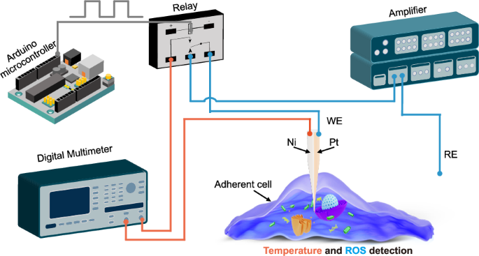
temperature. In the experiment, ROS detection and temperature measurement were selectively controlled using a single-pole-double-throw signal switching relay (G6EU-134P-US, Omron, Japan),
which is programmed by an Arduino microcontroller (Uno R3, Italy). The nanopipette operation was carried out using an automated micromanipulation system previously developed in our lab (Fig.
S1)32. The system can perform image-based detection of both the cell and nanoprobe tip, as well as precise three-dimensional positioning of the nanopipette tip in proximity to the cell of
interest, cell approaching, proximity detection between the nanopipette tip and the cell surface, and cell penetration, with a resolution of 50 nm. Each step of the nanoprobe fabrication
process was characterized, as shown in Fig. 2a. Initially, the nanoprobe tip had a diameter of approximately 100 nm after the pulling process (Fig. 2a i). After sputtering the Pt and Ni
layers, the outer diameter of the nanopipette increased to around 300 nm, and the Pt and Ni layers were connected at the tip of the nanoprobe (Fig. 2a ii). Following the coating of an Al2O3
insulation layer, the nanoprobe diameter expanded to approximately 500 nm (Fig. 2a iii). Energy-dispersive X-ray spectroscopy (EDS) analysis (Fig. 2b) confirmed that the Pt and Ni layers
were continuous and uniformly adhered to the nanopipette. The Pt and Ni layers at the tip were exposed using a FIB coupled with a Helium Ion Microscope (HIM) setup (Fig. 2c). The morphology
of the nanoprobe tip before and after FIB cutting is shown in Fig. 2d, e, and Fig. S2. The two metal layers were bridged at the tip, forming a nano-thermocouple capable of detecting
intracellular temperature with high precision. The small size of the nanoprobe enables high spatial resolution measurements. Meanwhile, it significantly reduces potential cell damage and
supports long-term intracellular analysis. ELECTROCHEMICAL RESPONSE OF THE NANOPROBE To characterize the electrochemical performance of the nanoprobe, the tip of the nanoprobe was placed in
a 1 M KCl solution containing 1 mM Ru(NH3)63+ before and after FIB milling. The cyclic voltammogram in Fig. 3a shows a limiting current of approximately 0.25 nA after FIB milling, while no
obvious current was observed before milling, indicating the steady effectiveness of the insulation layer. The nanoprobe was further characterized in 1 mM H2O2 and phosphate-buffered solution
(PBS) solution before intracellular ROS detection. Compared to PBS, an obvious current increase was observed in H2O2, proving the electrochemical response to H2O2 due to the Pt layer (Fig.
3b). Chronoamperometry experiments were conducted with an overpotential of 0.85 V (Ag/AgCl) to establish a calibration curve. The results showed that the response current for H2O2 increased
linearly with the target analyte concentrations in the range of 0.08–1.28 mM (Fig. 3c). Further experiments were conducted to evaluate the current readouts in the presence of common
disruptors, including various amino acids and ions. Our results demonstrate that the nanoprobes exhibit high selectivity and reliability for ROS detection under conditions that mimic the
cell environment (Fig. S3a). Additionally, we found that temperature variations have little impact on ROS detection (Fig. S3b). TEMPERATURE MEASUREMENT BY THE NANOPROBE Due to the different
thermoelectric properties of Ni and Pt, the Pt/Ni junction at the nanoprobe’s tip generates a thermoelectric potential in response to temperature variations between the tip (the hot point)
and the tail end (the cold point). The hot end functions as the temperature-sensing region, while the cold end serves as the temperature reference. The thermoelectric potential (_E_T) across
a thermocouple can be determined by the equation: $${E}_{{\rm{T}}}=\varDelta S({T}_{{\rm{h}}}-{T}_{{\rm{c}}})$$ where _ΔS_ denotes the thermoelectric sensitivity, _T_h represents the
temperature at the hot end (measured temperature), _T_c indicates the temperature at the cold end (room temperature)33. After coating the Pt and Ni layers on both sides of the nanopipette,
the two metals were bridged at the tip of the nanoprobe, as indicated by the _I_-_V_ curve in Fig. 3d. The resistance of the nanoprobe was 1.05 kΩ (_n_ = 5). We further evaluated the
nanoprobe’s sensitivity, repeatability, stability, and responsiveness to thermal changes. A calibration curve for temperature measurement using the nanoprobe was established in 10 mM PBS, as
shown in Fig. 3e. As the temperature of the solution increased, the _E_T values increased accordingly. A clear linear relationship was observed in the range of 294.15 K to 320.15 K (20–46
°C), with the equation _E_[µv] = 15.22 T–251.05, _R_2 = 0.99. The slope, representing the thermoelectric sensitivity of the sensor, showed excellent consistency and repeatability across five
independently prepared nanoprobes, with an average sensitivity of 15.02 µV/K (n = 5) (Fig. 3f). This experimental value is close to the theoretical sensitivity (15 µV/K), which is
consistent with the Seebeck coefficient of Ni and Pt34. The stability of the nanoprobe was also evaluated. The _E_T remained nearly constant when the nanoprobe was immersed in a 298 K PBS
solution for continuous monitoring for 3000 s (Fig. 3g). The sensitivity of the Pt/Ni nanoprobe showed negligible change over five days, demonstrating its exceptional stability (Fig. 3h).
The anti-interference capability of the nanoprobe was also tested by adding various metal ions, ROS, and amino acids to the solution during temperature measurement. As shown in Fig. 3i, no
discernible alterations were observed, underscoring the high selectivity of the nanoprobe for temperature. To study the influence of metal layer thickness, a 50 nm coating of both Pt and Ni
was applied on the two sides of the nanopipette. The corresponding _I_-_V_ curve is shown in Fig. S4a. The resistance of the nanoprobe (d = 50 nm) was 3.86 kΩ (n = 5) (Fig. S4 b). A
calibration curve for different temperatures was also established (Fig. S5a), with the correlation of _E_[µv] = 13.95 T–2.45, _R_2 = 0.99. The average sensitivity of five independent
nanoprobes is 13.38 µV/K (Fig. S5b), which is slightly lower than that of the nanoprobe with a 100 nm layer. This discrepancy could be attributed to the nanosized effects on electron
transport in the tip region, where the Seebeck coefficients of thin film material are influenced by both the film thickness and the electron mean free path35. The reversibility of the
nanosensor is crucial for real-time, continuous, and accurate monitoring. We observed temperature fluctuations within the range of 298-325 K. As depicted in Fig. S6a, the nanoprobe exhibited
a remarkably reversible temperature response as the tip was periodically inserted into hot water (325 K) and retracted into air (298 K). To evaluate the temporal response of the nanoprobe,
we monitored the temperature fluctuations caused by laser heating of the tip. As shown in Fig. 3j and Fig S6 b, a rapid temperature response was observed. The slight decrease in measured
voltage in Fig S6 b is attributed to the gradual shift in the laser spot’s position relative to the nanoprobe tip, which caused a reduction in the tip’s temperature over time. To investigate
the transient heat transport of the nanoprobe in a cytoplasmic environment, we conducted finite element simulations using COMSOL. Detailed descriptions of the modeling and implementation
are provided in the Supporting Information (Text S1, Fig. S7). Initially, the temperature of the nanoprobe was set to 300 K, while the cell temperature was 301 K. When the nanoprobe tip was
inserted 5 µm into the cell, the simulation showed that the nanoprobe reached the intracellular environment within 3.28 µs (Fig. 3k). We also evaluated the response times for the temperature
differences of 0.5 K and 2 K, which were found to be 2.36 µs and 4.19 µs, respectively (Fig. 3l, Fig. S8, Fig. S9). The development of a bifunctional nanoprobe is crucial due to its ability
to provide more comprehensive insights into cellular function while minimizing cell damage. Conventional approaches often require multiple separate nanoprobes or measurements, which can
disrupt the delicate cellular structure and interfere with the very processes being studied36. This can lead to inaccuracies, particularly when it is difficult to ensure that different
probes are measuring the same intracellular location. The bifunctional nanoprobe reduces the damage to the cell caused by insertion operations. A nanoprobe integrated with both functions
only requires a single insertion to obtain both signals, whereas using two separate nanoprobes would need two insertions. Although it has been reported that the penetration of nanoscale tips
into cells is minimally invasive, repeated penetrations can still affect cell viability and function37. Therefore, while obtaining multiple types of intracellular information, it is
important to minimize the interference caused by the measurements as much as possible. Besides, simultaneous detection improves spatial positioning accuracy. Although cells are small in
volume, their intracellular environment is highly complex. Even for the same biomolecule, such as ROS or Ca²+, or physical factors like temperature, there can be significant differences in
different regions within the cell22,38,39. Therefore, to obtain two or more signals from the same region, the nanoprobe would need to be repeatedly inserted into the same location within the
cell. However, repeated operations make it difficult to ensure consistency in the insertion position, leading to inaccurate signals. INTRACELLULAR SENSING UNDER SYNERGISTIC
PHOTOTHERMAL-/CHEMOTHERAPY Cancer cells typically exhibit higher ROS levels compared to normal cells due to increased metabolic activity and mitochondrial dysfunction. Cancer therapies,
including radiation, immunotherapy, and certain chemotherapeutic agents, can further escalate ROS levels within cancer cells40. Monitoring intracellular temperature and ROS concentration is
crucial for optimizing cancer therapies41. Strategies that modulate these factors, such as combining hyperthermia with ROS-inducing treatments, are actively studied to enhance cancer
therapy42,43. Ti3C2 MXene, a novel two-dimensional material, exhibits excellent biocompatibility and photothermal effects under NIR light. Using DOX as a model drug, Ti3C2@DOX shows high
drug loading capacity and pH-/thermo-sensitivity, where drug release of the nanosystem can be promoted in an acidic tumor microenvironment or by elevated temperature under 980 nm laser
irradiation44. Here, we studied changes in intracellular temperature and ROS during the photothermal-/chemotherapy of cancer cells using a MXene-based therapeutic nanosystem, as illustrated
in Fig. 4a. HeLa cells were incubated with Ti3C2@DOX nanocomposites at a concentration of 20 µg/mL for 10 hours. After incubation, extracellular Ti3C2@DOX was removed by washing with PBS.
The nanoprobe was robotically inserted into the cell. Upon cellular penetration, a significant and immediate increase in the electrochemical current was observed, as shown in Fig. 4b. This
spike indicates the successful interaction of the nanoprobe with the intracellular environment. In contrast, the control group which was not treated with Ti3C2@DOX (Fig S10), exhibited
negligible ROS levels, highlighting the effectiveness of Ti3C2@DOX in elevating ROS concentrations within cancer cells. These results also indicate that the detected signals arise from the
effects of the chemical agents, rather than mechanical stimulation caused by the insertion. Additionally, the experiment was extended to measure ROS levels in 19 cells. The ROS concentration
in the cells was determined based on Faraday law _n_ = _Q/ZF_, where _n_ is the amount of substance, _Q_ is the amount of charge (obtained by integrating the current over time), _F_ is the
Faraday constant (_F_ = 96485 C/mol), and _Z_ is the number of electrons transferred (in this case, _Z_ = 1). Given the average diameter of HeLa cells is approximately 10 µm, we were able to
calculate the corresponding ROS concentration in the cells. The results revealed that Ti3C2@DOX treatment consistently increased intracellular ROS concentration to approximately 0.44 mM, as
depicted in Fig. 4c. These findings provide evidence for the ROS-inducing capability of Ti3C2@DOX and its potential as an effective chemotherapeutic agent. As shown in Fig. 4c, the
insertion of the nanoprobes into cells does not show significant influence to the detected signals. However, the physical insertion of nanoprobes into cells can still potentially induce a
mechanical response. This mechanical stimulation, caused by the deformation or disruption of the cell membrane, could trigger various cellular responses, such as ion channel activation,
changes in membrane tension, or cytoskeletal reorganization45. In general, several strategies can be employed to decouple the effects of mechanical stimulation from chemical stimulation: 1)
Performing control experiments where the nanoprobes are inserted without introducing any chemical stimuli allows for the characterization of the mechanical response alone; 2) Use of
mechanically unresponsive cells (genetic knockouts of key mechanotransduction proteins, e.g., integrins, ion channels like PIEZO146); 3) Temporal analysis of signals: Mechanical responses
are often immediate, while chemical responses, particularly intracellular signaling cascades, may take longer to manifest47. The intracellular temperature was monitored during photothermal
therapy. A 980 nm laser with a power of 10 mW was then used to irradiate the cell, ensuring that the laser beam did not directly impact the nanoprobe tip. Upon activation of the laser, a
significant increase in thermoelectric voltage was observed, corresponding to a temperature rise of approximately 10 K. In contrast, when the probe was positioned outside the cell under the
same experimental conditions, the temperature increase was much smaller, around 3 K (Fig. 4d). Additionally, when the concentration of Ti3C2@DOX was reduced to 5 µg/mL, the intracellular
presence of Ti3C2@DOX was visibly lower compared to the higher concentration group (20 µg/mL), as evidenced by bright-field imaging (Fig. S11). Under these conditions, the cell temperature
increased by only about 4 K (Fig. 4e). These findings indicate that the observed increase in cell temperature is directly related to the presence of Ti3C2@DOX within the cells. Additionally,
we examined the temperature changes within the cells under varying laser intensities. As shown in Fig. 4f, there is a clear correlation between laser intensity and cell temperature, with
higher laser intensities leading to greater temperature increases. These results provide precise measurements of temperature changes at the single-cell level during photothermal therapy,
demonstrating that the temperature rise is primarily due to the amount of photothermal material inside the cells. This multifunctional nanoprobe is capable of accurately measuring both
intracellular temperature changes and ROS levels within cancer cells during photothermal and chemotherapy. The ability to monitor these parameters in real-time offers valuable insights into
the cellular responses to therapy, facilitating the optimization of treatment protocols and enhancing therapeutic efficacy. INTRACELLULAR ANALYSIS OF ZEBRAFISH EMBRYOS UNDER MITOCHONDRIAL
TOXIC STRESS We further employed our nanoprobe to investigate mitochondrial respiration and energy metabolism associated with mitochondrial toxicity in zebrafish embryos. FCCP, a potent
mitochondrial uncoupler, was utilized to induce mitochondrial damage by disrupting the proton gradient within the mitochondria48. The procedure of intracellular sensing is illustrated in
Fig. 5a, b. FCCP was added to the zebrafish embryo medium at a concentration of 60 µM to initiate the stimulation of mitochondrial stress (step i). The nanoprobe was robotically maneuvered
towards a zebrafish embryo. Notably, no significant current changes were observed when the embryo was gently deformed or when the nanoprobe tip was inserted into the perivitelline fluid
areas (step ii and step iii). However, a significant increase in current was detected (at 0.85 V versus Ag/AgCl electrodes) when the nanoprobe tip penetrated the embryo (step iv), as
demonstrated in Fig. 5c (red curve). This substantial current increase signifies the successful detection of elevated ROS levels within the FCCP-treated zebrafish embryos. In a control
experiment where no FCCP was added, no significant current response indicative of increased ROS levels was recorded in individual zebrafish embryos (Fig. 5d, gray curve). Statistical
analysis was performed using five FCCP-treated embryos. As depicted in Fig. 5e, the FCCP-treated group exhibited a noticeable signal increase compared to untreated zebrafish embryos,
confirming the mitochondrial stress-induced ROS production. These results validate the capability of our nanoprobe to quantitatively monitor intracellular ROS in real-time, even in complex,
three-dimensional biological systems such as zebrafish embryos. Sequentially, the nanoprobe was electrically switched to monitor intracellular temperature, and the resulting thermoelectric
potentials were recorded. As depicted in Fig. 5f, a significant elevation in intracellular temperature can be found in FCCP-treated embryos, whereas no notable change was detected in the
control group. The entire process of intracellular temperature change was continuously monitored. It was found that the intracellular thermal signal increased within 3 minutes of FCCP
addition and remained elevated for more than 10 minutes. Similar experiments were conducted on another five FCCP-treated embryos. Compared to the control group, the average temperature
increase of the FCCP-treated group was approximately 1.2 K, as shown in Fig. 5g. Compared to the fluorescent nanogel-based approach for detecting intracellular temperature, the
nanoprobe-based method developed in this study offers superior accuracy and enhanced temporal resolution in detecting intracellular thermal signals. By integrating with a high-precision
robotic nanomanipulation system, high spatiotemporal intracellular sensing in living cells can be achieved. These advancements highlight the nanoprobe’s potential for detailed and dynamic
intracellular analysis, paving the way for more precise and insightful studies of cellular responses under various physiological and pathological conditions. CONCLUSION In summary, this
study has successfully developed an innovative bifunctional nanoprobe capable of real-time monitoring of intracellular ROS and temperature. The nanoprobe, with a precisely engineered tip
size of approximately 500 nm, demonstrates remarkable sensitivity, detecting temperature variations from 20 to 46 °C (294.15 K to 320.15 K) and ROS levels from 0.08 to 1.28 mM. The nanoprobe
exhibits exceptional stability, selectivity, and response time, which are crucial for accurate and reliable intracellular measurements, particularly in dynamic cellular environments. In the
study of photothermal and chemotherapy on HeLa cells, the ROS concentration within the cells was observed to reach up to 0.4 mM during treatment. Notably, a positive correlation was found
between the elevation of cellular temperature and the concentration of photothermal materials within the cells, offering valuable insights into the mechanisms of these therapeutic
approaches. Further experiments conducted on zebrafish embryos demonstrated the nanoprobe’s effectiveness in monitoring fluctuations in intracellular temperature and ROS levels induced by
chemical stimuli, specifically FCCP. The FCCP-treated group showed a marked increase in signal compared to untreated zebrafish embryos, indicating ROS production and temperature elevation
induced by mitochondrial stress. This bifunctional nanoprobe provides real-time, simultaneous measurements of two critical cellular parameters, potentially enhancing disease diagnosis and
treatment strategies by providing more accurate and comprehensive data on cellular responses to various stimuli and therapeutic interventions. MATERIALS AND METHODS MATERIALS AND REAGENTS
H2O2 was purchased from Aladdin Reagent Co., Ltd. (Shanghai, China). FCCP was purchased from GLPBIO. The physiological PBS solution (10 mM, pH 7.4) containing 137 mM NaCl, 2.7 mM KCl, 10 mM
Na2HPO4, 2 mM KH2PO4 was obtained from Solarbio. Ru(NH3)6Cl3 was purchased from Sigma-Aldrich (USA). Zebrafish embryos were provided by Professor Dong Liu from the Department of Life Science
at the Southern University of Science and Technology. Ti3C2@DOX was synthesized following our previously established protocols49. Borosilicate glass capillaries (BF-100-50-10), were
acquired from Sutter Instrument Co. The CO2-based laser puller (P-2000 laser puller, Sutter Instrument) was made available by Professor Yi Li from the Department of Microelectronics at
Southern University of Science and Technology. All electrolyte solutions used in this study were prepared using ultrapure water with a resistivity of 18.2 mΩ cm. FABRICATION OF THE
NANOPROBES The nanoprobes were fabricated from borosilicate glass capillaries through a four-step process. The glass capillaries were first subjected to a plasma cleaning for 5 min to remove
any surface contaminations. The capillaries were then pulled into nanopipettes with tip diameters of approximately 100 nm using a P-2000 laser puller. The specific parameters for the
pulling process were as follows: HEAT: 325; FIL: 3; VEL:30; DEL: 250; and PUL: 150. In the third step, a 5 nm Ti adhesion layer was sputtered onto both sides of the nanopipette by rotating
the nanopipette 180° around its axis, using an electron beam evaporator (HHV, TF500, India). Subsequently, 100 nm layers of Pt and Ni were coated onto both sides of the nanopipette, ensuring
that the tip of the nanopipette was effectively bridged. This dual-metal coating is crucial for the nanoprobe’s functionality as a nano-thermocouple. Finally, a 100 nm layer of Al2O3 was
deposited over the entire nanopipette using atomic layer deposition (ALD), with a deposition rate of approximately 0.1 nm per cycle. This Al2O3 layer serves as an insulation barrier,
enhancing the probe’s durability and performance. The tip of the nanopipette was then precisely milled with focused ion beam helium ion microscopy (FIB-HIM, Orion Nanofab) to expose the
functional tip of the nanoprobe. CHARACTERIZATION OF THE NANOPROBES The morphological characteristics and elemental distribution of the nanoprobe were analyzed using a field-emission
scanning electron microscope (FE-SEM, ZEISS Merlin) equipped with an energy-dispersive spectrometer (EDS, Octane Pro) at an acceleration voltage of 5 kV. All bright field images were
captured with an inverted fluorescence microscope (Axio Observer 7, Zeiss, Germany). All extracellular current measurements were conducted using electrochemical stations: the CHI 760E (CHI
Instrumental, Inc., USA) and the Autolab PGSTAT302N (Metrohm Autolab, Netherlands). A copper wire was carefully attached to the Pt side of the nanoprobe and connected to the electrochemical
station. The electrochemical performance of the nanoprobe was characterized using cyclic voltammetry in a solution of 1 mM Ru(NH3)63+/1 M KCl before and after FIB milling. The scanning
potential was in the range of −0.6 to 0.4 V (versus Ag/AgCl), with a scan rate of 100 mV/s. To evaluate the nanoprobe’s performance in detecting ROS, 1 mM H2O2 and PBS solutions were
employed, with the scanning potential ranging from 0 to 0.8 V at a scan rate of 100 mV/s. Quantitative measurements of various H2O2 concentrations were performed using chronoamperometry at a
fixed potential of 0.85 V. Thermoelectric signals from the nanoprobes were recorded using a KEYSIGHT 34461 A digital multimeter. Two copper wires, attached to opposite sides of the
nanoprobe, were connected to the multimeter. Temperature-controlling experiments were performed using a water bath and calibrated by a commercial mercury thermometer. To assess the
temperature response time of the nanoprobe, a laser beam with a light spot diameter of 10 µm and a wavelength of 980 nm was used. For the selectivity test, the nanoprobe tip was immersed in
an electrochemical cell containing 10 mM PBS (pH 7.4) maintained at a constant temperature. The selectivity of the nanoprobe was evaluated against a panel of potential interferents,
including 1 mM H2O2, ascorbic acid (AA), L-cysteine (L-Cys), Uric Acid (UA), glutathione (GSH), L-arginine(L-Arg), as well as 10 mM glucose (Glu), physiologically relevant concentrations of
ions such as Ca2+, Na+, K+, and Cl−. CELLULAR UPTAKE OF TI3C2@DOX HeLa cells were cultured in Dulbecco’s Modified Eagle Medium (DMEM) supplemented with 10% fetal bovine serum, 100 IU/ml
penicillin, and 100 mg/ml streptomycin in a humidified incubator at 37 °C with 5% CO2. The cells were seeded on a 3 cm TC-treated dish and incubated overnight. Subsequently, the cells were
subjected to Ti3C2@DOX nanoparticles at different concentrations for at least 10 h, allowing sufficient time for cellular uptake and response. Prior to intracellular measurements, the cells
were washed three times with phosphate-buffered saline (PBS) to ensure the complete removal of extracellular Ti3C2@DOX nanoparticles. INTRACELLULAR ROS MEASUREMENT USING THE NANOPROBES All
intracellular current measurements were conducted using an Axopatch 700B low-noise amplifier and an Axon Digidata 1550B low-noise data acquisition system (Molecular Devices, Sunnyvale, CA).
An Ag/AgCl wire served as the reference/counter electrode in these experiments. The whole setup was enclosed in a Faraday cage that was properly grounded to minimize electromagnetic
interference. Signals were sampled at 1 kHz. The nanoprobe was securely fixed under the microscope using a specialized holder (Axon Instruments, Union City, CA). The motion control system
featured two independent micromanipulators (μMp-4, Sensapex, Finland), each offering four degrees of freedom (DOF). These micromanipulators provided a travel range of 20 mm, a motion
resolution of 5 nm, and a maximum speed of 5 mm/s for each degree of freedom. REFERENCES * Guillaume-Gentil, O. et al. Tunable single-cell extraction for molecular analyses. _Cell_ 166,
506–516 (2016). Article Google Scholar * Liu, J., Wen, J., Zhang, Z., Liu, H. & Sun, Y. Voyage inside the cell: Microsystems and nanoengineering for intracellular measurement and
manipulation. _Microsyst. Nanoeng._ 1, 20 (2015). Article Google Scholar * Dong, B. et al. Reversible Self-assembly of nanoprobes in live cells for dynamic intracellular pH imaging. _ACS
Nano._ 13, 1421–1432 (2019). Google Scholar * Qi, Y. T. et al. Homeostasis inside single activated phagolysosomes: quantitative and selective measurements of submillisecond dynamics of
reactive oxygen and nitrogen species production with a nanoelectrochemical Sensor. _J. Am. Chem. Soc._ 139, 13055–13062 (2022). Google Scholar * Singhal, R. et al. Multifunctional
carbon-nanotube cellular endoscopes. _Nat. Nanotechnol._ 6, 57–64 (2011). Article Google Scholar * Aramesh, M. et al. Localized detection of ions and biomolecules with a force-controlled
scanning nanopore microscope. _Nat. Nanotechnol._ 14, 791–798 (2019). Article Google Scholar * Zhang, Y. et al. Fast and sensitive GCaMP calcium indicators for imaging neural populations.
_Nature_ 615, 884–891 (2023). Article Google Scholar * Wang, X. et al. Intracellular manipulation and measurement with multipole magnetic tweezers. _Sci. Robot._ 4, eaav6180 (2019).
Article Google Scholar * Schuerle, S. et al. Robotically controlled microprey to resolve initial attack modes preceding phagocytosis. _Sci. Robot._ 2, eaah6094 (2017). Article Google
Scholar * Liu, H. J., Usprech, J. F., Parameshwar, R. K., Sun, Y. & Simmons, C. A. Combinatorial screen of dynamic mechanical stimuli for predictive control of MSC
mechano-responsiveness. _Sci. Adv._ 7, eabe7204 (2021). Article Google Scholar * Sakaguchi, K. et al. Cell-based microfluidic device utilizing cell sheet technology. _Cyborg. Bionic.
Syst._ 2022, 9758187 (2022). Article Google Scholar * Zhang, Y. et al. High-resolution label-free 3D mapping of extracellular pH of single living cells. _Nat. Commun._ 10, 5610 (2019).
Article Google Scholar * Hanif, S. et al. Organic cyanide decorated SERS active nanopipettes for quantitative detection of hemeproteins and Fe3+ in single cells. _Anal. Chem._ 89,
2522–2530 (2017). Article Google Scholar * Hu, P., Zhang, Y., Wang, D., Qi, G. & Jin, Y. Glutathione content detection of single cells under ingested doxorubicin by functionalized
glass nanopores. _Anal. Chem._ 93, 4240–4245 (2021). Article Google Scholar * Wu, W. T. et al. Large-scale synthesis of functionalized nanowires to construct nanoelectrodes for
intracellular sensing. _Angew. Chem. Int. Ed._ 60, 19337–19343 (2021). Article Google Scholar * Zhu, H. et al. Metabolomic profiling of single enlarged lysosomes. _Nat. Methods_ 18,
788–798 (2021). Article Google Scholar * Pan, R., Xu, M., Jiang, D., Burgess, J. D. & Chen, H. Y. Nanokit for single-cell electrochemical analyses. _Proc. Natl. Acad. Sci._ 113,
11436–11440 (2016). Article Google Scholar * Guo, Z., Ai, N., Ge, W. & Xu, Q. Design of an automated robotic microinjection system for batch injection of zebrafish embryos and larvae.
_Microsyst. Nanoeng._ 10, 20 (2024). Article Google Scholar * Li, Y. et al. Direct electrochemical measurements of reactive oxygen and nitrogen species in nontransformed and metastatic
human breast cells. _J. Am. Chem. Soc._ 139, 13055–13062 (2017). Article Google Scholar * Sies, H. & Jones, D. P. Reactive oxygen species (ROS) as pleiotropic physiological signalling
agents. _Nat. Rev. Mol. Cell. Biol._ 21, 363–383 (2020). Article Google Scholar * Lennicke, C. & Cocheme, H. M. Redox metabolism: ROS as specific molecular regulators of cell signaling
and function. _Mol. Cell_ 81, 3691–3707 (2021). Article Google Scholar * Pak, V. V. et al. Ultrasensitive genetically encoded indicator for hydrogen peroxide identifies roles for the
oxidant in cell migration and mitochondrial function. _Cell Metabolism_ 31, 642–653 (2020). Article Google Scholar * Qi, Y. T. et al. Nanosensor detection of reactive oxygen and nitrogen
species leakage in frustrated phagocytosis of nanofibres. _Nat. Nanotechnol._ 19, 524–533 (2024). Article Google Scholar * Knapp, B. D. & Huang, K. C. The effects of temperature on
cellular physiology. _Annu. Rev. Biophys._ 51, 499–526 (2022). Article Google Scholar * Uchiyama, S., Gota, C., Tsuji, T. & Inada, N. Intracellular temperature measurements with
fluorescent polymeric thermometers. _Chem. Commun._ 53, 10976–10992 (2017). Article Google Scholar * Okabe, K. et al. Intracellular temperature mapping with a fluorescent polymeric
thermometer and fluorescence lifetime imaging microscopy. _Nat. Commun._ 3, 705 (2012). Article Google Scholar * Lemieux, H. & Blier, P. U. Exploring thermal sensitivities and
adaptations of oxidative phosphorylation pathways. _Metabolites_ 12, 360 (2022). Article Google Scholar * Xie, H. et al. Polydopamine-modified 2D iron (II) immobilized MnPS3 nanosheets for
multimodal imaging-guided cancer synergistic photothermal-chemodynamic therapy. _Adv. Sci._ 11, e2306494 (2024). Article Google Scholar * Overchuk, M., Weersink, R. A., Wilson, B. C.
& Zheng, G. Photodynamic and photothermal therapies: synergy opportunities for nanomedicine. _ACS Nano_ 17, 7979–8003 (2023). Article Google Scholar * Wu, L. et al. Combined cellular
thermometry reveals that salmonella typhimurium warms macrophages by inducing a pyroptosis-like phenotype. _J. Am. Chem. Soc._ 144, 19396–19409 (2022). Article Google Scholar * Liu, K. et
al. Spatial analysis of reactive oxygen species in a 3D cell model using a sensitive nanocavity electrode. _Anal. Chem._ 94, 13287–13292 (2022). Article Google Scholar * Hu, W., Ma, Y.,
Zhan, Z., Hussain, D. & Hu, C. Robotic intracellular electrochemical sensing for adherent cells. _Cyborg. Bionic. Syst._ 2022, 9763420 (2022). Article Google Scholar * Wang, F., Han,
Y. & Gu, N. Cell temperature measurement for biometabolism monitoring. _ACS Sens_ 6, 290–302 (2021). Article Google Scholar * Moffat, R. J. Notes on using thermocouples. _Electron.
Cool._ 3, 12–15 (1997). Google Scholar * Zhang, X., Choi, H., Datta, A. & Li, X. Design, fabrication and characterization of metal embedded thin film thermocouples with various film
thicknesses and junction sizes. _J. Micromech. Microeng._ 16, 900–905 (2006). Article Google Scholar * Elnathan, R. et al. Biointerface design for vertical nanoprobes. _Nat. Rev. Mater._
7, 953–973 (2022). Article Google Scholar * Song, J. et al. Ultrasmall nanopipette: toward continuous monitoring of redox metabolism at subcellular level. _Angew. Chem. Int. Ed._ 57,
13226–13230 (2018). Article Google Scholar * Zhou, H. et al. Accurate visualization of metabolic aberrations in cancer cells by temperature mapping with quantum coherence modulation
microscopy. _ACS. Nano_ 17, 8433–8441 (2023). Article Google Scholar * Bagur, R. & Hajnoczky, G. Intracellular Ca2+ sensing: its role in calcium homeostasis and signaling. _Mol. Cell_
66, 780–788 (2017). Article Google Scholar * Dong, A. et al. NIR-II responsive inorganic 2D nanomaterials for cancer photothermal therapy: recent advances and future challenges. _Adv.
Funct. Mater._ 31, 2101625 (2021). Article Google Scholar * Ding, H., Liu, K., Zhao, X., Su, B. & Jiang, D. Thermoelectric nanofluidics probing thermal heterogeneity inside single
cells. _J. Am. Chem. Soc._ 145, 22433–22441 (2023). Article Google Scholar * Chang, M. Y., Hou, Z. Y., Wang, M., Li, C. X. & Lin, J. Recent advances in hyperthermia therapy-based
synergistic immunotherapy. _Adv. Mater._ 33, 2004788 (2020). Article Google Scholar * Zhou, W. et al. Micro-/nano-structures on biodegradable magnesium@PLGA and their cytotoxicity,
photothermal, and anti-tumor effects. _Small Methods_ 5, e2000920 (2021). Article Google Scholar * Liu, G. et al. Surface modified Ti3C2 MXene nanosheets for tumor targeting
photothermal/photodynamic/chemo synergistic therapy. _ACS Appl. Mater. Interfaces_ 9, 40077–40086 (2017). Article Google Scholar * Vining, K. H. & Mooney, D. J. Mechanical forces
direct stem cell behaviour in development and regeneration. _Nat. Rev. Mol. Cell. Biol._ 18, 728–742 (2017). Article Google Scholar * Nourse, J. L. & Pathak, M. M. How cells channel
their stress: Interplay between Piezo1 and the cytoskeleton. _Semin. Cell Dev. Biol._ 71, 3–12 (2017). Article Google Scholar * Ruan, Y. F. et al. An integrated photoelectrochemical
nanotool for intracellular drug delivery and treatment effect evaluation. _Angew. Chem. Int. Ed. Engl._ 60, 25762–25765 (2021). Article Google Scholar * Zhou, C., Zhao, W. X., You, F. T.,
Geng, Z. X. & Peng, H. S. Highly stable and luminescent oxygen nanosensor based on ruthenium-containing metallopolymer for real-time imaging of intracellular oxygenation. _ACS Sens_ 4,
984–991 (2019). Article Google Scholar * Yang, M. et al. MXBOTs: biodegradable Ti3C2 MXene-based microrobots for targeted delivery and synergistic chemo-photothermal therapy. _ACS
Materials Lett_ 6, 1801–1810 (2024). Article Google Scholar Download references ACKNOWLEDGEMENTS This work was supported by the National Key Technologies R&D Program of China (Grant
No. 2023YFC2415900) and Guangdong Basic and Applied Basic Research Foundation (No. 2024A1515011915, No. 2022A1515010098). This work was partly supported by the Science, Technology, and
Innovation Commission of Shenzhen Municipality under grant no. ZDSYS20200811143601004 and in part by the Stable Support Plan Program of Shenzhen Natural Science Fund under grant
20220815104331001. The authors acknowledge the assistance of the SUSTech Core Research Facilities. AUTHOR INFORMATION AUTHORS AND AFFILIATIONS * Shenzhen Key Laboratory of Biomimetic
Robotics and Intelligent Systems, Department of Mechanical and Energy Engineering, Southern University of Science and Technology, Shenzhen, 518000, China Yanmei Ma, Weikang Hu, Jian Hu,
Muyang Ruan, Ming Yang, Yi Zhang, Hanhan Xie & Chengzhi Hu * Department of Electrical and Electronic Engineering, Southern University of Science and Technology, Shenzhen, 518000, China
Jie Hu Authors * Yanmei Ma View author publications You can also search for this author inPubMed Google Scholar * Weikang Hu View author publications You can also search for this author
inPubMed Google Scholar * Jian Hu View author publications You can also search for this author inPubMed Google Scholar * Muyang Ruan View author publications You can also search for this
author inPubMed Google Scholar * Jie Hu View author publications You can also search for this author inPubMed Google Scholar * Ming Yang View author publications You can also search for this
author inPubMed Google Scholar * Yi Zhang View author publications You can also search for this author inPubMed Google Scholar * Hanhan Xie View author publications You can also search for
this author inPubMed Google Scholar * Chengzhi Hu View author publications You can also search for this author inPubMed Google Scholar CONTRIBUTIONS Yanmei Ma: Methodology, Investigation,
Formal analysis, Data curation, Writing - review & editing; Weikang Hu: Methodology, Investigation, Formal analysis, Data curation; Jian Hu: Simulation, Formal analysis; Muyang Ruan:
Formal analysis, Data curation; Characterization; Jie Hu: Data curation; Ming Yang: Methodology; Yi Zhang: Data curation; Hanhan Xie: Data curation; Chengzhi Hu: Supervision, Funding
acquisition, Project administration, Writing - review & Editing, Funding acquisition. CORRESPONDING AUTHOR Correspondence to Chengzhi Hu. ETHICS DECLARATIONS CONFLICT OF INTEREST The
authors declare no competing interests. SUPPLEMENTARY INFORMATION SUPPLEMENTAL MATERIAL RIGHTS AND PERMISSIONS OPEN ACCESS This article is licensed under a Creative Commons Attribution 4.0
International License, which permits use, sharing, adaptation, distribution and reproduction in any medium or format, as long as you give appropriate credit to the original author(s) and the
source, provide a link to the Creative Commons licence, and indicate if changes were made. The images or other third party material in this article are included in the article’s Creative
Commons licence, unless indicated otherwise in a credit line to the material. If material is not included in the article’s Creative Commons licence and your intended use is not permitted by
statutory regulation or exceeds the permitted use, you will need to obtain permission directly from the copyright holder. To view a copy of this licence, visit
http://creativecommons.org/licenses/by/4.0/. Reprints and permissions ABOUT THIS ARTICLE CITE THIS ARTICLE Ma, Y., Hu, W., Hu, J. _et al._ Bifunctional nanoprobe for simultaneous detection
of intracellular reactive oxygen species and temperature in single cells. _Microsyst Nanoeng_ 10, 171 (2024). https://doi.org/10.1038/s41378-024-00814-1 Download citation * Received: 01
August 2024 * Revised: 08 September 2024 * Accepted: 20 September 2024 * Published: 19 November 2024 * DOI: https://doi.org/10.1038/s41378-024-00814-1 SHARE THIS ARTICLE Anyone you share the
following link with will be able to read this content: Get shareable link Sorry, a shareable link is not currently available for this article. Copy to clipboard Provided by the Springer
Nature SharedIt content-sharing initiative