- Select a language for the TTS:
- UK English Female
- UK English Male
- US English Female
- US English Male
- Australian Female
- Australian Male
- Language selected: (auto detect) - EN
Play all audios:
ABSTRACT OBJECTIVE: A better understanding of the processes influencing energy expenditure could provide new therapeutic strategies for reducing obesity. As the metabolic activity of the
brown adipose tissue (BAT) and skeletal muscle is an important determinant of overall energy expenditure and adiposity, we investigated the role of genes that could influence cellular
bioenergetics in these two tissues. DESIGN: We screened for genes that are induced in both the BAT and skeletal muscle during acute adaptive thermogenesis in the mouse by microarray. We used
C57BL/6J mice as well as the primary and immortalized brown adipocytes and C2C12 myocytes to validate the microarray data. Further characterization included gene expression, mitochondrial
density, cellular respiration and substrate utilization. We also used a Hybrid Mouse Diversity Panel to assess _in vivo_ effects on obesity and body fat content. RESULTS: We identified the
transcription factor _Zbtb16_ (also known as _Plzf_ and _Zfp14_) as being induced in both the BAT and skeletal muscle during acute adaptive thermogenesis. _Zbtb16_ overexpression in brown
adipocytes led to the induction of components of the thermogenic program, including genes involved in fatty acid oxidation, glycolysis and mitochondrial function. Enhanced _Zbtb16_
expression also increased mitochondrial number, as well as the respiratory capacity and uncoupling. These effects were accompanied by decreased triglyceride content and increased
carbohydrate utilization in brown adipocytes. Natural variation in _Zbtb16_ mRNA levels in multiple tissues across a panel of >100 mouse strains was inversely correlated with body weight
and body fat content. CONCLUSION: Our results implicate Zbtb16 as a novel determinant of substrate utilization in brown adipocytes and of adiposity _in vivo_. SIMILAR CONTENT BEING VIEWED BY
OTHERS EPAC1 ENHANCES BROWN FAT GROWTH AND BEIGE ADIPOGENESIS Article Open access 09 January 2024 _EGR1_ LOSS-OF-FUNCTION PROMOTES BEIGE ADIPOCYTE DIFFERENTIATION AND ACTIVATION
SPECIFICALLY IN INGUINAL SUBCUTANEOUS WHITE ADIPOSE TISSUE Article Open access 28 September 2020 GRAF1 DEFICIENCY LEADS TO DEFECTIVE BROWN ADIPOSE TISSUE DIFFERENTIATION AND THERMOGENIC
RESPONSE Article Open access 20 November 2024 INTRODUCTION The increasing prevalence of obesity worldwide reflects changes in lifestyle, including a combination of increased food intake and
reduced physical activity.1, 2, 3 This combination contributes to the accumulation of an energy surplus in the form of adipose tissue. Because obesity develops when energy intake exceeds
energy expenditure, increasing energy expenditure is an attractive strategy to reduce body weight and fat storage. There has been considerable interest in modulating thermogenesis,
particularly in the skeletal muscle and brown adipose tissue (BAT), as a target for the treatment of obesity.4, 5, 6 Skeletal muscle is one of the central tissues involved in shivering
thermogenesis and also contributes to non-shivering thermogenesis.5, 7, 8 Skeletal muscle is responsible for at least 30% of the basal metabolic energy expenditure and 80% of whole-body
glucose uptake.9 Because skeletal muscle accounts for approximately 30–40% of total body mass, it may contribute substantially to adaptive thermogenesis in humans.5 Recently, the existence
of BAT in humans has been reappraised and there is good evidence that brown fat depots are active in adults and are capable of energy dissipation through adaptive thermogenesis.10, 11, 12,
13 Furthermore, body-mass index and percentage of body fat are inversely correlated with the BAT in humans12, suggesting that the BAT contributes to metabolic rate and is an important
regulator of body fat accumulation. Further evidence for a shared physiological function between brown adipocytes and myocytes is the fact that they are derived from a common cell
lineage.14, 15 Increased heat production through decreased coupling between fatty acid oxidation and ATP synthesis has been observed during both diet-induced and cold-induced adaptive
thermogenesis.16, 17 During adaptive thermogenesis, a significant amount of energy stored as fat and glycogen is converted to heat instead of being efficiently converted to ATP, suggesting
that induction of mitochondrial respiration or the level of uncoupling activity could promote caloric dissipation. Because of its potential for energy dissipation, it is of great interest to
identify the genes that orchestrate the changes occurring during adaptive thermogenesis and to determine their mechanisms of action. One key factor in adaptive thermogenesis of the BAT is
the mitochondrial _uncoupling protein-1_ (_Ucp1_). _Ucp1_ is responsible for enabling the proton leak in mitochondria that dissipates energy produced through oxidative metabolism to generate
heat.17, 18, 19 Previous systematic studies have identified genes that are induced in BAT after cold exposure in mouse, rat and squirrel,20, 21, 22, 23, 24, 25, 26 and a recent
transcriptome-profiling study identified several genes that are induced in the skeletal muscle and BAT in mice.27 We hypothesized that understanding the function of genes promoting energy
expenditure would reveal new targets for modulation of energy balance. Genes differentially expressed in both the BAT and skeletal muscle could be of great importance because they are
indicative of a general thermogenic response not limited to the BAT-specific uncoupling or skeletal muscle-specific shivering. We performed transcriptional profiling of the BAT and skeletal
muscle during acute cold exposure in mice, a model of adaptive thermogenesis. We identified genes that were induced in both the BAT and skeletal muscle following cold exposure. One of the
significantly upregulated genes is the transcription factor _Zbtb16_, which belongs to the POZ/domain and Krüpel zinc finger family and generally acts as a repressor.28, 29, 30 Here, we
demonstrate that _Zbtb16_ levels influence cellular bioenergetics _in vitro_ and inversely correlate with adiposity _in vivo_. MATERIALS AND METHODS MICE Animal studies were performed under
approved UCLA animal research protocols and according to guidelines established in the Guide for Care and Use of Laboratory Animals. C57BL/6J mice were maintained in 12-h light/dark
conditions and fed a regular chow diet (Purina 50010, Lab Diet, Richmond, IN, USA). Cold exposure was performed as described.31 After euthanasia, tissues were collected and frozen until use.
MICROARRAY ANALYSIS Total RNA was isolated from mouse tissues by extraction with TRIzol (Invitrogen, Carlsbad, CA, USA). Samples were then hybridized to Illumina 24K BeadChips using
standard protocols (The Southern California Genotyping Consortium at UCLA). Differentially expressed genes were identified by applying a Student’s _t_-test and fold-change thresholds. The
microarray data can be accessed from the NCBI Gene Expression Omnibus (GEO) database. CELL CULTURE AND OVER-EXPRESSION Primary brown adipocytes were cultured as described previously.31 C2C12
cells (ATCC, Manassas, VA, USA) were grown in complete DMEM (Dulbecco’s modified Eagle’s medium; 10% fetal bovine serum, 25 mM glucose, 1 mM pyruvate and 1 mM glutamine). For
differentiation, serum was switched to 2% horse serum for 6 days. The immortalized brown adipocyte cell line was a gift from Bruce Spiegelman and was cultured as described.32 Mouse Zbtb16
cDNA was cloned into pcDNA3.1/V5-His vector (Invitrogen) and transfected with BioT (Bioland, La Palma, CA, USA). Empty vector was used as control. For transduction, an adenovirus was
constructed from the Zbtb16 cDNA and the AdEasy XL Adenoviral Vector System (Stratagene, Santa Clara, CA, USA). Viral particles were purified with Adeno-X Maxi Purification Kit (Clontech,
Mountain View, CA, USA) and titered with Adeno-X Rapid Titer Kit (Clontech). Transduction (MOI 50) was performed at day 3 post-confluence and gene expression analyzed at day 6. QUANTITATIVE
REAL-TIME (QRT-PCR) GENE EXPRESSION ANALYSES Total RNA was isolated from mouse tissues by extraction with TRIzol (Invitrogen). For human tissues, a cDNA panel was purchased from Clontech.
cDNA synthesis, real-time PCR, and qRT-PCR were performed as described previously.33 Values are expressed in arbitrary units, normalized to housekeeping genes. All primer sequences used in
this study are presented in Supplementary Table S1. IMMUNOBLOTTING Cells were lysed in lysis buffer containing protease inhibitor cocktail (Roche, Indianapolis, IN, USA). Proteins (50
μg/lane) were resolved by 7% Tris-Acetate pre-cast gel (Invitrogen), transferred to nitrocellulose membrane and incubated for 3 h with 5% milk TBS-T (Tris-buffered saline with Tween 20),
overnight with primary antibody in 1% milk TBS-T, then 1 h with a horseradish peroxidase-conjugated secondary antibody in 1% milk TBS-T. ECL Plus (GE Healthcare, Piscataway, NJ, USA) was
used for chemiluminescent detection. Antibodies included ZBTB16 (OP128, Calbiochem, Billerica, MA, USA), UCP1 (662045, Calbiochem), β-ACTIN (AC-15, Sigma, Dallas, TX, USA) and LAMIN A/C
(sc-6215, Santa Cruz Biotechnology, Santa Cruz, CA, USA). TRIGLYCERIDE (TAG) CONTENTS AND LIPOLYSIS ASSAY Cells were homogenized by sonification and TAG contents determined with L-Type
triglyceride M kit (Waco Chemicals, Richmond, VA, USA). Lipolysis experiments from cell medium were performed after 10 h incubation with the Adipolysis Assay Kit (AB100, Millipore,
Billerica, MA, USA). TAG contents and glycerol concentrations were normalized to protein. CELLULAR OXYGEN CONSUMPTION AND EXTRACELLULAR ACIDIFICATION RATES Cellular metabolic rates were
measured using a XF24 Analyzer (Seahorse Bioscience, Billerica, MA, USA). Immediately before the measurement, cells were washed with unbuffered DMEM as described.34 Plates were placed into
the XF24 instrument for measurement of oxygen consumption and extracellular acidification (ECAR) rates. Mixing, waiting and measurement times were 3, 2, and 3 min for C2C12 and 5, 2 and 1.5
min for brown adipocyte cells, respectively. The measures were normalized per protein. Test compounds were obtained from Sigma and injected during the assay at the following final
concentration: 0.75 or 0.5 μM oligomycin, 0.5 or 0.4 μM FCCP (carbonyl cyanide 4-(trifluoromethoxy)phenylhydrazone), 0.75 or 0.15 μM rotenone/myxothiazol (differentiated brown adipocyte
cells or C2C12, respectively), 25 mM glucose, 100 μM 2,4-dinitrophenol, 50 mM Na-oxamate, and 200 mM 2-deoxyglucose. The basal and percentage of oxygen consumption rate and ECAR levels as
well as area under the curve were obtained with the XF24 Analyzer software. MITOCHONDRIAL DNA CONTENT Total genomic DNA from the BAT tissue was isolated by phenol/chloroform/isoamyl alcohol
extraction. Mitochondrial and nuclear DNA were amplified by RT-PCR with 25 ng of DNA and primers in the D-Loop region and Tert gene, respectively (sequences in Supplementary Table S1).
SUBSTRATE UTILIZATION WITH BIOLOG MICROPLATES PM-M TOX1 microplates (Biolog, Hayward, CA, USA), consisting of eight different substrates in replicate, were used. Differentiated brown
adipocyte cells were seeded at 60 000 cells per well in complete medium without pyruvate, glucose or phenol red. After 24 h, Biolog Redox Dye MA was added and rates measured every hour at
590 nm to detect the metabolic activity. Time 0 was subtracted for each time point. The reductase activity (NADH) is proportional to the energy produced from each substrate. HYBRID MOUSE
DIVERSITY PANEL Mouse strains, RNA isolation and expression profiling were described previously.35 Briefly, at 16 weeks of age, 4–6 mice per strain were assessed for body composition using
an EchoMRI instrument (EchoMRI, Houston, TX, USA). Mice were fasted overnight before plasma and tissue collection. Tissues were weighed and then snap frozen in liquid nitrogen until RNA,
cRNA and cDNA were prepared. Biotin-labeled cRNA was generated from the cDNA and used to probe by using Affymetrix Mouse Genome HT_MG430A arrays (4–6 mice per strain). Expression data (probe
1442025_a_at) and phenotypic traits are presented in Supplementary Table S2. STATISTICAL ANALYSIS The data are expressed as the mean±s.d., except for the XF24 Seahorse Bioscience analysis
where s.e. was used. Unpaired Student’s _t_-test was used to compare the difference between the groups. RESULTS IDENTIFYING GENES DIFFERENTIALLY REGULATED IN ADAPTIVE THERMOGENESIS We
assayed the transcriptomes of mice exposed to cold using gene expression microarrays to identify genes differentially expressed during thermogenesis. In the BAT, 124 genes were upregulated
and 176 were downregulated at least twofold in response to cold exposure. In the skeletal muscle (quadriceps), 69 genes were upregulated and 67 were downregulated at least twofold in
response to cold exposure. We detected 11 genes that were altered by cold exposure in both the BAT and skeletal muscle, 5 of which were coordinately upregulated and 6 downregulated (Table
1). The transcription factor _Zbtb16_ (also known as _Plzf_ and _Zfp145_) exhibited a robust induction in both the BAT (5.6-fold) and skeletal muscle (4.8-fold). _Zbtb16_ has been associated
with white adipogensis36 but has not previously been implicated in the BAT or muscle metabolism. We selected this gene to further characterize its function. VALIDATION OF _ZBTB16_ INDUCTION
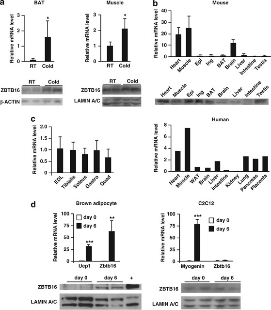
IN ADAPTIVE THERMOGENESIS We first confirmed that _Zbtb16_ expression is consistently upregulated by cold exposure at the mRNA and protein levels in an independent group of mice. As shown
in Figure 1a, _Zbtb16_ mRNA levels were increased 15.3-fold in the BAT and 2.1-fold in the muscle as detected by qPCR. _Zbtb16_ mRNA induction by cold was observed after 4 or 8 h and whether
mice were fasted or not during the cold exposure (data not shown). Western blot analysis confirmed a concomitant increase in ZBTB16 protein levels (Figure 1a, bottom inserts). Thus, the
upregulation of _Zbtb16_ during adaptive thermogenesis was validated at both the transcript and protein levels. TISSUE EXPRESSION PATTERN OF _ZBTB16_ IN MOUSE AND HUMAN We then investigated
whether the BAT and skeletal muscle were the most biologically relevant tissues for studying _Zbtb16_ function by comparing mRNA expression levels in mouse and human tissue panels by qPCR.
The highest _Zbtb16_ expression levels were detected in the skeletal muscle and heart for both mouse and human (Figure 1b). In contrast to what Mikkelsen, _et al._36 reported, we detected a
lower expression level in the mouse BAT than in the skeletal muscle. However, the levels of ZBTB16 protein measured by immunoblot in the white adipose tissue and BAT were similar to those of
the heart and skeletal muscle (Figure 1b, insert). Within the muscle, the expression of _Zbtb16_ was similar in fast twitch (extensor digitorum longus and tibialis), slow twitch (soleus)
and mixed (quadriceps and gastronecmius) muscles (Figure 1c), suggesting that Zbtb16 may function across muscle fiber types. EXPRESSION OF _ZBTB16_ DURING BROWN ADIPOCYTE AND MYOCYTE
DIFFERENTIATION We then studied Zbtb16 function _in vitro_ using a mouse brown adipocyte cell line32 and the mouse C2C12 myocyte cell line. We first assessed _Zbtb16_ expression dynamics
during differentiation of brown adipocyte precursors into mature brown adipocytes. _Zbtb16_ expression was dramatically upregulated 60-fold during brown adipocyte differentiation, whereas
_Ucp1_ showed a 30-fold induction (Figure 1d). By contrast, Zbtb16 mRNA levels did not change during differentiation of C2C12 myoblasts to myotubes, whereas myogenin, used as control, was
dramatically induced. The increased mRNA levels in brown adipocytes were mirrored by a substantial increase in ZBTB16 protein levels (Figure 1d, bottom inserts). The induction of _Zbtb16_
during brown adipogenesis is consistent with a role for this protein in mature brown adipocytes. _ZBTB16_ EXPRESSION ACTIVATES THE BROWN ADIPOCYTE GENE EXPRESSION PROGRAM To begin to
elucidate the effect of _Zbtb16_ induction during thermogenesis, we overexpressed Zbtb16 in brown adipocyte cells using an adenoviral vector carrying _Zbtb16_ cDNA. We achieved roughly
sixfold overexpression of _Zbtb16_ mRNA (Figure 2a), which led to alterations in expression of genes involved in brown adipocyte differentiation and mitochondrial function. Indeed, compared
with cells infected with a control lacZ-containing adenovirus, cells overexpressing _Zbtb16_ exhibited significant upregulation of brown fat-enriched markers such as _Ucp1_, _Ppargc1a_,
_Ppara_ and _Prdm16_ (_PR domain containing 16_; Figure 2b). Enhanced _Zbtb16_ expression also stimulated the expression of genes promoting fatty acid oxidation, such as _Fabp3 (fatty
acid-binding protein 3_), _Fabp5_ (also known as _Mal1_) and _Cpt1b_ (_carnitine palmitoyltransferase 1b_). _Zbtb16_ expression also elevated expression levels of mitochondrial gene markers
such as _Tfam_ (_mitochondrial transcription factor A_), _Elovl3_ (_elongation of very long chain fatty acids-like 3_), _Cidea_ (_cell death-inducing DFFA-like effector a_), _Cox7a1_
(_cytochrome c oxidase_, _subunit VIIa 1_) and _Cox8b_ (_cytochrome c oxidase_, _subunit VIIIb_). To confirm these effects observed in an immortalized brown adipocyte cell line, we performed
similar studies in primary brown adipocytes. Zbtb16 adenoviral expression induced UCP1 protein levels (Figure 2c) and increased expression of brown fat, fatty acid oxidation and
mitochondrial genes (Figure 2d). Thus, _Zbtb16_ expression promotes a gene expression signature characteristic of active brown adipocytes and enhanced fatty acid oxidation. ZBTB16 ENHANCES
MITOCHONDRIAL RESPIRATION AND MITOCHONDRIAL BIOGENESIS The effect of Zbtb16 on mitochondrial gene expression described above suggested that it may promote changes in brown adipocyte
energetics. To assess this, we measured cellular respiration in brown adipocytes by real-time detection of oxygen consumption. Using a XF24 Seahorse Bioscience instrument, we determined the
level of different components of mitochondrial respiration contributing to the oxygen consumption using specific effectors. First, we determined the basal and coupling state of mitochondrial
respiration with sequential injections of oligomycin and rotenone/myxothiazol. Oligomycin (F1F0-ATP synthase inhibitor) allows the measurement of ATP-linked oxygen consumption (ATP
turnover) and rotenone/myxothiazol (complex I and III inhibitor, respectively) by blocking mitochondrial respiration infers the non-mitochondrial respiration and the mitochondrial uncoupling
(difference between oligomycin and rotenone/myxothiazol injections). Cells were ‘activated’ 2 h before the first measurement with 10 nM of the adrenoceptor agonist CL316,243. In both the
immortalized and primary brown adipocytes, _Zbtb16_ overexpression did not alter basal mitochondrial respiration, but showed higher mitochondrial uncoupling at the expense of coupling
respiration (Figures 3a and b). C2C12 cells can also be ‘activated’ by beta-adrenergic stimulation when they are differentiated in myotubes.37 When we pretreated the myotubes for 1 h with
100 nM isoproterenol, we also observed a significant increase in mitochondrial uncoupling owing to _Zbtb16_ expression (Figure 3c). We also assessed mitochondrial reserve capacity by
injecting the electron transport uncoupler FCCP. Brown adipocytes (primary and immortalized) and C2C12 cells over-expressing _Zbtb16_ exhibited a higher FCCP response, indicating more
mitochondrial respiratory capacity (Figure 3d). Altogether, these results demonstrate that Zbtb16 is able to promote proton leak during mitochondrial respiration and increase mitochondrial
respiration capacity. The enhanced expression of mitochondrial gene markers and mitochondrial respiration capacity suggested a potential effect of Zbtb16 on mitochondrial biogenesis. To
assess mitochondrial density, we measured the relative abundance of mitochondrial-encoded gene _D-loop_ to the nuclear-encoded telomerase reverse transcriptase (_Tert_) by qPCR. _Zbtb16_
expression resulted in increased mitochondrial DNA by threefold in brown adipocytes and by 1.7-fold in C2C12 myotubes (Figure 3e). We next asked whether the increased mitochondrial
respiration owing to _Zbtb16_ overexpression affects lipolysis or lipid content in brown adipocytes. Lipolysis rates in brown adipocytes were not affected by _Zbtb16_ expression, whereas
forskolin elicited the expected increase (Figure 3f). However, intracellular TAG content was decreased by 40% in _Zbtb16_ overexpressing cells (Figure 3g). Together, these data demonstrate
that _Zbtb16_ promotes biogenesis and respiration of mitochondria, and that higher expression of _Zbtb16_ reduces cellular TAG levels in brown adipocytes. ZBTB16 INCREASES GLUCOSE OXIDATION
We then investigated whether _Zbtb16_ affects substrate utilization. Control brown adipocytes or Zbtb16 overexpressing cells were plated in Biolog microplates in which each well contains one
of eight specific energy substrates. All the eight substrates were metabolized by brown adipocytes (Figure 4a and Supplementary Figure S1). Overexpression of _Zbtb16_ specifically increased
utilization of glucose (glycolysis pathway) and xylitol (pentose phosphate pathway), while metabolism of other substrates was not affected by _Zbtb16_. The preference in carbohydrate
utilization suggests that _Zbtb16_ may influence glucose metabolism. Therefore, we assayed the expression of genes controlling glycolysis in response to _Zbtb16_ overexpression using qPCR.
Levels of _Glut4_ mRNA were not changed, but _Hexkin2_ (_hexokinase II_) and _Pkm2_ (_pyruvate kinase_) expression levels were increased by _Zbtb16_ (Figure 4b). In addition, expression of
_Pdk4_ was decreased. Increased glucose utilization and the differential expression of glycolysis-specific enzymes support a role for _Zbtb16_ in glucose consumption. We then assessed the
effects of _Zbtb16_ overexpression on glucose metabolism by quantitating a measure of glycolysis, the ECAR. Addition of glucose or the mitochondrial uncoupler 2,4-dinitrophenol increased
ECAR in differentiated brown adipocytes. This response was amplified in cells with increased _Zbtb16_ expression, suggesting a greater ability to increase glycolysis (Figure 4c). We
therefore exposed the brown adipocytes to different modulators of glycolysis and measured the ECAR response. Cells overexpressing _Zbtb16_ had a greater response to treatment with oligomycin
which inhibits mitochondrial ATP production, suggesting an increased glycolytic capacity when ATP demand increases (Figure 4d). When treated with the glucose analog 2-deoxyglucose, which
inhibits glycolysis, the effect was slightly less pronounced in cells expressing _Zbtb16_ (Figure 4d). We also performed experiments with Na-oxamate, an inhibitor of lactate dehydrogenase,
which catalyzes the conversion of pyruvate to lactic acid. Na-oxamate did not significantly inhibit ECAR in brown adipocytes (∼7%), suggesting that the final step of glycolysis does not
account for a substantial amount of glycolysis detected by the ECAR and that anerobic glycolysis is not very active in these cells. Nevertheless, this modest inhibition was abrogated when
_Zbtb16_ was overexpressed (Figure 4d). Together, these results suggest that _Zbtb16_ promotes glucose oxidation, consistent with the increased mitochondrial respiration and lower _Pdk4_
expression. ZBTB16 EXPRESSION CORRELATES WITH BODY WEIGHT, FAT MASS AND DIABETES _IN VIVO_ As described above, our data indicate that _Zbtb16_ is involved in the adaptive thermogenesis
response and promotes mitochondrial respiration and carbohydrate utilization in brown adipocytes. To assess the role of Zbtb16 on energy balance _in vivo_, we assessed whether genetic
variations in _Zbtb16_ gene expression levels are correlated with clinical traits. For these studies, we examined _Zbtb16_ expression levels in a Hybrid Mouse Diversity Panel.35 This
resource consists of more than 100 highly diverse inbred and recombinant inbred mouse strains, which allow the analysis of association between gene expression levels and metabolic traits
such as plasma lipids, plasma hormones and different body fat parameters.35, 38 We investigated the correlations between _Zbtb16_ expression levels and obesity traits in two tissues
available in this panel that are most relevant to our study: as the BAT and skeletal muscle were not available in the hybrid mouse diversity panel, we examined the white adipose tissue and
heart. The most robust associations for _Zbtb16_ were inverse correlations with body weight and fat mass (measured by nuclear magnetic resonance and weight) in both the tissues (Table 2 and
Supplementary Figure S2). The inverse correlation for fat mass included gonadal, retroperitoneal, femoral and mesenteric fat pads. This finding demonstrates that common, natural variations
in _Zbtb16_ expression levels are correlated with body fat content in the mouse. Given the association of Zbtb16 mRNA levels with body weight and fat mass, we also assessed whether _ZBTB16_
expression levels are associated with fat mass in humans as well. Data mining from previously reported microarray analyses revealed that in visceral adipose tissue of obese diabetic women,
_ZBTB16_ expression was significantly lower than age- and body-mass index-matched normal glucose-tolerant controls (_t_-test _P_-value=0.0056; GEO accession GDS3665). This result
demonstrates that in humans, _ZBTB16_ expression levels are associated with glucose levels, suggesting a complex interrelationship between _ZBTB16_ expression, glucose and obesity.
DISCUSSION Manipulation of cellular bioenergetics is an attractive approach to increase cellular energy expenditure with the ultimate goal to decrease obesity at the whole body level.
Mitochondrial respiration, primarily in the BAT and skeletal muscle, can be modified in order to respond to physical activity, diet or ambient temperature. In the current work, we identified
_Zbtb16_ as a novel gene involved in the cold response in both the BAT and skeletal muscle. _Zbtb16_ was among a few genes that were transcriptionally induced by several-fold in both the
BAT and skeletal muscle during acute cold exposure. Previous works on Zbtb16 have shown potential involvement in cancer and development. The human _ZBTB16_ gene has been implicated in acute
promyelocytic leukemia, through chromosomal translocation and fusion to the retinoic receptor alpha.39, 40 Mice lacking _Zbtb16_ function exhibit limb defects and loss of spermatogonial stem
cells.41, 42 Interestingly, Zbtb16 is induced during differentiation of 3T3-L1 preadipocytes.36 The role of Zbtb16 as a transcription factor provides a potential mechanism by which it may
influence thermogenesis, through effects on expression of other genes. Indeed, we found that induction of _Zbtb16_ expression in brown adipocytes enhances the expression of many genes of the
thermogenic program, such as genes involved in fatty acid oxidation and mitochondrial respiration. This effect was accompanied by an increase in mitochondrial content and maximal
respiration capacity, and a decrease in intracellular TAG levels, consistent with a role for _Zbtb16_ in fatty acid metabolism in brown adipocytes. When differentiated brown adipocytes or
myotubes were activated by adrenoceptor agonists, _Zbtb16_ increased mitochondrial uncoupling, in agreement with the increased _Ucp1_ gene expression. Another aspect of the role of _Zbtb16_
is its function in glucose metabolism. _Zbtb16_ expression drives the expression of glycolysis-related genes (_Hexkn2_, _Pkm2_), and is able to increase glycolytic capacity. Consistent with
this role, _Zbtb16_ expression increases carbohydrate utilization _in vitro_. The glucose utilization seems to be directed to mitochondrial oxidation as observed by the decrease in _Pdk4_
expression and lower Na-oxamate-ensitive ECAR levels. Altogether, these results indicate that Zbtb16 promotes oxidative metabolism. Ultimately, the goal is to identify genes that influence
energy metabolism in BAT that translates to whole-body effects on energy balance and adiposity. To evaluate the potential effect of variations in _Zbtb16_ expression levels _in vivo_, we
assessed the relationship between _Zbtb16_ expression levels and body mass and fat pad mass across a set of >100 mouse strains for which gene expression and body fat traits have been
determined.35 In this panel, we observed a striking association between _Zbtb16_ mRNA levels in two different tissues and body fat parameters. Higher levels of _Zbtb16_ expression were
associated with a decrease in body fat storage in four major white adipose tissue depots and overall body weight. We also detected a correlation between decreased _ZBTB16_ expression levels
in human visceral adipose tissue and diabetic state among obese women. These results are consistent with a role for _Zbtb16_ in increasing mitochondrial respiration and substrate
utilization. Interestingly, a congenic rat strain harboring a mutation in _Zbtb16_ affected total body weight and adiposity, but it was not possible to rule out other mutations in the
congenic strain.43 Although global loss of _Zbtb16_ function in mice41 and humans44, 45 causes skeletal defects, more subtle variation in Zbtb16 expression levels observed among individuals
may be a determinant of adiposity. In the future, cell-type–specific knockout or transgenic models could be useful in determining further the role of _Zbtb16_ in adipose tissue function,
energy balance and obesity. ACCESSION CODES ACCESSIONS GENBANK/EMBL/DDBJ * GDS3665 REFERENCES * Wyatt S, Winters KP, Dubbert PM . Overweight and obesity: prevalence, consequences, and causes
of a growing public health problem. _Am J Med Sci_ 2006; 331: 166–174. Article Google Scholar * Flegal KM, Carroll MD, Ogden CL, Curtin LR . Prevalence and trends in obesity among US
adults, 1999-2008. _JAMA_ 2010; 303: 235–241. Article CAS Google Scholar * Hamilton MT, Hamilton DG, Zderic TW . Role of low energy expenditure and sitting in obesity, metabolic syndrome,
type 2 diabetes, and cardiovascular disease. _Diabetes_ 2007; 56: 2655–2667. Article CAS Google Scholar * Costford S, Gowing A, Harper ME . Mitochondrial uncoupling as a target in the
treatment of obesity. _Curr Opin Clin Nutr Metab Care_ 2007; 10: 671–678. Article CAS Google Scholar * Wijers SL, Saris WH, van Marken Lichtenbelt WD . Recent advances in adaptive
thermogenesis: potential implications for the treatment of obesity. _Obes Rev_ 2009; 10: 218–226. Article CAS Google Scholar * Tseng YH, Cypess AM, Kahn CR . Cellular bioenergetics as a
target for obesity therapy. _Nat Rev Drug Discov_ 2010; 9: 465–482. Article CAS Google Scholar * Clarke IJ, Henry BA . Targeting energy expenditure in muscle as a means of combating
obesity. _Clin Exp Pharmacol Physiol_ 2010; 37: 121–124. Article CAS Google Scholar * van den Berg SA, van Marken Lichtenbelt W, Willems van Dijk K, Schrauwen P . Skeletal muscle
mitochondrial uncoupling, adaptive thermogenesis and energy expenditure. _Curr Opin Clin Nutr Metab Care_ 2011; 14: 243–249. Article Google Scholar * de Lange P, Moreno M, Silvestri E,
Lombardi A, Goglia F, Lanni A . Fuel economy in food-deprived skeletal muscle: signaling pathways and regulatory mechanisms. _FASEB J_ 2007; 21: 3431–3441. Article CAS Google Scholar *
Nedergaard J, Bengtsson T, Cannon B . Unexpected evidence for active brown adipose tissue in adult humans. _Am J Physiol Endocrinol Metab_ 2007; 293: E444–E452. Article CAS Google Scholar
* Saito M, Okamatsu-Ogura Y, Matsushita M, Watanabe K, Yoneshiro T, Nio-Kobayashi J _et al_. High incidence of metabolically active brown adipose tissue in healthy adult humans: effects of
cold exposure and adiposity. _Diabetes_ 2009; 58: 1526–1531. Article CAS Google Scholar * van Marken Lichtenbelt WD, Vanhommerig JW, Smulders NM, Drossaerts JM, Kemerink GJ, Bouvy ND _et
al_. Cold-activated brown adipose tissue in healthy men. _N Engl J Med_ 2009; 360: 1500–1508. Article CAS Google Scholar * Cypess AM, Lehman S, Williams G, Tal I, Rodman D, Goldfine AB
_et al_. Identification and importance of brown adipose tissue in adult humans. _N Engl J Med_ 2009; 360: 1509–1517. Article CAS Google Scholar * Gesta S, Tseng YH, Kahn CR .
Developmental origin of fat: tracking obesity to its source. _Cell_ 2007; 131: 242–256. Article CAS Google Scholar * Timmons JA, Wennmalm K, Larsson O, Walden TB, Lassmann T, Petrovic N
_et al_. Myogenic gene expression signature establishes that brown and white adipocytes originate from distinct cell lineages. _Proc Natl Acad Sci U S A_ 2007; 104: 4401–4406. Article CAS
Google Scholar * Cannon B, Nedergaard J . Metabolic consequences of the presence or absence of the thermogenic capacity of brown adipose tissue in mice (and probably in humans). _Int J Obes
(Lond)_ 2010; 34 (Suppl 1): S7–16. Article CAS Google Scholar * Cannon B, Nedergaard J . Brown adipose tissue: function and physiological significance. _Physiol Rev_ 2004; 84: 277–359.
Article CAS Google Scholar * Nicholls DG . A history of UCP1. _Biochem Soc Trans_ 2001; 29: 751–755. Article CAS Google Scholar * Klaus S, Casteilla L, Bouillaud F, Ricquier D . The
uncoupling protein UCP: a membraneous mitochondrial ion carrier exclusively expressed in brown adipose tissue. _Int J Biochem_ 1991; 23: 791–801. Article CAS Google Scholar * Puigserver
P, Wu Z, Park CW, Graves R, Wright M, Spiegelman BM . A cold-inducible coactivator of nuclear receptors linked to adaptive thermogenesis. _Cell_ 1998; 92: 829–839. Article CAS Google
Scholar * Daikoku T, Shinohara Y, Shima A, Yamazaki N, Terada H . Specific elevation of transcript levels of particular protein subtypes induced in brown adipose tissue by cold exposure.
_Biochim Biophys Acta_ 2000; 1457: 263–272. Article CAS Google Scholar * Adams SH, Chui C, Schilbach SL, Yu XX, Goddard AD, Grimaldi JC _et al_. BFIT, a unique acyl-CoA thioesterase
induced in thermogenic brown adipose tissue: cloning, organization of the human gene and assessment of a potential link to obesity. _Biochem J_ 2001; 360: 135–142. Article CAS Google
Scholar * Yu XX, Lewin DA, Forrest W, Adams SH . Cold elicits the simultaneous induction of fatty acid synthesis and beta-oxidation in murine brown adipose tissue: prediction from
differential gene expression and confirmation _in vivo_. _FASEB J_ 2002; 16: 155–168. Article Google Scholar * Watanabe M, Yamamoto T, Kakuhata R, Okada N, Kajimoto K, Yamazaki N _et al_.
Synchronized changes in transcript levels of genes activating cold exposure-induced thermogenesis in brown adipose tissue of experimental animals. _Biochim Biophys Acta_ 2008; 1777: 104–112.
Article CAS Google Scholar * Yan J, Burman A, Nichols C, Alila L, Showe LC, Showe MK _et al_. Detection of differential gene expression in brown adipose tissue of hibernating arctic
ground squirrels with mouse microarrays. _Physiol Genomics_ 2006; 25: 346–153. Article CAS Google Scholar * Navet R, Mathy G, Douette P, Dobson RL, Leprince P, De Pauw E _et al_.
Mitoproteome plasticity of rat brown adipocytes in response to cold acclimation. _J Proteome Res_ 2007; 6: 25–33. Article CAS Google Scholar * Anunciado-Koza RP, Zhang J, Ukropec J,
Bajpeyi S, Koza RA, Rogers RC _et al_. Inactivation of the mitochondrial carrier SLC25A25 (ATP-Mg2+/Pi transporter) reduces physical endurance and metabolic efficiency in mice. _J Biol Chem_
2011; 286: 11659–111671. Article CAS Google Scholar * Filipponi D, Hobbs RM, Ottolenghi S, Rossi P, Jannini EA, Pandolfi PP _et al_. Repression of kit expression by Plzf in germ cells.
_Mol Cell Biol_ 2007; 27: 6770–6781. Article CAS Google Scholar * Schefe JH, Menk M, Reinemund J, Effertz K, Hobbs RM, Pandolfi PP _et al_. A novel signal transduction cascade involving
direct physical interaction of the renin/prorenin receptor with the transcription factor promyelocytic zinc finger protein. _Circ Res_ 2006; 99: 1355–1366. Article CAS Google Scholar * Li
JY, English MA, Ball HJ, Yeyati PL, Waxman S, Licht JD . Sequence-specific DNA binding and transcriptional regulation by the promyelocytic leukemia zinc finger protein. _J Biol Chem_ 1997;
272: 22447–2255. Article CAS Google Scholar * Vergnes L, Chin R, Young SG, Reue K . Heart-type fatty acid-binding protein is essential for efficient brown adipose tissue fatty acid
oxidation and cold tolerance. _J Biol Chem_ 2011; 286: 380–390. Article CAS Google Scholar * Uldry M, Yang W, St-Pierre J, Lin J, Seale P, Spiegelman BM . Complementary action of the
PGC-1 coactivators in mitochondrial biogenesis and brown fat differentiation. _Cell Metab_ 2006; 3: 333–341. Article CAS Google Scholar * Phan J, Peterfy M, Reue K . Lipin expression
preceding peroxisome proliferator-activated receptor-gamma is critical for adipogenesis _in vivo_ and _in vitro_. _J Biol Chem_ 2004; 279: 29558–29564. Article CAS Google Scholar * Wu M,
Neilson A, Swift AL, Moran R, Tamagnine J, Parslow D _et al_. Multiparameter metabolic analysis reveals a close link between attenuated mitochondrial bioenergetic function and enhanced
glycolysis dependency in human tumor cells. _Am J Physiol Cell Physiol_ 2007; 292: C125–C136. Article CAS Google Scholar * Bennett BJ, Farber CR, Orozco L, Kang HM, Ghazalpour A, Siemers
N _et al_. A high-resolution association mapping panel for the dissection of complex traits in mice. _Genome Res_ 2010; 20: 281–290. Article CAS Google Scholar * Mikkelsen TS, Xu Z, Zhang
X, Wang L, Gimble JM, Lander ES _et al_. Comparative epigenomic analysis of murine and human adipogenesis. _Cell_ 2010; 143: 156–169. Article CAS Google Scholar * Pearen MA, Ryall JG,
Maxwell MA, Ohkura N, Lynch GS, Muscat GE . The orphan nuclear receptor, NOR-1, is a target of beta-adrenergic signaling in skeletal muscle. _Endocrinology_ 2006; 147: 5217–5227. Article
CAS Google Scholar * Schadt EE, Lamb J, Yang X, Zhu J, Edwards S, Guhathakurta D _et al_. An integrative genomics approach to infer causal associations between gene expression and disease.
_Nat Genet_ 2005; 37: 710–717. Article CAS Google Scholar * McConnell MJ, Licht JD . The PLZF gene of t (11;17)-associated APL. _Curr Top Microbiol Immunol_ 2007; 313: 31–48. CAS PubMed
Google Scholar * Costoya JA, Pandolfi PP . The role of promyelocytic leukemia zinc finger and promyelocytic leukemia in leukemogenesis and development. _Curr Opin Hematol_ 2001; 8:
212–217. Article CAS Google Scholar * Barna M, Hawe N, Niswander L, Pandolfi PP . Plzf regulates limb and axial skeletal patterning. _Nat Genet_ 2000; 25: 166–172. Article CAS Google
Scholar * Costoya JA, Hobbs RM, Barna M, Cattoretti G, Manova K, Sukhwani M _et al_. Essential role of Plzf in maintenance of spermatogonial stem cells. _Nat Genet_ 2004; 36: 653–659.
Article CAS Google Scholar * Seda O, Liska F, Krenová D, Kazdová L, Sedová L, Zima T _et al_. Differential linkage of triglyceride and glucose levels on rat chromosome 4 in two
segregating rat populations. _Folia Biol (Praha)_ 2003; 49: 223–226. CAS Google Scholar * Wieczorek D, Koster B, Gillessen-Kaesbach G . Absence of thumbs, A/hypoplasia of radius,
hypoplasia of ulnae, retarded bone age, short stature, microcephaly, hypoplastic genitalia, and mental retardation. _Am J Med Genet_ 2002; 108: 209–213. Article Google Scholar * Fischer S,
Kohlhase J, Böhm D, Schweiger B, Hoffmann D, Heitmann M _et al_. Biallelic loss of function of the promyelocytic leukaemia zinc finger (PLZF) gene causes severe skeletal defects and genital
hypoplasia. _J Med Genet_ 2008; 45: 731–737. Article CAS Google Scholar Download references ACKNOWLEDGEMENTS We thank Dr Bruce Spiegelman for the kind gift of the mouse brown adipocyte
cell line and Dr Andrea Hevener for providing cDNA for mouse skeletal muscle subtypes. This work was supported by the American Heart Association Grant 09BGIA2080363 (to LV), National
Institutes of Health P01 HL28481 (to KR), and the National Center for research Resources Grant S10RR026744 (to KR). AUTHOR INFORMATION Author notes * C L Plaisier Present address: 6Current
address: Institute for Systems Biology, Seattle, WA 98109, USA, * B J Bennett Present address: 7Current address: Department of Genetics, University of North Carolina, Chapel Hill, NC 28081,
USA, AUTHORS AND AFFILIATIONS * Department of Human Genetics, David Geffen School of Medicine, University of California, Los Angeles, CA, USA C L Plaisier, B J Bennett, A J Lusis, K Reue
& L Vergnes * Department of Applied Genomics, Bristol Myers-Squibb, Pennington, NJ, USA A He & B Guan * Department of Medicine, University of California, Los Angeles, CA, USA A J
Lusis & K Reue * Department of Microbiology, Immunology and Molecular Genetics, David Geffen School of Medicine, University of California, Los Angeles, CA, USA A J Lusis * Department of
Molecular Biology Institute, University of California, Los Angeles, CA, USA A J Lusis & K Reue Authors * C L Plaisier View author publications You can also search for this author
inPubMed Google Scholar * B J Bennett View author publications You can also search for this author inPubMed Google Scholar * A He View author publications You can also search for this author
inPubMed Google Scholar * B Guan View author publications You can also search for this author inPubMed Google Scholar * A J Lusis View author publications You can also search for this
author inPubMed Google Scholar * K Reue View author publications You can also search for this author inPubMed Google Scholar * L Vergnes View author publications You can also search for this
author inPubMed Google Scholar CORRESPONDING AUTHOR Correspondence to L Vergnes. ETHICS DECLARATIONS COMPETING INTERESTS The authors declare no conflict of interest. ADDITIONAL INFORMATION
Supplementary Information accompanies the paper on the Nutrition and Diabetes website SUPPLEMENTARY INFORMATION SUPPLEMENTARY FIGURE S1 (PDF 57 KB) SUPPLEMENTARY FIGURE S2 (PDF 112 KB)
SUPPLEMENTARY TABLE S1 (DOC 44 KB) SUPPLEMENTARY TABLE S2 (XLS 57 KB) RIGHTS AND PERMISSIONS This work is licensed under the Creative Commons Attribution-NonCommercial-No Derivative Works
3.0 Unported License. To view a copy of this license, visit http://creativecommons.org/licenses/by-nc-nd/3.0/ Reprints and permissions ABOUT THIS ARTICLE CITE THIS ARTICLE Plaisier, C.,
Bennett, B., He, A. _et al._ Zbtb16 has a role in brown adipocyte bioenergetics. _Nutr & Diabetes_ 2, e46 (2012). https://doi.org/10.1038/nutd.2012.21 Download citation * Received: 16
August 2012 * Accepted: 18 August 2012 * Published: 17 September 2012 * Issue Date: September 2012 * DOI: https://doi.org/10.1038/nutd.2012.21 SHARE THIS ARTICLE Anyone you share the
following link with will be able to read this content: Get shareable link Sorry, a shareable link is not currently available for this article. Copy to clipboard Provided by the Springer
Nature SharedIt content-sharing initiative KEYWORDS * Zbtb16 * Plzf * brown adipose tissue * adaptive thermogenesis * bioenergetics