
- Select a language for the TTS:
- UK English Female
- UK English Male
- US English Female
- US English Male
- Australian Female
- Australian Male
- Language selected: (auto detect) - EN
Play all audios:
ABSTRACT Cleavage of eukaryotic translation initiation factor 4G (eIF4G) by enterovirus proteases during infection leads to the shutoff of cellular cap-dependent translation, but does not
affect the initiation of cap-independent translation of mRNAs containing an internal ribosome entry site (IRES). Death-associated protein 5 (DAP5), a structural homolog of eIF4G, is a
translation initiation factor specific for IRES-containing mRNAs. Coxsackievirus B3 (CVB3) is a positive single-stranded RNA virus and a primary causal agent of human myocarditis. Its RNA
genome harbors an IRES within the 5′-untranslated region and is translated by a cap-independent, IRES-driven mechanism. Previously, we have shown that DAP5 is cleaved during CVB3 infection.
However, the protease responsible for cleavage, cleavage site and effects on the translation of target genes during CVB3 infection have not been investigated. In the present study, we
demonstrated that viral protease 2A but not 3C is responsible for DAP5 cleavage, generating 45- and 52-kDa N- (DAP5-N) and C-terminal (DAP5-C) fragments, respectively. By site-directed
mutagenesis, we found that DAP5 is cleaved at amino acid G434. Upon cleavage, DAP5-N largely translocated to the nucleus at the later time points of infection, whereas the DAP5-C largely
remained in the cytoplasm. Overexpression of these DAP5 truncates demonstrated that DAP5-N retained the capability of initiating IRES-driven translation of apoptosis-associated p53, but not
the prosurvival Bcl-2 (B-cell lymphoma 2) when compared with the full-length DAP5. Similarly, DAP5-N expression promoted CVB3 replication and progeny release; on the other hand, DAP5-C
exerted a dominant-negative effect on cap-dependent translation. Taken together, viral protease 2A-mediated cleavage of DAP5 results in the production of two truncates that exert
differential effects on protein translation of the IRES-containing genes, leading to enhanced host cell death. SIMILAR CONTENT BEING VIEWED BY OTHERS COXSACKIEVIRUS INFECTION INDUCES A
NON-CANONICAL AUTOPHAGY INDEPENDENT OF THE ULK AND PI3K COMPLEXES Article Open access 04 November 2020 NOROVIRUS MLKL-LIKE PROTEIN INITIATES CELL DEATH TO INDUCE VIRAL EGRESS Article 29
March 2023 DIRECT CLEAVAGE OF CASPASE-8 BY HERPES SIMPLEX VIRUS 1 TEGUMENT PROTEIN US11 Article Open access 19 July 2022 MAIN Coxsackievirus B3 (CVB3), a primary cause of viral myocarditis,
is associated with sudden, unexpected death,1 dilated cardiomyopathy and heart failure.2 The CVB3 genome consists of a single open reading frame, which is translated to a polyprotein, and
subsequently processed by viral proteases 2A and 3C. Similar to other picornaviruses, the CVB3 genome is linked to a small viral peptide, Vpg, rather than a 7-methyl guanosine cap structure
at its 5′ terminus. Thus, CVB3 RNA is translated by a cap-independent mechanism and is driven by an internal ribosome entry site (IRES) harbored in the 5′ untranslated region (5′-UTR).3, 4
Viral proteases actively suppress multiple host cell activities that help the virus evade host defense mechanisms, promote viral replication and promote host cell apoptosis. For example,
enterovirus proteases can cleave eukaryotic translation initiation factors 4GI (eIF4GI)5, 6, 7 and eIF4GII.8, 9 Picornavirus proteases have also been reported to cleave other canonical
translation initiation factors in the cap-dependent translation initiation complex, such as eIF5B10 and eIF4A.11 Additionally, viral protease 3C has been demonstrated to cleave Ras-GAP SH3
domain-binding protein 1 (G3BP1), a key nucleating protein in stress granule formation, at late time points of infection.12, 13 G3BP1 cleavage causes stress granule disassembly12 and leads
to the release of translation initiation factors that may be hijacked for viral polyprotein translation. Accumulating evidence implies that viral proteases have crucial roles in modulating
viral and host gene expression through cleavage of various host protein factors involved in mRNA transcription and cap-dependent translation. Death-associated protein 5 (DAP5) is a
eukaryotic translation initiation factor that preferentially initiates cap-independent translation.14 This 97-kDa protein is homologous with the central region of canonical translation
initiation factor eIF4GI. DAP5 contains binding sites for eIF4A and eIF3, but lacks an eIF4E site, the mRNA cap-binding protein.14 DAP5 initiates translation of proteins specifically
expressed during development, cell cycle regulation and endoplasmic reticulum (ER) stress conditions, where global cap-dependent translation is compromised.15, 16 It has been reported that
DAP5 regulates IRES-driven translation of Bcl-2 (B-cell lymphoma 2), p53, XIAP (X-linked inhibitor of apoptosis protein), CDK1 (cyclin-dependent kinase 1), c-Myc and other IRES-containing
genes under conditions of stress, growth and apoptosis.17, 18, 19 DAP5 mRNA also contains an IRES in its 5′-UTR, and is proficient in promoting its own translation, generating a positive
feedback loop for its expression.14, 15 Additionally, DAP5 may undergo post-translational modifications, such as cleavage by caspases during apoptosis, generating a truncated, yet functional
86-kDa form of DAP5 that is more potent and efficient at translation initiation.14 The role of DAP5 in initiation of IRES-driven translation has been studied in various model systems but
not in the setting of viral infection. In the present study, we demonstrated that DAP5 is cleaved during CVB3 infection by protease 2A but not 3C. Further, we identified amino-acid reside
G434 as the site of 2A cleavage. Once cleaved, DAP5 N-terminal truncate (DAP5-N) largely translocates into the nucleus, whereas the DAP5 C-terminal truncate (DAP5-C) remains primarily within
the cytoplasm. Our data indicate that DAP5-N retains IRES-mediated translation; however, it differentially regulates the IRES-containing genes involved in cell growth and apoptosis compared
with the wild type (WT) DAP5. Exogenous expression of DAP5-N or DAP5-C induces more cell death when compared with the WT DAP5. Further, viral capsid protein 1 (VP1) as well as viral titer
is higher in DAP5-N-overexpressing cells during the late phase of infection (7 h post infection (hpi)) compared with that in WT (WT DAP5) or an uncleavable point mutant of DAP5 (glycine 434
to glutamic acid-mutated DAP5 (G434E DAP5)). Thus, viral protease 2A cleavage of DAP5 produces a cleavage product that robustly contributes to viral replication and viral-induced apoptosis,
facilitating CVB3 progeny release. RESULTS DAP5 IS CLEAVED DURING CVB3 INFECTION IN TISSUE CULTURE AND IN MOUSE HEART 
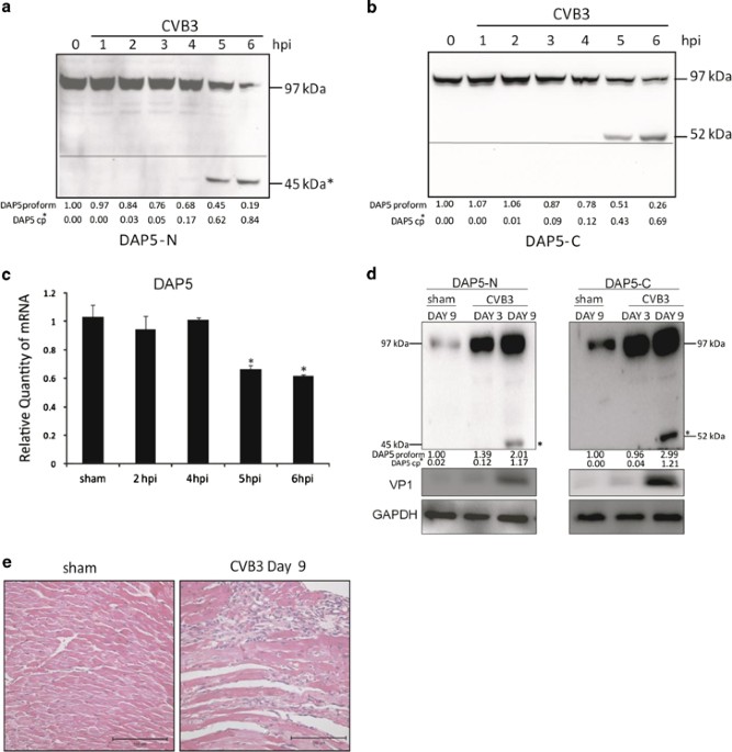
During CVB3 infection, we observed cleavage of DAP5 indicated by
accumulation of ~45-kDa DAP5-N and ~52-kDa DAP5-C at 5 hpi in HeLa cells (Figures 1a and b). This finding suggests that DAP5 is not degraded during CVB3 infection, but cleaved into truncated
fragments. However, at later time points, the sum of proform and cleavage products does not equal the total level of DAP5 early in infection, potentially because of altered expression of
DAP5. To verify this, quantitative RT-PCR (q-RT-PCR) was performed. DAP5 transcription was downregulated at 5 and 6 hpi (Figure 1c). To investigate whether the cleavage of DAP5 also occurs
_in vivo_, a CVB3-infected myocarditis mouse model was used. Mouse heart tissue was harvested at 3 and 9 days post infection (dpi). Cleavage of DAP5 to 45-kDa DAP5-N and 52-kDa DAP5-C
fragments were observed (Figure 1d). In contrast to _in vitro_ DAP5 expression, DAP5 was upregulated relative to controls in mouse hearts. This may be because of a lower percentage of
CVB3-infected cells in the myocardium compared with that in tissue culture cells. This may also explain the relatively low abundance of DAP5 cleavage products _in vivo_ versus _in vitro_, as
viral infection is required for cleavage. Additionally, we found that at 9 dpi, heart tissue showed immune cell infiltration, that is, myocarditis (Figure 1e). Our findings indicate that
the cleavage of DAP5 occurs both _in vitro_ and _in vivo_. VIRAL PROTEASE 2A BUT NOT 3C IS RESPONSIBLE FOR DAP5 CLEAVAGE DURING CVB3 INFECTION To determine if viral proteases are responsible
for cleavage of DAP5, plasmids expressing WT FLAG-tagged DAP5 and protease 2A or 3C via IRES-driven cap-independent translation were co-transfected into HeLa cells, and cleavage was
determined by western blot. Our laboratory previously showed that caspases are unable to cleave DAP5 into 45- and 52-kDa fragments7 and that transient transfection of plasmids expressing
CVB3 2A or 3C does not induce cleavage of DAP5. This was likely due to the use of a cap-dependent classical expression plasmid, which would be inactivated once the synthesized protease
cleaved eIF4G and other canonical translation initiation factors. As 2A processivity of eIF4G may be higher compared with 2A processivity of DAP5, the loss of 2A expression may have
mitigated any detectable result. To verify this, we co-transfected HeLa cells with WT DAP5 and pIRES-2A or pIRES-3C. The 45-kDa DAP5-N products were only observed in the 2A-expressing cells,
suggesting that viral protease 2A is responsible for the cleavage of DAP5 during CVB3 infection (Figure 2a). To confirm that these plasmids expressed active 2A and 3C, the cleavage of
endogenous, known substrates of 2A and 3C was measured. Figure 2b shows that endogenous DAP5 was cleaved by 2A but not 3C. Other known substrates, eIF5B and eIF4G,10, 20, 21 were cleaved
either by 2A or 3C (Figures 2c and d). Additionally, the cleavage of eIF4G occurred at 24h, compared with 48 h after transfection for DAP5, suggesting that 2A processivity of eIF4G is in
fact faster than 2A processivity of DAP5. GLYCINE 434 IS THE SITE OF DAP5 CLEAVAGE BY 2A DURING CVB3 INFECTION According to the molecular mass of the cleavage products DAP5-N and DAP5-C, we
proposed that the cleavage site is located upstream of the middle region of DAP5 protein (Figure 3a). Further, we narrowed down the potential DAP5 cleavage sites by using the reported 2A
attack sites as a guide.22 Additionally, bioinformatics prediction using the NetPicoRNA 1.0 software (http://www.cbs.dtu.dk/services/NetPicoRNA/) (data not shown) was performed. Based on
this available information, we proposed that glycine 434 might be the site for DAP5 cleavage. For verification, a point mutation changing G434E was constructed by site-directed mutagenesis
(Figure 3b) in the FLAG-tagged DAP5 construct, referred to as G434E DAP5. Cells expressing WT DAP5 showed the 45-kDa cleavage product at the 6 hpi, whereas cells expressing G434E-DAP5
remained uncleaved (Figure 3c). DAP5-N TRANSLOCATES TO THE NUCLEUS DURING ECTOPIC 2A OVEREXPRESSION OR CVB3 INFECTION To determine if the subcellular localization of the DAP5 cleavage
products differs from that of WT DAP5, FLAG-tagged DAP5-N (from N terminus to G434) and HA-tagged DAP5-C (from G434 to the C terminus) expression plasmids (Figure 4a) were generated.
Following transfection, lysates were subjected to nuclear and cytoplasmic fractionation. WT DAP5 (100%) and DAP5-C (87%) remained mainly in the cytoplasm (Figure 4b). However, a significant
portion of DAP5-N localized to the nucleus (45%). Further confirmation was provided by immunostaining. The ectopic expression of the cleavage products resulted in nuclear translocation only
for DAP5-N (Figure 4c). Next, we determined whether ectopic expression of viral protease 2A could cause cleavage of DAP5 and subsequent nuclear translocation of the N-terminal portion of
DAP5. Figure 4d demonstrates that in WT DAP5/pIRES-2A-transfected cells the N-terminal FLAG tag portion of WT DAP5 translocated to the nucleus, whereas the C-terminal HA portion remained in
the cytoplasm. However, in G434E DAP5/pIRES-2A-transfected cells the majority of the DAP5 proteins were distributed in the cytoplasm, similar to cells expressing WT DAP5 co-transfected with
empty vector. Last, WT DAP5-transfected cells show DAP5 nuclear translocation beginning at 5 hpi with native CVB3 (Figure 4e). These results demonstrate that the viral protease 2A cleavage
of DAP5 at G434 during CVB3 infection results in a partial nuclear translocation of the N-terminal DAP5. DAP5-N ENHANCES VP1 PRODUCTION, WHILE SIRNA KNOCKDOWN OF DAP5 SUPPRESSES VP1
PRODUCTION We sought to determine the effect of DAP5 cleavage on CVB3 replication. Cleavage percentages for each time point were calculated relative to proform and normalized to _β_-actin
(Figure 5d). WT DAP5-transfected samples show DAP5 cleavage at 5 hpi (Figure 5a), whereas G434E DAP5-transfected samples show only full-length DAP5 with anti-FLAG antibody; anti-DAP5 does
detect a small amount of cleaved endogenous DAP5 without FLAG tag (Figure 5a). Compared with empty vector-transfected cells, WT DAP5 induced 1.72-fold VP1 expression, whereas G434E DAP5
showed 0.48-fold expression (Figure 5e) at 7 hpi. This prompted us to examine the effect of DAP5-N and DAP5-C overexpression during CVB3 infection. Figure 5b demonstrates that ectopically
expressed DAP5-N during CVB3 infection induces 2.5-fold VP1 translation compared with cells overexpressing DAP5-C, which remained equal to the vector (Figure 5f). These data suggest that
viral protease 2A cleavage of DAP5 and subsequent generation of the N-terminal truncated form aid in viral replication. Data were further confirmed by knocking down DAP5-specific small
interfering RNA (siDAP5) (Figure 5c), indicating that at 5 and 7 hpi, siRNA treatment induced 3.12- and 3.97-fold VP1 expression, respectively (Figure 5g). DAP5-N ENHANCES VIRAL PARTICLE
FORMATION, WHILE SIRNA KNOCKDOWN OF DAP5 DECREASES VIRAL PARTICLE FORMATION Given that WT DAP5 and DAP5-N enhance VP1 production compared with uncleavable G434E and DAP5-C, we sought to
determine their effect on viral titer by viral plaque assay, using the supernatant of the cells described in Figure 5. WT DAP5-transfected cells show significantly more virus (data at 5 and
7 hpi: vector 5.33 × 10⁵ and 2.74 × 10⁶ plaque-forming units (pfu), WT 106 and 1.3 × 10⁷ pfu, G434E 4.97 × 10⁵ and 3.95 × 10⁶ pfu; Figures 6a and b), indicating that cleavage of DAP5
enhances viral replication and progeny release. A similar increase was seen with DAP5-N (6.46 × 10⁵ and 4.90 × 10⁷ pfu). DAP5-C suppressed virus titer at 5 hpi (3.12 × 10⁵ hpi) but not at 7
hpi (2.68 × 10⁶ pfu; Figures 6c and d). Furthermore, siDAP5 significantly reduced viral particle formation (siDAP5 1.34 × 10⁵ and 1.28 × 10⁶ pfu at 5 and 7 hpi, scrambled 1.66 × 10⁶ and 5.33
× 107 pfu; Figures 6e and f), indicating a significant role of DAP5 in viral particle formation and release during CVB3 infection. DAP5-N AND DAP5-C DIFFERENTIALLY REGULATE TRANSLATION OF
P53 AND BCL-2 AND RESULT IN APOPTOTIC CELL DEATH Previous studies have identified a role of DAP5 in the translation of IRES-containing genes _Bcl-2_ and _p53_.17, 18 Overexpression of
WT-DAP5 and DAP5-N enhanced translation of p53 and Bcl-2, whereas DAP5-C increased p53 expression and decreased Bcl-2 expression (relative to control, p53: WT-DAP5 1.91, DAP5-N 1.73, DAP5-C
1.42; Bcl-2: WT-DAP5 2.33, DAP5-N 1.39, DAP5-C 0.54; Figures 7a and c). To study the mechanism(s) by which DAP5 and its cleavage products mediate cap-independent translation, phosphorylated
(Ser209) eIF4E and total eIF4E were quantified (Figures 7a and d). To our surprise, overexpression of WT-DAP5, DAP5-N and DAP5-C significantly reduced eIF4E phosphorylation (7.94-, 8.68- and
10.27-fold relative to vector) compared with the total protein, indicating an inhibitory effect on cap-dependent translation. Differential alterations of p53 and Bcl-2 translation by DAP5
fragments contributed to the overall induction of apoptotic cell death as indicated by cleavage of procaspase-3, cleavage of poly-ADP-ribose polymerase (PARP; Figures 7b and e) and a
significant reduction in cell survival (Figure 7f) when compared with vector or WT DAP5. DAP5-N AND DAP5-C DIFFERENTIALLY ALTER TRANSLATION BUT NOT TRANSCRIPTION OF IRES-CONTAINING GENES
_P53_ AND _BCL-2_ To directly demonstrate an effect of DAP5 cleavage products on IRES-driven translation of p53, Bcl-2 and CVB3 mRNA, bicistronic luciferase reporters were constructed by
insertion of the 5′-UTR of these genes or the CVB3 genome between _Renilla_ and firefly reporter genes (Figure 8a). WT DAP5 and DAP5-C enhanced translation of p53, Bcl-2, and CVB3 genome,
whereas DAP5-N only significantly enhanced translation of p53 and CVB3 genome, not Bcl-2 (Figure 8b), consistent with data in Figure 7a. To rule out transcriptional changes, q-RT-PCR was
performed to measure Bcl-2 and p53 using _β_-actin, a non-IRES-containing mRNA, as a baseline (Figure 8c). No significant differences in mRNA levels were observed among these groups,
indicating that DAP5-N and DAP5-C do not directly affect transcription of p53 or Bcl-2 mRNA. DISCUSSION Enterovirus proteases, such as CVB3 2A, cleave translation initiation factors,5, 6, 7,
10, 20, 23, 24, 25, 26 leading to shutoff of cellular translation. DAP5, an eIF4G homolog responsible for IRES-mediated translation, was found to be cleaved by CVB3 protease 2A. The role of
DAP5 in initiation of mRNA translation of IRES-containing genes in cellular stress conditions has been studied in different model systems.14, 15, 19, 27, 28 However, proteolytic
modifications of DAP5 by viral proteases and subsequent functional changes have not been previously explored. By site-directed mutagenesis, FMKSQ↓G434LSQ was identified as the cleavage site
of 2A protease, consistent with cleavage recognition sites reported for other substrates of 2A.22, 29 Intriguingly, cleavage of DAP5 caused partial nuclear translocation of the N-terminal
cleavage product, whereas the C-terminal cleavage product remained primarily in the cytoplasm. Cleavage and the subsequent subcellular redistribution of the truncated proteins resulted in
altered DAP5 function, analogous to that reported for caspase-3 cleavage of DAP5.14 Cleavage alters the cellular environment in a way that is advantageous for viral replication. This occurs
_in vitro_ and _in vivo_ as demonstrated here. Knockdown of DAP5 during CVB3 infection resulted in significant reductions in VP1 levels and viral titer. Overexpression of DAP5-N resulted in
higher levels of both VP1 and virus production. Upon cleavage, DAP5-N is still functional and has a role in differentially regulating translation. In addition, DAP5-N undergoes nuclear
translocation upon cleavage during late CVB3 infection (a bioinformatically predicted nuclear localization signal is present), although the function following nuclear localization remains
unknown. DAP5 reportedly translocates to the nucleus during all-_trans_ retinoic acid (ATRA)-induced terminal differentiation in leukemia cells, resulting in the inhibition of
phosphoinositide 3-kinase/protein kinase B (PI3K/Akt) prosurvival pathway.30 PI3K/Akt pathway suppression via dephosphorylation of Akt31 occurs concurrently with DAP5 nuclear translocation
(6 hpi) during CVB3 infection, corresponding with the induction of apoptosis. This is similar to our finding showing induction of apoptosis, as evidenced by activation of procaspase-3 and
reduction of cell survival following cleavage and nuclear translocation of DAP5-N. Future studies may identify the nuclear function of DAP5-N and explore the effect of DAP5 cleavage on
immune cell infiltration and viral pathogenesis. In this study, CVB3 2A protease cleavage of DAP5 produced two fragments of DAP5 protein: the 45-kDa DAP5-N remained active in IRES-driven
translation initiation and favored the translation of proapoptotic genes (e.g. _p53_) and of CVB3, whereas the 52-kDa DAP5-C inhibited global translation and indirectly facilitated
IRES-driven translation by dephosphorylation of eIF4E, the cap-binding translation initiation factor. Preferential enhancement by DAP5-N of p53 but not Bcl-2 may be attributed to
DAP5-N-induced caspase activation, limiting the expression of Bcl-2 and thereby inducing apoptosis.32 DAP5-C induced nonspecific, cap-independent translation of Bcl-2, p53 and CVB3 likely
through competition with eIF4E for MAP kinase signal-integrating kinase 1 (MNK1) phosphorylation, thus inhibiting eIF4E from participating in translation initiation.33, 34, 35 Changes in
function may be explained based on the structural differences in each DAP5 fragment. DAP5 shares structural homology with the middle region of eIF4GI, termed the MIF4G domain. This 30-kDa
MIF4G domain mediates protein–protein interactions with eIF4A and eIF3 and also exhibits RNA- and DNA-binding capabilities.36 It has been shown to interact directly with the IRES element of
the encephalomyocarditis virus (EMCV) RNA37, 38 and allows eIF4G to recruit the ribosome to the EMCV RNA in a cap-independent manner by interacting with eIF3 and the RNA simultaneously. As
DAP5-N retains the eIF4A- and eIF3-binding domains, it is competent for initiation of IRES-driven translation. This result is in line with previous reports that the C-terminal fragment of
eIF4G drives IRES-containing mRNA translation.39, 40 This study showed that primary cleavage of eIF4G by picornavirus proteases generates an 18-kDa N-terminal polypeptide and a large
C-terminal fragment. The C-terminal fragment possesses eIF4A- and eIF3-binding domains, is responsible for ribosome binding via eIF3 and, in complex with eIF4A, forms part of the RNA
helicase apparatus.39, 40 This is further supported by another _in vitro_ translation study using rabbit reticulocyte lysates,41 and recently, the interaction sites between DAP5, MIF4G and
eIF4A have been identified by crystal structural analysis.42, 43 These data suggest that eIF4A- and eIF3-binding domains on DAP5-N are critical for cap-independent translation.
DAP5-N-mediated translation of IRES-containing genes differs from WT DAP5. DAP5-N initiates translation of IRES-containing genes that are proapoptotic, as indicated by our results showing
the enhancement of p53 IRES translation, cleavage of procaspase-3 and a significant reduction in cell survival rate. Furthermore, DAP5-C likely contributes to the enhancement of cell death
by reduction of Bcl-2 and enhancement of IRES-mediated p53. The function of DAP5-C may also relate to its structural features. It is documented that the C-terminal region of DAP5 shares
structural homology with eIF4G. These homologous regions include MA3 (or mini-protein) domain and an MNK1-binding motif. However, the MA3 domain of DAP5 does not support eIF4A binding,44
leaving DAP5 with a single eIF4A interaction domain on the DAP5-N cleavage product, compared with two eIF4A-binding domains on protease 2A cleaved eIF4G.45 CVB3 and all type I viral IRES
translation requires cleaved eIF4G (or DAP5) and eIF4A45 as eIF4G (and DAP5) bring eIF4A into the translation initiation complex. As such, eIF4G has a stoichiometric advantage (i.e. higher
avidity) over DAP5.46 In combination with the higher affinity of 2A to eIF4G over DAP5,47 this may explain the relatively low changes in VP1 expression when DAP5-C is overexpressed. Although
DAP5-C induces dephosphorylation of eIF4E, its effect on enhancement of viral translation may be modest, given that eIF4E is downregulated by microRNA 141 during enteroviral infection48 and
eIF4E dephosphorylation occurs early during infection.34 However, the generation of DAP5-C, along with 4EBP1,49 may partially explain the dephosphorylation of eIF4E later during infection,
where DAP5-C competes with eIF4E for MNK1 phosphorylation. In addition, the C-terminal regions of both DAP5 and eIF4G contain two aromatic and acidic boxes (AA boxes), also known as eIF5C or
W2 domains.50, 51 Notably, both eIF4G and DAP5 interact with MNK1, which phosphorylates eIF4E, using the AA-box motif, when eIF4E is in the pretranslation initiation complex.35 In contrast,
DAP5, but not eIF4G, binds to eIF2_β_ through the AA box motif.52, 53 Therefore, DAP5-C is incapable of direct interaction with IRES mRNA owing to the lack of eIF4A-binding domain; however,
it likely indirectly enhances IRES-driven translation by competing with eIF4E. Thus, DAP5-C may have a dominant-negative effect on cap-dependent translation and indirectly enhance
cap-independent translation. Taken together, this study has revealed for the first time that DAP5 is cleaved by protease 2A during infection, resulting in altered subcellular distribution
and activity. The DAP5 cleavage products differentially regulate IRES-containing genes, promoting the translation of proapoptotic genes as well as viral genome, leading to enhanced viral
replication and cell apoptosis. MATERIALS AND METHODS VIRUS, CELLS, ANIMALS AND PLASMIDS This study was carried out in strict accordance with the recommendations in the Guide to the Care and
Use of Experimental Animals – Canadian Council on Animal Care. All protocols were approved by the Animal Care Committee, University of British Columbia (protocol number: A11-0052). CVB3
(CG) strain was obtained from Dr. Charles Gauntt (University of Texas Health Science Center, San Antonio, TX, USA) and propagated in HeLa cells (ATCC, Manassas, VA, USA). Virus stock was
isolated from cells by three freeze–thaw cycles, followed by centrifugation to remove cell debris, and stored at −80oC. The titer of virus stock was determined by plaque assay as described
in a later section. 4-Week old male A/J mice were purchased from Jackson Laboratories (Bar Harbor, ME, USA). Mice were infected with 105 pfu of CVB3 or sham infected with PBS (Sigma, St.
Louis, MO, USA) by intraperitoneal inoculation. HeLa cells were grown in Dulbecco’s modified Eagle’s medium (DMEM) supplemented with 10% fetal bovine serum (FBS) (Clontech, Palo Alto, CA,
USA). FLAG-DAP5/97 plasmid (WT DAP5) was generously provided by Dr. Martin Holcik (Children’s Hospital of Eastern Ontario, Ottawa, ON, Canada).15 pIRES-2A was produced as described
previously.12 Cell lystates were prepared at 24 and/or 48 h after transfection for western blot analysis using the appropriate antibody, described below. Two plasmids (pcDNA3-FLAG-DAP5N and
pcDNA3-DAP5C-HA), expressing the N- and C-terminal cleavage products of DAP5, respectively, were constructed by a PCR-mediated method. Briefly, cDNA fragments encoding the N- or C-terminal
region of DAP5 were synthesized by PCR using the up- and downstream primers containing _Xho_I/_Bam_HI and _Xho_I/_Eco_RI restriction sites, respectively. The fragments were cloned into
pcDNA3.1-5′-FLAG and pcDNA3.1-3′-HA vector at the above-mentioned restriction sites, respectively. The uncleavable DAP5 plasmid pcDNA3.1-FLAG-DAP5/G434E was constructed commercially (TopGene
Technologies, Montreal, QC, Canada) by site-directed mutagenesis of the WT pcDNA3.1-FLAG-DAP5 plasmid to change nucleotides G to E, which resulted in the change from glycine at 434 to
glutamic Acid. HeLa cells were transfected with WT DAP5 or G434E DAP5 and subsequently infected with CVB3 at 48 h after transfection. Cell lysates were analyzed by western blot using a FLAG
antibody. The bicistronic luciferase reporter plasmids (C49-CVB3-5′-UTR, C49-Bcl2-5′-UTR, C49-p53-5′-UTR) were constructed by inserting the corresponding 5′-UTRs between the _Renilla_ and
the firefly luciferase coding regions on C49 vector, a kind gift from Dr. Joanna Floros’s laboratory (Hershey, PA, USA). The CVB3 and Bcl-2 5′-UTRs were amplified by PCR using specific
primers and the plasmids we have as templates. The p53 5′-UTR18 was synthesized by RT-PCR and cloned into the C49 vector. All the 5′-UTRs are flanked by _Eco_RI digesting sites matching the
cloning site on the vector. TRANSFECTION AND VIRUS INFECTION HeLa cells (2 × 10⁵/well) at 70–80% confluence in 6-well plates were washed with PBS and overlaid for 24 h with transfection
complex containing 2 _μ_g of plasmid DNA and 10 _μ_l of Lipofectamine 2000 (Invitrogen, Waltham, MA, USA) or 10 _μ_M scramble siRNA or DAP5 siRNA (Santa Cruz Biotechnology Inc., Dallas, TX,
USA) and 5 _μ_l Oligofectamine (Invitrogen) reagent per well. The transfection medium was then replaced with DMEM containing 10% FBS and the incubation was continued for 24 h before viral
infection, or 48 h for plasmid DNA or 48 h for all siRNA experiments before harvesting. For viral infection, cells were washed with PBS and infected in 500 _μ_l of serum-free DMEM at an MOI
of 10 or sham-infected with PBS for the indicated time points in hours (hpi). WESTERN BLOT ANALYSIS Western blots were performed using standard protocols as described previously.54 Mouse
heart tissue was sectioned from the apical tissue and homogenized in MOSLB lysis buffer. The lysates were centrifuged at 13 000 × _g_ for 20 min and the supernatants were collected for
measuring the protein concentration using the Bradford Assay (Bio-Rad Laboratories, Mississauga, ON, Canada). Cells were washed two times in cold PBS after transfection or infection at
different time points mentioned above. Cells were lysed in lysis buffer (0.025 M Tris-HCl, pH 8.0, 137 mM NaCl, 10% glycerol, 1 mM EDTA, 1 mM EGTA, 1% Triton X-100 and protease inhibitor
cocktail) on ice for 20 min. Supernatants containing proteins were isolated by centrifugation at 13 000 × _g_ at 4 °C for 15 min. Nuclear and cytoplasmic proteins were isolated using the
NePER Kit according to the manufacturer’s instructions (Thermo Scientific, Waltham, MA, USA). WT DAP5, DAP5-N or DAP5-C was transfected into HeLa cells for 48 h in the absence of CVB3
infection, followed by nuclear protein extraction to separate the nuclear and cytoplasmic fractions. Histone-1 was used as a nuclear purity control and _β_-actin was used as a loading
control, which is present in both the nucleus and cytoplasm.55, 56 Proteins were separated by 10% SDS-polyacrylamide gel electrophoresis and transferred onto nitrocellulose membranes
(Amersham GE, Buckinghamshire, UK). Membranes were blocked in 5% skim milk in Tris-buffered saline 10% Tween (TBST) buffer for 1 h and subsequently incubated with one of the following
primary antibodies against DAP5-N (Thermo Scientific), DAP5-C (Cell Signaling Technologies, Danvers, MA, USA), Oct-8 (FLAG), HA and p53 (Santa Cruz Biotechnology Inc.), Bcl-2 (Cell Signaling
Technologies), caspase-3 (Cell Signaling Technologies), PARP (Santa Cruz Biotechnology Inc.), phospho-eIF4E (Ser209) (Santa Cruz Biotechnology Inc.), total eIF4E (Santa Cruz Biotechnology
Inc.), VP1 (Dako, Santa Clara, CA, USA) and _β_-actin (Sigma) at 4 °C overnight. Membranes were then washed in TBST three times for 10 min each, followed by incubation with the appropriate
secondary antibody (goat anti-mouse or donkey anti-rabbit) conjugated to horseradish peroxidase (Amersham, Piscataway, NJ, USA) and visualized using the enhanced chemiluminescence method as
per the manufacturer’s instructions (Amersham). QUANTITATIVE RT-PCR Cellular mRNAs were extracted and isolated using RNeasy Mini Kit (Qiagen, Venlo, Netherlands) according to the
manufacturer’s instructions. Reverse transcription of RNAs was performed using SuperScript III First-Strand cDNA Synthesis System for RT-PCR according to the manufacturer’s instructions
(Invitrogen). cDNA was measured by q-RT-PCR using the QuantiTect SYBR Green PCR Kit (Qiagen). Primers were designed from previous publications; for p53,18 Bcl-217 and DAP5,17 glyceraldehyde
3-phosphate dehydrogenase (GAPDH) mRNA levels served as an endogenous control. All real-time q-RT-PCR experiments were performed in triplicate with no-template as a negative control.
BICISTRONIC LUCIFERASE REPORTER ASSAY HeLa cells were seeded in 24-well plates and co-transfected with C49-CVB3-5′-UTR, C49-Bcl2-5′-UTR or C49-p53-5′-UTR and plasmid expressing pcDNA3.1
vector, WT DAP5, DAP5-N- or DAP5-C as described above. At 48 h after transfection, firefly and _Renilla_ luciferase activities were measured using the Dual-Glo luciferase analysis system
(Promega, Madison, WI, USA) according to the manufacturer’s protocol. Briefly, the transfected cells were lysed with 100 _μ_l/well of passive lysis buffer (Promega), and then 20 _μ_l of the
lysates were mixed with 100 _μ_l of LAR II luciferase assay substrate. The firefly luciferase activity was measured immediately using a Tecan GENios fluorescence reader to detect the
intensity of signal. One hundred microliters of Stop & Glo solution (Promega) was then added and the _Renilla_ luciferase activity was detected using the same reader. All the assays were
performed in triplicates. IMMUNOCYTOCHEMISTRY AND CONFOCAL MICROSCOPY HeLa cells proliferating on glass coverslips in a 6-well plate at ~70% confluence were co-transfected with pIRES-2A and
plasmid (FLAG-DAP5-HA) expressing WT DAP5 containing an N-terminal FLAG tag and a C-terminal HA tag or G434E DAP5 (containing an N-terminal FLAG tag) and then immunostained the N- and
C-terminal regions of DAP5 with FLAG and HA antibody, respectively, at 48 h after co-transfection or subjected to CVB3 infection at the indicated time points and subsequently immunostained
as described previously.57 Briefly, cells were fixed with 4% paraformaldehyde, permeabilized in methanol/acetone (50:50) at −20 °C for 20 min and stained with an anti-FLAG or anti-HA primary
antibody (Santa Cruz Biotechnology Inc.). Slides were washed and stained with a goat anti-rabbit IgG (H+L) labeled with Alexa Fluor 488 or 594 (Invitrogen). Nuclei were stained with 4′,
6′-diamidine-2′-phenylindole dihydrochloride (DAPI) (Vector Laboratories, Burlingame, CA, USA). Cells were observed with a Leica SP2 AOBS confocal microscope (Leica Microsystems, Wetzlar,
Germany). MTS ASSAY Cell viability was analyzed by using a 3-(4-5-dimethylthiazol-2-yl)-5-(-3-carboxymethoxyphenyl)-2_H_-tetrazolium salt (MTS) assay kit following the manufacturer’s
instructions (Promega). Briefly, HeLa cells were transfected with a plasmid expressing WT, G434E, N-terminal or C-terminal DAP5 for 48 h. Cells were then incubated with MTS solution for 2 h.
Absorbency of formazan was measured at 492 nm using enzyme-linked immunosorbent assay plate reader. The absorbency of vector-transfected cells was defined as 100% survival (control). The
remaining data were converted to the percentage of control. VIRAL PLAQUE ASSAY Viral titers were determined as described previously.58 Briefly, HeLa cells were seeded into 6-well plates (8 ×
105 cells per well) and incubated at 37 °C for 20 h to a confluence of ~0%, then washed with PBS and overlaid with 500 _μ_l of virus serially diluted in cell culture medium. Virus was
obtained by centrifugation (4000 × _g_) of freeze-thawed cell suspensions as described above. After a viral-adsorption period of 60 min at 37 °C, the supernatant was removed, the cells
overlaid with 2 ml of sterilized soft Bacto-Agar minimal essential medium, cultured at 37 °C for 72 h, fixed with Carnoy’s fixative for 30 min and stained with 1% crystal violet. The plaques
were counted and viral pfu/ml calculated. STATISTICAL ANALYSIS Student’s _t-_test was used to analyze the data. Results are expressed as means±S.D of three independent experiments. A
_P_-value <0.05 was considered statistically significant. ABBREVIATIONS * 5′-UTR: 5′-untranslated region * AA boxes: aromatic and acidic boxes * Akt: protein kinase B * ATRA: all-_trans_
retinoic acid * Bcl-2: B-cell lymphoma 2 * CDK1: cyclin-dependent kinase 1 * CVB3: coxsackievirus B3 * dpi: days post infection * DAP5: death-associated protein 5 * DAP5-C: death-associated
protein 5 C-terminal truncate * DAP5-N: death-associated protein 5 N-terminal truncate * eIF: eukaryotic translation initiation factor * EMCV: encephalomyocarditis virus * ER: endoplasmic
reticulum * G3BP1: Ras-GAP SH3 domain-binding protein 1 * G434E DAP5: glycine 434 to glutamic acid-mutated death-associated protein 5 * GAPDH: glyceraldehyde 3-phosphate dehydrogenase * hpi:
hours post infection * IRES: internal ribosome entry site * MNK1: MAP kinase signal-integrating kinase 1 * PARP: poly-ADP-ribose polymerase * PI3K: phosphoinositide 3-kinase * pfu:
plaque-forming units * scr: scrambled small interfering RNA * siDAP5: death-associated protein 5-specific small interfering RNA * VP1: viral capsid protein 1 * WT: wild type * XIAP: X-linked
inhibitor of apoptosis protein REFERENCES * Martino TA, Liu P, Sole MJ . Viral infection and the pathogenesis of dilated cardiomyopathy. _Circ Res_ 1994; 74: 182–188. Article CAS PubMed
Google Scholar * Feldman AM, McNamara D . Myocarditis. _N Engl J Med_ 2000; 343: 1388–1398. Article CAS PubMed Google Scholar * Klump WM, Bergmann I, Muller BC, Ameis D, Kandolf R .
Complete nucleotide sequence of infectious Coxsackievirus B3 cDNA: two initial 5' uridine residues are regained during plus-strand RNA synthesis. _J Virol_ 1990; 64: 1573–1583. CAS
PubMed PubMed Central Google Scholar * Yang D, Wilson JE, Anderson DR, Bohunek L, Cordeiro C, Kandolf R _et al_. _In vitro_ mutational and inhibitory analysis of the _cis_-acting
translational elements within the 5' untranslated region of coxsackievirus B3: potential targets for antiviral action of antisense oligomers. _Virology_ 1997; 228: 63–73. Article CAS
PubMed Google Scholar * Lamphear BJ, Kirchweger R, Skern T, Rhoads RE . Mapping of functional domains in eukaryotic protein synthesis initiation factor 4G (eIF4G) with picornaviral
proteases. Implications for cap-dependent and cap-independent translational initiation. _J Biol Chem_ 1995; 270: 21975–21983. Article CAS PubMed Google Scholar * Lamphear BJ, Rhoads RE .
A single amino acid change in protein synthesis initiation factor 4G renders cap-dependent translation resistant to picornaviral 2A proteases. _Biochemistry_ 1996; 35: 15726–15733. Article
CAS PubMed Google Scholar * Chau DH, Yuan J, Zhang H, Cheung P, Lim T, Liu Z _et al_. Coxsackievirus B3 proteases 2A and 3C induce apoptotic cell death through mitochondrial injury and
cleavage of eIF4GI but not DAP5/p97/NAT1. _Apoptosis_ 2007; 12: 513–524. Article CAS PubMed Google Scholar * Svitkin YV, Gradi A, Imataka H, Morino S, Sonenberg N . Eukaryotic initiation
factor 4GII (eIF4GII), but not eIF4GI, cleavage correlates with inhibition of host cell protein synthesis after human rhinovirus infection. _J Virol_ 1999; 73: 3467–3472. CAS PubMed
PubMed Central Google Scholar * Gradi A, Svitkin YV, Imataka H, Sonenberg N . Proteolysis of human eukaryotic translation initiation factor eIF4GII, but not eIF4GI, coincides with the
shutoff of host protein synthesis after poliovirus infection. _Proc Natl Acad Sci USA_ 1998; 95: 11089–11094. Article CAS PubMed PubMed Central Google Scholar * de Breyne S, Bonderoff
JM, Chumakov KM, Lloyd RE, Hellen CU . Cleavage of eukaryotic initiation factor eIF5B by enterovirus 3C proteases. _Virology_ 2008; 378: 118–122. Article CAS PubMed Google Scholar *
Belsham GJ, McInerney GM, Ross-Smith N . Foot-and-mouth disease virus 3C protease induces cleavage of translation initiation factors eIF4A and eIF4G within infected cells. _J Virol_ 2000;
74: 272–280. Article CAS PubMed PubMed Central Google Scholar * Fung G, Ng CS, Zhang J, Shi J, Wong J, Piesik P _et al_. Production of a dominant-negative fragment due to G3BP1 cleavage
contributes to the disruption of mitochondria-associated protective stress granules during CVB3 infection. _PLoS One_ 2013; 8: e79546. Article PubMed PubMed Central Google Scholar *
Fung G, Shi J, Deng H, Hou J, Wang C, Hong A _et al_. Cytoplasmic translocation, aggregation, and cleavage of TDP-43 by enteroviral proteases modulate viral pathogenesis. _Cell Death Differ_
2015; 58: 1–11. Google Scholar * Henis-Korenblit S, Strumpf NL, Goldstaub D, Kimchi A . A novel form of DAP5 protein accumulates in apoptotic cells as a result of caspase cleavage and
internal ribosome entry site-mediated translation. _Mol Cell Biol_ 2000; 20: 496–506. Article CAS PubMed PubMed Central Google Scholar * Lewis SM, Cerquozzi S, Graber TE, Ungureanu NH,
Andrews M, Holcik M . The eIF4G homolog DAP5/p97 supports the translation of select mRNAs during endoplasmic reticulum stress. _Nucleic Acids Research_ 2008; 36: 168–178. Article CAS
PubMed Google Scholar * Yamanaka S, Zhang XY, Maeda M, Miura K, Wang S, Farese RV Jr. _et al_. Essential role of NAT1/p97/DAP5 in embryonic differentiation and the retinoic acid pathway.
_EMBO J_ 2000; 19: 5533–5541. Article CAS PubMed PubMed Central Google Scholar * Marash L, Liberman N, Henis-Korenblit S, Sivan G, Reem E, Elroy-Stein O _et al_. DAP5 promotes
cap-independent translation of Bcl-2 and CDK1 to facilitate cell survival during mitosis. _Mol Cell_ 2008; 30: 447–459. Article CAS PubMed Google Scholar * Weingarten-Gabbay S, Khan D,
Liberman N, Yoffe Y, Bialik S, Das S _et al_. The translation initiation factor DAP5 promotes IRES-driven translation of p53 mRNA. _Oncogene_ 2014; 33: 611–618. Article CAS PubMed Google
Scholar * Henis-Korenblit S, Shani G, Sines T, Marash L, Shohat G, Kimchi A . The caspase-cleaved DAP5 protein supports internal ribosome entry site-mediated translation of death proteins.
_Proc Natl Acad Sci USA_ 2002; 99: 5400–5405. Article CAS PubMed PubMed Central Google Scholar * Joachims M, Van Breugel PC, Lloyd RE . Cleavage of poly(A)-binding protein by
enterovirus proteases concurrent with inhibition of translation _in vitro_. _J Virol_ 1999; 73: 718–727. CAS PubMed PubMed Central Google Scholar * Carthy CM, Granville DJ, Watson KA,
Anderson DR, Wilson JE, Yang D _et al_. Caspase activation and specific cleavage of substrates after coxsackievirus B3-induced cytopathic effect in HeLa cells. _J Virol_ 1998; 72: 7669–7675.
CAS PubMed PubMed Central Google Scholar * Wong J, Zhang J, Yanagawa B, Luo Z, Yang X, Chang J _et al_. Cleavage of serum response factor mediated by enteroviral protease 2A contributes
to impaired cardiac function. _Cell Res_ 2012; 22: 360–371. Article CAS PubMed Google Scholar * Kerekatte V, Keiper BD, Badorff C, Cai A, Knowlton KU, Rhoads RE . Cleavage of
poly(A)-binding protein by coxsackievirus 2A protease _in vitro_ and _in vivo_: another mechanism for host protein synthesis shutoff? _J Virol_ 1999; 73: 709–717. CAS PubMed PubMed Central
Google Scholar * Kahvejian A, Svitkin YV, Sukarieh R, M'Boutchou MN, Sonenberg N . Mammalian poly(A)-binding protein is a eukaryotic translation initiation factor, which acts via
multiple mechanisms. _Genes Dev_ 2005; 19: 104–113. Article CAS PubMed PubMed Central Google Scholar * Kuyumcu-Martinez NM, Van Eden ME, Younan P, Lloyd RE . Cleavage of poly(A)-binding
protein by poliovirus 3C protease inhibits host cell translation: a novel mechanism for host translation shutoff. _Mol Cell Biol_ 2004; 24: 1779–1790. Article CAS PubMed PubMed Central
Google Scholar * Cui W, Tao J, Wang Z, Ren M, Zhang Y, Sun Y _et al_. Neuregulin1beta1 antagonizes apoptosis via ErbB4-dependent activation of PI3-kinase/Akt in APP/PS1 transgenic mice.
_Neurochem Res_ 2013; 38: 2237–2246. Article CAS PubMed Google Scholar * Warnakulasuriyarachchi D, Cerquozzi S, Cheung HH, Holcik M . Translational induction of the inhibitor of
apoptosis protein HIAP2 during endoplasmic reticulum stress attenuates cell death and is mediated via an inducible internal ribosome entry site element. _J Biol Chem_ 2004; 279: 17148–17157.
Article CAS PubMed Google Scholar * Nevins TA, Harder ZM, Korneluk RG, Holcik M . Distinct regulation of internal ribosome entry site-mediated translation following cellular stress is
mediated by apoptotic fragments of eIF4G translation initiation factor family members eIF4GI and p97/DAP5/NAT1. _J Biol Chem_ 2003; 278: 3572–3579. Article CAS PubMed Google Scholar *
Blom N, Hansen J, Blaas D, Brunak S . Cleavage site analysis in picornaviral polyproteins: discovering cellular targets by neural networks. _Protein Sci_ 1996; 5: 2203–2216. Article CAS
PubMed PubMed Central Google Scholar * Ozpolat B, Akar U, Zorrilla-Calancha I, Vivas-Mejia P, Acevedo-Alvarez M, Lopez-Berestein G . Death-associated protein 5 (DAP5/p97/NAT1) contributes
to retinoic acid-induced granulocytic differentiation and arsenic trioxide-induced apoptosis in acute promyelocytic leukemia. _Apoptosis_ 2008; 13: 915–928. Article CAS PubMed Google
Scholar * Zhang HM, Qiu Y, Ye X, Hemida MG, Hanson P, Yang D . P58(IPK) inhibits coxsackievirus-induced apoptosis via the PI3K/Akt pathway requiring activation of ATF6a and subsequent
upregulation of mitofusin 2. _Cell Microbiol_ 2014; 16: 411–424. Article CAS PubMed Google Scholar * Hanson PJ, Zhang HM, Hemida MG, Ye X, Qiu Y, Yang D . IRES-dependent translational
control during virus-induced endoplasmic reticulum stress and apoptosis. _Front Microbiol_ 2012; 3: 92. Article CAS PubMed PubMed Central Google Scholar * Montero H, Garcia-Roman R,
Mora SI . eIF4E as a control target for viruses. _Viruses_ 2015; 7: 739–750. Article CAS PubMed PubMed Central Google Scholar * Kleijn M, Vrins CL, Voorma HO, Thomas AA .
Phosphorylation state of the cap-binding protein eIF4E during viral infection. _Virology_ 1996; 217: 486–494. Article CAS PubMed Google Scholar * Walsh D, Mathews MB, Mohr I . Tinkering
with translation: protein synthesis in virus-infected cells. _Cold Spring Harbor Perspect Biol_ 2013; 5: a012351. Article Google Scholar * Ponting CP . Novel eIF4G domain homologues
linking mRNA translation with nonsense-mediated mRNA decay. _Trends Biochem Sci_ 2000; 25: 423–426. Article CAS PubMed Google Scholar * Pestova TV, Shatsky IN, Hellen CU . Functional
dissection of eukaryotic initiation factor 4F: the 4A subunit and the central domain of the 4G subunit are sufficient to mediate internal entry of 43S preinitiation complexes. _Mol Cell
Biol_ 1996; 16: 6870–6878. Article CAS PubMed PubMed Central Google Scholar * Lomakin IB, Hellen CU, Pestova TV . Physical association of eukaryotic initiation factor 4G (eIF4G) with
eIF4A strongly enhances binding of eIF4G to the internal ribosomal entry site of encephalomyocarditis virus and is required for internal initiation of translation. _Mol Cell Biol_ 2000; 20:
6019–6029. Article CAS PubMed PubMed Central Google Scholar * Rogers Jr GW, Richter NJ, Lima WF, Merrick WC . Modulation of the helicase activity of eIF4A by eIF4B, eIF4H, and eIF4F. _J
Biol Chem_ 2001; 276: 30914–30922. Article CAS PubMed Google Scholar * Oberer M, Marintchev A, Wagner G . Structural basis for the enhancement of eIF4A helicase activity by eIF4G.
_Genes Dev_ 2005; 19: 2212–2223. Article CAS PubMed PubMed Central Google Scholar * Ohlmann T, Rau M, Pain VM, Morley SJ . The C-terminal domain of eukaryotic protein synthesis
initiation factor (eIF) 4G is sufficient to support cap-independent translation in the absence of eIF4E. _EMBO J_ 1996; 15: 1371–1382. Article CAS PubMed PubMed Central Google Scholar *
Frank F, Virgili G, Sonenberg N, Nagar B . Crystallization and preliminary X-ray diffraction analysis of the MIF4G domain of DAP5. _Acta Crystallogr Sec F_ 2010; 66 (Pt 1): 15–19. Article
CAS Google Scholar * Virgili G, Frank F, Feoktistova K, Sawicki M, Sonenberg N, Fraser CS _et al_. Structural analysis of the DAP5 MIF4G domain and its interaction with eIF4A. _Structure_
2013; 21: 517–527. Article CAS PubMed PubMed Central Google Scholar * Imataka H, Sonenberg N . Human eukaryotic translation initiation factor 4G (eIF4G) possesses two separate and
independent binding sites for eIF4A. _Mol Cell Biol_ 1997; 17: 6940–6947. Article CAS PubMed PubMed Central Google Scholar * de Breyne S, Yu Y, Unbehaun A, Pestova TV, Hellen CU .
Direct functional interaction of initiation factor eIF4G with type 1 internal ribosomal entry sites. _Proc Natl Acad Sci USA_ 2009; 106: 9197–9202. Article CAS PubMed PubMed Central
Google Scholar * Padrick SB, Deka RK, Chuang JL, Wynn RM, Chuang DT, Norgard MV _et al_. Determination of protein complex stoichiometry through multisignal sedimentation velocity
experiments. _Anal Biochem_ 2010; 407: 89–103. Article CAS PubMed PubMed Central Google Scholar * Etchison D, Milburn SC, Edery I, Sonenberg N, Hershey JW . Inhibition of HeLa cell
protein synthesis following poliovirus infection correlates with the proteolysis of a 220,000-dalton polypeptide associated with eucaryotic initiation factor 3 and a cap binding protein
complex. _J Biol Chem_ 1982; 257: 14806–14810. CAS PubMed Google Scholar * Ho BC, Yu SL, Chen JJ, Chang SY, Yan BS, Hong QS _et al_. Enterovirus-induced miR-141 contributes to shutoff of
host protein translation by targeting the translation initiation factor eIF4E. _Cell Host Microbe_ 2011; 9: 58–69. Article CAS PubMed Google Scholar * Li X, Li Z, Zhou W, Xing X, Huang
L, Tian L _et al_. Overexpression of 4EBP1, p70S6K, Akt1 or Akt2 differentially promotes Coxsackievirus B3-induced apoptosis in HeLa cells. _Cell Death Dis_ 2013; 4: e803–e809. Article CAS
PubMed PubMed Central Google Scholar * Asano K, Krishnamoorthy T, Phan L, Pavitt GD, Hinnebusch AG . Conserved bipartite motifs in yeast eIF5 and eIF2Bepsilon, GTPase-activating and
GDP-GTP exchange factors in translation initiation, mediate binding to their common substrate eIF2. _EMBO J_ 1999; 18: 1673–1688. Article CAS PubMed PubMed Central Google Scholar *
Bellsolell L, Cho-Park PF, Poulin F, Sonenberg N, Burley SK . Two structurally atypical HEAT domains in the C-terminal portion of human eIF4G support binding to eIF4A and Mnk1. _Structure_
2006; 14: 913–923. Article CAS PubMed Google Scholar * Lee SH, McCormick F . p97/DAP5 is a ribosome-associated factor that facilitates protein synthesis and cell proliferation by
modulating the synthesis of cell cycle proteins. _EMBO J_ 2006; 25: 4008–4019. Article CAS PubMed PubMed Central Google Scholar * Liberman N, Gandin V, Svitkin YV, David M, Virgili G,
Jaramillo M _et al_. DAP5 associates with eIF2beta and eIF4AI to promote internal ribosome entry site driven translation. _Nucleic Acids Res_ 2015; 43: 3764–3775. Article CAS PubMed
PubMed Central Google Scholar * Liu Z, Zhang HM, Yuan J, Lim T, Sall A, Taylor GA _et al_. Focal adhesion kinase mediates the interferon-gamma-inducible GTPase-induced phosphatidylinositol
3-kinase/Akt survival pathway and further initiates a positive feedback loop of NF-kappaB activation. _Cell Microbiol_ 2008; 10: 1787–1800. Article CAS PubMed PubMed Central Google
Scholar * Hofmann WA, de Lanerolle P . Nuclear actin: to polymerize or not to polymerize. _J Cell Biol_ 2006; 172: 495–496. Article CAS PubMed PubMed Central Google Scholar * McDonald
D, Carrero G, Andrin C, de Vries G, Hendzel MJ . Nucleoplasmic beta-actin exists in a dynamic equilibrium between low-mobility polymeric species and rapidly diffusing populations. _J Cell
Biol_ 2006; 172: 541–552. Article CAS PubMed PubMed Central Google Scholar * Sall A, Zhang HM, Qiu D, Liu Z, Yuan J, Liu Z _et al_. Pro-apoptotic activity of mBNIP-21 depends on its
BNIP-2 and Cdc42GAP homology (BCH) domain and is enhanced by coxsackievirus B3 infection. _Cell Microbiol_ 2010; 12: 599–614. Article CAS PubMed Google Scholar * Yuan J, Liu Z, Lim T,
Zhang H, He J, Walker E _et al_. CXCL10 inhibits viral replication through recruitment of natural killer cells in coxsackievirus B3-induced myocarditis. _Circ Res_ 2009; 104: 628–638.
Article CAS PubMed Google Scholar Download references ACKNOWLEDGEMENTS We thank Dr. Adi Kimchi (Weizmann Institute of Science, Rehovot, Israel) and Dr. Martin Holcik (Apoptosis Research
Centre, Children’s Hospital of Eastern Ontario) for providing us the plasmid Bcl-2 luciferase reporter and WT-FLAG-DAP5, respectively. We also thank Angela Chang and Erika Jang for their
technical help (University of British Columbia). This work was supported by a grant-in-aid from the Canadian Institutes of Health Research (MOP-125995). Dr. Maged Hemida is a recipient of
the CIHR-IMPACT and Heart and Stroke Foundation of Canada postdoctoral fellowship. XY is supported by a UGF Award from the University of British Columbia. AUTHOR INFORMATION AUTHORS AND
AFFILIATIONS * Department of Pathology and Laboratory Medicine, University of British Columbia, Vancouver, BC, Canada P J Hanson, X Ye, Y Qiu, H M Zhang, M G Hemida, T Lim, G Fung, D J
Granville & D Yang * The Centre for Heart and Lung Innovation, St Paul’s Hospital, Vancouver, BC, Canada P J Hanson, X Ye, Y Qiu, H M Zhang, M G Hemida, F Wang, T Lim, A Gu, B Cho, H
Kim, G Fung, D J Granville & D Yang Authors * P J Hanson View author publications You can also search for this author inPubMed Google Scholar * X Ye View author publications You can also
search for this author inPubMed Google Scholar * Y Qiu View author publications You can also search for this author inPubMed Google Scholar * H M Zhang View author publications You can also
search for this author inPubMed Google Scholar * M G Hemida View author publications You can also search for this author inPubMed Google Scholar * F Wang View author publications You can
also search for this author inPubMed Google Scholar * T Lim View author publications You can also search for this author inPubMed Google Scholar * A Gu View author publications You can also
search for this author inPubMed Google Scholar * B Cho View author publications You can also search for this author inPubMed Google Scholar * H Kim View author publications You can also
search for this author inPubMed Google Scholar * G Fung View author publications You can also search for this author inPubMed Google Scholar * D J Granville View author publications You can
also search for this author inPubMed Google Scholar * D Yang View author publications You can also search for this author inPubMed Google Scholar CORRESPONDING AUTHORS Correspondence to D J
Granville or D Yang. ETHICS DECLARATIONS COMPETING INTERESTS The authors declare no conflict of interest. ADDITIONAL INFORMATION Edited by A Willis RIGHTS AND PERMISSIONS Reprints and
permissions ABOUT THIS ARTICLE CITE THIS ARTICLE Hanson, P., Ye, X., Qiu, Y. _et al._ Cleavage of DAP5 by coxsackievirus B3 2A protease facilitates viral replication and enhances apoptosis
by altering translation of IRES-containing genes. _Cell Death Differ_ 23, 828–840 (2016). https://doi.org/10.1038/cdd.2015.145 Download citation * Received: 09 April 2015 * Revised: 17
September 2015 * Accepted: 25 September 2015 * Published: 20 November 2015 * Issue Date: May 2016 * DOI: https://doi.org/10.1038/cdd.2015.145 SHARE THIS ARTICLE Anyone you share the
following link with will be able to read this content: Get shareable link Sorry, a shareable link is not currently available for this article. Copy to clipboard Provided by the Springer
Nature SharedIt content-sharing initiative