- Select a language for the TTS:
- UK English Female
- UK English Male
- US English Female
- US English Male
- Australian Female
- Australian Male
- Language selected: (auto detect) - EN
Play all audios:
ABSTRACT The point-of-care testing (POCT) is having increasing role on modern health care systems due to a possibility to perform tests for patients conveniently and immediately. POCT
includes lot of disposable devices because of the environment they are often used. For a disposable system to be reasonably utilized, it needs to be high in quality but low in price. Optics
based POCT systems are interesting approach to be developed and here we describe a low-cost fabrication process for microlens arrays for microscopy. Lens arrays having average lens diameter
of 222 μm with 300 μm lens pitch were fabricated. The lenses were characterized to have standard deviation of 0.06 μm in height and 4.61 μm in diameter. The resolution limit of 3.9μm is
demonstrated with real images and the images were compared with ones made with glass and polycarbonate lens arrays. The image quality is at the same level than with the glass lenses and the
manufacturing costs are very low, thus making them suitable for POCT applications. SIMILAR CONTENT BEING VIEWED BY OTHERS A SOFT AND TRANSPARENT CONTACT LENS FOR THE WIRELESS QUANTITATIVE
MONITORING OF INTRAOCULAR PRESSURE Article 03 May 2021 EVALUATION OF PIGMENT DISTRIBUTION IN CONTACT LENSES WITH IRIS PATTERNS BY MULTIPROBE ANALYSIS METHODS Article Open access 25 July 2023
IN VIVO, CONTACTLESS, CELLULAR RESOLUTION IMAGING OF THE HUMAN CORNEA WITH POWELL LENS BASED LINE FIELD OCT Article Open access 29 September 2024 INTRODUCTION Point-of-care testing (POCT)
is medical testing at or near the location of a patient. This distinguishes it from traditional testing. Earlier all the samples were taken to a laboratory for testing, because of the need
for large devices. With the development of the technology, smaller and portable equipment are now available; therefor the testing can be taken to the patient. POCT consists of fast and smart
diagnostics devices which give immediate response and therefore test and therapeutic turnaround time is reduced. POCT is usually pleasant for the patient and because of its many benefits1
also doctors have an interest to use it more2. When the samples can be tested near the source, they are not transported and, for example tubs for blood collection are not needed anymore.
Because of the environment where the POCTs are utilized, many of the instruments and components need to be disposable, because sterilization is not always possible or practical. Disposable
devices3 and small tests4 for POCT have been developed and demonstrated and some of them do include optics, but still little focus is given to disposable optics itself. One useful POCT
device is a small portable microscope, which is very useful in health diagnostics in developing countries, where many globally important parasitic infections such as schistosomiasis or
malaria are often under- or misdiagnosed. Conventional microscopy is good for this type of diagnostics, but it is expensive and the samples need to be taken to a laboratory for analysis,
which causes unnecessary delay. Therefore there is a need for portable, robust and easy-to-use tools5. These types of small microscopes require also small optics and micro lens arrays (MLA)
are perfect for this. Microlenses are also useful components for other optical applications, as they can be used for variety different purposes. For example, as single lenses they can be
used to improve coupling to an optical fiber6, or as a lens array to improve luminance in a display7 and what is really interesting, to capture and display 3D-images, which can be observed
without any special glasses8. They have also been used with conventional microscopes to capture light fields that enable new exiting imaging methods9. The semi microscopic imaging, using
microlens arrays as optics, has been shown to be a technique that allows capturing images of objects being as small as five micrometres in size. The typical egg sizes of parasites causing
schistosomiasis range between 75 and 150 μm. An imaging technique has been demonstrated earlier to be capable of detecting such eggs in an automated manner using a mobile computing
platform10. However, the microlens arrays enabling required imaging quality has been so far limited to expensive glass lenses. As a material, polymer is much more affordable than glass and
therefore it is applied a lot in research of MLA fabrication. Problems with MLA optics arise when examining for example stool samples, where many highly infective diseases and viruses can be
found11. The risk of contamination of the instrumentation and thus other samples, is always present12 and expensive MLAs can be difficult to keep clean and sterile. For this reason
disposable lens arrays would be practical. When the fabrication costs are low enough, this is also a valid option. For a consumer, proper lens array made of glass costs about 300 €. In order
to make lens matrix based POCT systems feasible, the target price for a disposable lens array should be less than 5 euros. With these prices disposability is a proper alternative for an
awkward cleaning process which is not always practical when working outside proper facilities. Many different fabrication methods for polymer micro lens arrays have been developed, such as
special drop-on-demand devices13 and variety of different moulding techniques14,15,16. For imaging purposes the lens needs to be as smooth as possible for high-quality images. Hot embossing
is a simple method for polymer lenses, but it requires high-quality mould for proper images increasing the fabrication costs and making it difficult to tune the lens properties. This type of
issues do not exist when inkjet printing is used; the shape of the lens is formed by surface tension and thus the lens surface is as smooth as the printed material because of the principle
of minimum energy. And when photolithographic patterning is used on the substrate to define the wanted lens placement, the lens shape can be tuned more precisely compared to a plane
drop-on-demand printing. In addition, production yield increases with the patterning. In this work a process is presented for a low-cost inkjet printed micro lens array. Profile of the
printed lenses is measured and the resolution is demonstrated with real images. The images are compared to ones made with a glass and a hot embossed polymer lens arrays. MATERIALS AND
METHODS FABRICATION PROCESS FOR MLA A glass substrate (AF32ECO from Schott), thickness of 125 μm is cleaned with acetone and isopropanol, after which a thin layer (4.5 μm) of negative
photoresist (SU-8 3005) is spin coated with parameters presented in Table 1. A pattern is exposed onto the substrate with μPG101 micro pattern generator (Heidelberg Instruments). The pattern
consists of 600 holes sized of 200 μm in diameter with 100 μm space between. Total area of the MLA is 8.9 mm × 5.9 mm. The exposed resist is heated on a hotplate for 1 min at 65 °C and 2
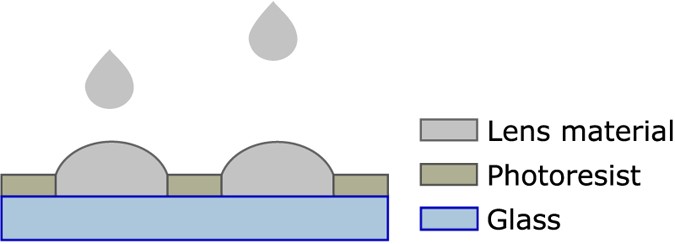
min at 95 °C after which it is developed in mr_Dev 600 Developer for one min and rinsed with IPA. Inkjet printer is used to deposit the lenses into the holes of the resist layer. Figure 1
presents the idea of the process. The used material (InkEpo from Micro Resist Technology) is negative photoresist in liquid form and is designed especially for optical purposes and is
therefore highly transparent to visible light. The printer is drop-on-demand type (Dimatix 2800 series) and the droplet volume of the used cartridge is 10 pl. One nozzle is used and the
settings for the nozzle are presented in Fig. 2. The nozzle voltage (equals Voltage level 100% in the Fig. 2) is 24 V. The “pools” are filled with 45 drops, drop by drop, until the liquid
surface rises just above the resist layer edge to form a convex surface, a lens. For this application the radius of curvature for a lens is needed large, thus the lens is printed flat. After
printing, the lenses are hardened with a normal procedure for photoresist, by pre-baking them at 85 °C for 10 min, then exposing at a wavelength of 365 nm for 10 s and finally baking at 95
°C for 5 min. IMAGING SYSTEM Direct imaging, using microlens arrays is a modified light field imaging technique that can be used for capturing images of semi microscopic targets17. Figure 3
illustrates the imaging concept. The system consists of a camera sensor, a microlens array, a sample holder and a light source with an aperture. The microlens array is positioned in front of
the sensor so that each lens images the light source aperture without overlap on the camera sensor. The sample is positioned between the light source and the microlens array so that sharp
images of the object are obtained. The RAW images resemble the focused light field camera data18 and the image reconstruction can be done by the patch mosaicking approach or alternatively by
the reparametrized back-projection19. The main advantages compared with conventional microscopes are the simplicity of the system and the scalability of the imaging area. While the system
allows imaging target area of the camera sensor, the resolution is limited by Nyquist sampling theory and the sensor pixel size, as there is no magnification. However, the achieved
resolution is useful for example for the detection of stained tumour cells or certain parasitic forms10. RESULTS For the finished MLA profile measurements are done and the shape of the
lenses is studied. The imaging properties are tested with microscope targets. MLA CHARACTERIZATION Lens characterization is limited to profile measurements, which shows the uniformity of the
lens array. Other properties (such as focal length) are not studied because of the demonstration-nature of this paper. The process steps still can be optimized, after which it would be more
meaningful for thorough characterization. Profile measurement for the printed lenses is made with Veeco Dektak 8 surface profiler. Four areas of 1 mm × 1 mm were examined; the stylus scans
the surface of the defined area and forms a 3D-image (Fig. 4a.). The shape of the lenses look uniform and in Fig. 4b the vertical and horizontal profiles are presented together. All together
36 lenses are measured and average height is 12.4 μm with standard deviation of 0.06 μm. Average diameter is 222 μm with standard deviation of 4.61 μm. The small inconsistences in the
curves are due to the Dektak’s feature of mathematically calculate the space between the measurement points. The graph also shows a small difference between the vertical and horizontal pitch
of the lenses, because there has been a small distortion for the patterning when the pattern file is transformed in the lithography device. With different machinery this can be avoided. The
focal length was not measured due to lack of appropriate equipment, but from the measured dimensions of the lens, the approximate focal length was calculated from lensmaker’s equation,
equation (1), which is simplified for plano-convex lens: IMAGING BY MLA Inkjet printed microlenses’ imaging properties are tested using microscope targets. The lighting is normalized by
dividing the image data with an image of the light source aperture. Based on USAF resolution target (Edmun Optics, R209441-11069) the maximum obtainable resolution is 128 line pairs per
millimetre or 3.9 μm line width (Fig. 5a). This is on the same level as demonstrated previously with glass microlenses17. The reconstructed images of a roundworm (Ascaris) transverse section
show clear improvement in an image quality with the inkjet manufactured lenses compared with the hot embossed polycarbonate lenses (Fig. 5b). Slight variation in light intensity can be
observed in the result images. While this is not observed with the glass reference the overall image quality is at the same level than with the glass reference17. DISCUSSION The disposable
optics has a great potential when considering the vast range of diagnostic and testing applications optics is utilized in. Many POCTs are based on chemical reactions, such as glucose
meters20, which people with diabetes use. Optics based POCTs are needed when studying, for example tissue structures and cells, or when parasites in samples need to be identified. This can
be done inside21 or outside of human body, thus disposable devices or parts are handy to prevent cross-contamination of samples and to maintain patient safety, even though many devices can
be used over and over again. One more suitable application for these low-cost micro lenses could be in drug delivery, where the laser focused through the lenses creates small holes in the
skin, which allow more effective absorption for the drug. The presented process for MLAs is simple and easily tuneable for different sized and shaped lenses. Although, the inkjet printed
lenses could be printed on a flat substrate with just a hydrophobic patterning, our process opens other possibilities for structures and shapes. Layers of different materials can be printed
on top of each other with a high precision (such as different filters, like colour filter, with which colour images can be made), which is difficult for moulded lenses. Because of the
patterning, the shape of the lens is not limited only to a circle, as with other drop-on-demand type processes. An ellipse, a polygonal or any shape is possible with the photolithography. As
a disposable component, the price is a key factor. For us the total price of one fabricated MLA rounded up approximately to 4 € excluding working hours. Fabrication in proper production
line decreases the cost even more, when the materials are used more efficiently. The lenses are now printed on a glass substrate which, in sense on recycling, is challenging. The glass can
be eliminated by printing the lenses on a PET substrate22, which would also decrease the prince of the MLA even more. The width of the lenses is 20 μm more than the original lithography
pattern made on the glass, because the material slightly spreads over the patterning, but this can be eliminated with a hydrophobic treatment on the patterning. It would make the shape of
the lens even more easily tuneable, as there is only the height that is changing according to the amount of the printed material. In this demonstration, as the distance between the MLA and
the sample is adjustable, the small change in the focal length, due to the spreading, is not significant. This type of MLAs could also be applied in far-field imaging. The parameters and
materials need to be changed, but the process itself would be the same. With thicker and non-transparent patterning, the lenses form image on the sensor without overlap. The development of
disposable optics is still at early stage, but because of all the positive effects it can have on point-of-care-testing, it is worth to give more focus to it in the future. CONCLUSION Our
study shows that low-cost fabrication method for micro lens array can produce lenses with quality of a same level than glass lenses. The height variation of the lenses is only 60 nm and the
images produced with the lenses compete with those obtained with the glass reference lenses. Because of the low fabrication costs, the presented MLA is also a valid option to be used as a
disposable optics component. ADDITIONAL INFORMATION HOW TO CITE THIS ARTICLE: Vilmi, P. _et al._ Disposable optics for microscopy diagnostics. _Sci. Rep._ 5, 16957; doi: 10.1038/srep16957
(2015). REFERENCES * Larsson, A., Greig-Pylypczuk, R. & Huisman, A. The state of point-of-care testing: a european perspective. Ups. J. Med. Sci. 120, 1–10 (2015). Article Google
Scholar * Howick, J. et al. Current and future use of point-of-care tests in primary care: an international survey in Australia, Belgium, The Netherlands, the UK and the USA. BMJ Open 4,
e005611 (2014). Article Google Scholar * Wippermann, F. C., Beckert, E., Dannberg, P., Messerschmidt, B. & Seyffert, G. Disposable low cost video endoscopes for straight and oblique
viewing direction with simplified integration. Proc. SPIE 7556, Design and Quality for Biomedical Technologies III 7556, 755607 (2010). Article Google Scholar * Ge, S. et al. A disposable
immunosensor device for point-of-care test of tumor marker based on copper-mediated amplification. Biosens. Bioelectron. 43, 425–431 (2013). Article CAS Google Scholar * Linder, E. et al.
On-chip imaging of schistosoma haematobium eggs in urine for diagnosis by computer vision. Plos. Neglect. Trop. D. 7, 1–9 (2013). Article Google Scholar * Ryu, Y. et al. Lensed
fiber-optic design for efficient photon collection in scattering media. Biomed. Opt. Express 6, 191–210 (2015). Article Google Scholar * Liu, K. H., Chen, M. F., Pan, C. T., Chang, M. Y.
& Huang, W. Y. Fabrication of various dimensions of high fill-factor micro-lens arrays for OLED package. Sensor. Actuat. A-Phys. 159, 126–134 (2010). Article CAS Google Scholar * Wei,
J., Wang, S., Zhao, Y. & Jin, F. Hierarchical prediction structure for subimage coding and multithreaded parallel implementation in integral imaging. Appl. Opt. 50, 1707–1716 (2011).
Article ADS Google Scholar * Levoy, M., Zhang, Z. & McDowall, I. Recording and controlling the 4D light field in a microscope. J. Microsc. 235, 144–162 (2009). Article CAS
MathSciNet Google Scholar * Varjo, S. & Hannuksela, J. A mobile imaging system for medical diagnostics. ACIVS Proc., Lecture Notes in Computer Science 8192, 215–226 (2013). Article
Google Scholar * Tao, Z. et al. Molecular Characterization of Enteroviruses Including a New Type EV-C99 Isolated from Xinjiang Students in Shandong, China in 2011. Sci. Rep. 4, 6564 (2014).
Article CAS Google Scholar * Belizario, V. Y., Jr., Erfe, J. M., Naig, J. R. A. & Chua, P. L. C. Evidence of increasing risk of schistosomiasis among school-age children in
municipality of Calatrava, Province of Negros Occidental, Philippines. Asian Pac. J. Trop. Med. 8, 373–377 (2015). Article Google Scholar * Zhu, X., Zhu, L., Chen, H., Yang, M. &
Zhang, W. Fabrication of multi-scale micro-lens arrays on hydrophobic surfaces using a drop-on-demand droplet generator. Opt. Laser Technol. 66, 156–165 (2015). Article CAS ADS Google
Scholar * Kim, C. et al. Fabrication of a fused silica based mold for the microlenticular lens array using a femtosecond laser and a CO2 laser. Opt. Mater. Express 4, 2233–2240 (2014).
Article CAS ADS Google Scholar * Zhang, X., Que, W., Javed, H. M. A. & Wei, W. Elliptical concave microlens arrays built in the photosensitive TiO2/ormosils hybrid films. Opt.
Commun. 330, 12–18 (2014). Article CAS ADS Google Scholar * Yoon, J. S., Lim, S. H., Kim, J. H., Yoo, Y. & Choi, D. A study on the fabrication of microlens array based on the volume
shrinkage of the photoresist solution during evaporation. Opt. Commun. 332, 70–74 (2014). Article CAS ADS Google Scholar * Varjo, S., Hannuksela, J. & Silven, O. Direct imaging with
printed microlens arrays. Pattern Recognition (ICPR), 21st Int. Conf. 1355–1358 (2012). * Georgiev, T. & Lumsdaine, A. Focused plenoptic camera and rendering. J. Electron. Imaging 19(2),
021106-1–021106-11 (2010). ADS Google Scholar * Isaksen, A., McMillian, L. & Gortler, S. J. Dynamically reparameterized light fields. Proc. SIGGRAPH'00, 297–306 (2000). * Jansen,
R. T. P. & Slingerland, R. J. SKML-Quality Mark for point-of-care test (POCT) glucose meters and glucose meters for home-use. Clin. Chem. Lab. Med. 48, 1021–1027 (2010). Article CAS
Google Scholar * Buderus, S., Sonderkoetter, H., Fleischhack, G. & Lentze, M. J. Diagnostic and Therapeutic Endoscopy in Children and Adolescents with Cancer. Pediatr. Hematol. Oncol.
29, 450–460 (2012). Article Google Scholar * Vilmi, P., Myllyla, R. & Fabritius, T. Inkjet printed micro lens array on patterned substrate. Proc. SPIE 8613, Advanced Fabrication
Technologies for Micro/nano Optics and Photonics VI 8613, 861317 (2013). Article Google Scholar Download references AUTHOR INFORMATION AUTHORS AND AFFILIATIONS * Department of Electrical
Engineering, Optoelectronics and Measurement Techniques Laboratory, University of Oulu, Finland Pauliina Vilmi, Rafal Sliz & Tapio Fabritius * Department of Computer Science and
Engineering, The Center for Machine Vision Research, University of Oulu, Finland Sami Varjo & Jari Hannuksela Authors * Pauliina Vilmi View author publications You can also search for
this author inPubMed Google Scholar * Sami Varjo View author publications You can also search for this author inPubMed Google Scholar * Rafal Sliz View author publications You can also
search for this author inPubMed Google Scholar * Jari Hannuksela View author publications You can also search for this author inPubMed Google Scholar * Tapio Fabritius View author
publications You can also search for this author inPubMed Google Scholar CONTRIBUTIONS P.V. wrote the main manuscript, S.V. wrote the imaging system part of the manuscript and prepared
Figure 5a,b, R.S. made the profile measurements and prepared Figure 4a,b, J.H. contributed to imaging system design and T.F. contributed to lens fabrication design. All authors reviewed the
manuscript. ETHICS DECLARATIONS COMPETING INTERESTS The authors declare no competing financial interests. RIGHTS AND PERMISSIONS This work is licensed under a Creative Commons Attribution
4.0 International License. The images or other third party material in this article are included in the article’s Creative Commons license, unless indicated otherwise in the credit line; if
the material is not included under the Creative Commons license, users will need to obtain permission from the license holder to reproduce the material. To view a copy of this license, visit
http://creativecommons.org/licenses/by/4.0/ Reprints and permissions ABOUT THIS ARTICLE CITE THIS ARTICLE Vilmi, P., Varjo, S., Sliz, R. _et al._ Disposable optics for microscopy
diagnostics. _Sci Rep_ 5, 16957 (2015). https://doi.org/10.1038/srep16957 Download citation * Received: 13 July 2015 * Accepted: 22 October 2015 * Published: 20 November 2015 * DOI:
https://doi.org/10.1038/srep16957 SHARE THIS ARTICLE Anyone you share the following link with will be able to read this content: Get shareable link Sorry, a shareable link is not currently
available for this article. Copy to clipboard Provided by the Springer Nature SharedIt content-sharing initiative