- Select a language for the TTS:
- UK English Female
- UK English Male
- US English Female
- US English Male
- Australian Female
- Australian Male
- Language selected: (auto detect) - EN
Play all audios:
ABSTRACT STUDY DESIGN: A retrospective study. OBJECTIVES: Diffusion tensor imaging (DTI) reflects pathological change in the spinal cord more sensitively than conventional magnetic resonance
imaging (MRI). Electrophysiological examination enables quantitative assessment of spinal cord function. Few studies have addressed the correlation between intraoperative spinal cord-evoked
potentials (SCEPs) and DTI. The purpose of this study was to examine whether DTI is an objective index for the diagnosis of the segmental level of dysfunction in cervical spondylotic
myelopathy (CSM). SETTING: Yamaguchi University Graduate School of Medicine, Japan. METHODS: Using 3.0-Tesla MRI, DTI values for the apparent diffusion coefficient (ADC) and fractional
anisotropy (FA) were measured at the disc level C2/C3 through C6/C7 in 11 normal subjects and 10 subjects with CSM. Subjects with CSM were divided into two groups based on the extent of
compression according to conventional MRI: single level (_n_=3) and multilevel (_n_=7). Intraoperative SCEPs were measured in subjects with CSM. For each group, the ADC and FA values were
compared with SCEPs with respect to the segmental levels of dysfunction. RESULTS: For all three subjects with single-level compression and six of seven with multilevel compression, the
maximal ADC value was observed at the segmental level of dysfunction as per the SCEP. Minimum FA values were observed at those sites in two of three patients with single-level compression
and in only two of seven with multi-level compression. CONCLUSION: Our results suggest that ADC might serve as a supplementary diagnostic indicator of the segmental levels of dysfunction in
CSM. SIMILAR CONTENT BEING VIEWED BY OTHERS APPLICATION OF THE T1W/T2W MAPPING TECHNIQUE FOR SPINAL CORD ASSESSMENT IN PATIENTS WITH DEGENERATIVE CERVICAL MYELOPATHY Article 02 November 2023
RELIABILITY OF PRE-OPERATIVE DIFFUSION TENSOR IMAGING PARAMETER MEASUREMENTS OF THE CERVICAL SPINE IN PATIENTS WITH CERVICAL SPONDYLOTIC MYELOPATHY Article Open access 15 October 2020
TRACT-SPECIFIC MAGNETIZATION TRANSFER RATIO PROVIDES INSIGHTS INTO THE SEVERITY OF DEGENERATIVE CERVICAL MYELOPATHY Article 01 October 2024 INTRODUCTION Cervical spondylotic myelopathy (CSM)
is diagnosed using magnetic resonance imaging (MRI) and neurological examination. However, the neurological findings are not always typical, and clinical symptoms of CSM may differ between
patients. Determination of the segmental levels of spinal cord involvement in CSM can be difficult, particularly in cases involving multi-level compression. In addition, the presence of
high-signal intensity lesions on T2-weighted MRI of the cervical spinal cord does not necessarily correspond to myelopathy. Spinal cord-evoked potentials (SCEPs) measured by
electrophysiological examination are useful for investigating the functional integrity of the spinal cord, even in cases involving compression at several levels. Diffusion tensor MR imaging
(DTI) is an advanced MRI technique that measures the diffusion of water molecules in tissues. Objective and quantitative assessment of microstructural change is possible by means of DTI.
Useful DTI parameters include the apparent diffusion coefficient (ADC) and fractional anisotropy (FA). The ADC reflects the diffusive strength of water molecules, regardless of
directionality. ADC values thus increase in compressed lesions of the spinal cord.1 FA measures the directional diffusion of water molecules, which is prescribed by axon structure. Normal
white matter has abundant nerve fibers, whereas gray matter has few. Thus, the FA values of the gray matter are smaller than those of white matter. Decreased FA values reflect a loss of
anisotropic diffusion of water molecules, indicating damage and degeneration of tract fibers in compressed sites of spinal cord.2 Several studies report the use of DTI for the diagnosis of
early-stage CSM and indicate a correlation between DTI values and prognosis following surgery.3, 4 However, these studies have not investigated the correlation between segmental levels of
dysfunction and the values of DTI parameters. The aim of present study was to consider whether DTI parameters are an objective indicator of segmental level involvement in CSM as determined
by means of SCEPs. We compared the results of intraoperative SCEPs, a quantitative measure of spinal cord function, with FA and ADC values. MATERIALS AND METHODS The study was approved by
the Institutional Review Board of Yamaguchi University Hospital and adhered to the tenets of the Declaration of Helsinki. SUBJECTS NORMAL SUBJECTS Eleven normal subjects, three men and eight
women, with a mean age of 68.6 years (range 56–87) were enrolled. All considered themselves to be healthy, and none had a history of neck injuries, neurological disorders. The absence of
cervical spinal cord compression was confirmed on MRI for all subjects. SUBJECTS WITH CERVICAL SPONDYLOTIC MYELOPATHY Ten consecutive patients with clinical signs and symptoms of chronic
cervical spinal cord compression (six men and four women; mean age, 71.8 years; range, 60–82) were enrolled in this study. CLINICAL ASSESSMENT Diagnosis of myelopathy was established based
on the presence of hyperreflexia, upper extremity sensory disturbance and motor examination of muscles.5 The findings on neurological examination of each CSM subject were shown in Table 1.
In addition, to evaluate the severity of CSM we assess muscle strength, gait disorder and bladder function. The Japanese Orthopedic Association scoring system for cervical myelopathy was
employed (Table 2). The Japanese Orthopedic Association score quantifies neurological impairment by evaluating upper extremity function (4 points), lower extremity function (4 points),
sensibility (6 points) and urinary bladder function (3 points). Preoperative Japanese Orthopedic Association scores ranged from 5.5 to 12 points with an average of 8.75 in this series. IMAGE
PROCEDURES All examinations were performed between 2010 and 2013 using a magnetic resonance imager (Achieva 3.0-T; Philips Medical System, Eindhoven, the Netherlands) equipped with a
16-channel neurovascular coil. For anatomical and diagnostic imaging of the spine, T1- and T2-weighted images were acquired in the sagittal and axial planes. DTI data were obtained using an
spin-echo–type single-shot echo-planar imagingsequence with the following parameters: echo time, 69 ms; repetition time, 9079 ms; number of slices, 30; interslice gap, 0 mm; band width,
1711.8 Hz per pixel; voxel size, 1.79 × 1.42 × 4.00 mm; acquisition matrix, 112 × 140; and NEX, 4. Images were obtained using _b_ values of 0 and 700 s mm−2. A Philips MRI workstation was
used to reconstruct ADC and FA maps from DTI data. Three eigenvalues (λ1,λ2 and λ3) were measured by averaging all selected pixels in the region of interest. ADC mean= (λ1+λ2+λ3)/3=λ and FA
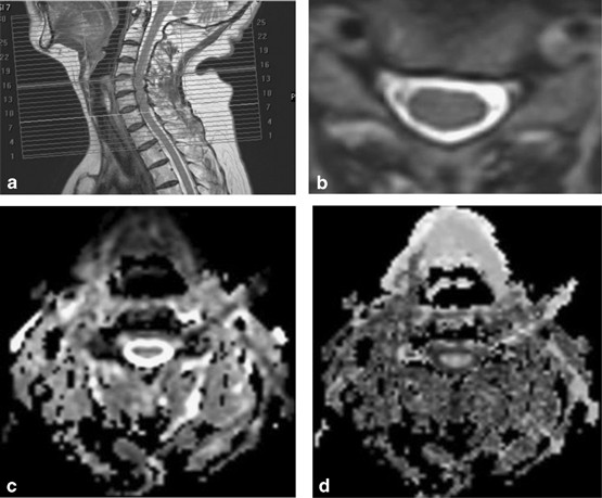
was calculated with the following formula: These parameters were measured at five disc levels (from C2/C3 to C6/C7) on axial sections that gave stable values. Because DTI is not capable of
capturing an image of plural stacks at one time, the image at each level were made parallel to C4/C5 disc. (Figure 1) REGION OF INTEREST SETTING AND MEASUREMENT After the appropriate axial
slice was selected using the sagittal T2-weighted images for anatomic reference, a region of interest (ROI) was drawn manually on the spinal cord by an orthopedist. The ROI included both
white and gray matter, excluding any cerebrospinal fluid contribution (Figure 2). The shape and size of each ROI varied according to the shape of the spinal cord, especially in patients with
CSM. INTRAOPERATIVE SPINAL CORD-EVOKED POTENTIALS We measured three different kinds of SCEPs: SCEPs after median nerve stimulation, transcranial electric stimulations, and spinal cord
stimulation SCEPs were recorded intraoperatively. The median nerves were stimulated (square wave pulse, 0.2 ms duration, 3 Hz rate) at wrist with the cathode placed proximally. The stimulus
intensity was set at 1.5 times for producing the thumb twitch in an awakened condition. Transcranial electric stimulation was delivered as square pulses of 0.2 ms duration and at an
intensity of 100 mA through needle electrodes (13R25, length 8 mm, diameter 0.8 mm, Dantec, Skovlunde, Denmark) placed on skull. The anode was placed 7 cm laterally to the right of the
vertex on line joining the external auditory meatus. The cathode was placed on the opposite side. Spinal cord stimulation-SCEPs were delivered by an epidural catheter electrode(UKG-100-2PM,
diameter 0.8 mm, length 900 mm, Unique Medical corporation, Japan) inserted into the dorsal epidural space from the C7-T1 interlaminar space. Square wave pulse (0.2 ms duration, 3 Hz rate)
were delivered at an intensity of 15–20 mA. Before laminoplasty, all SCEPs were recorded intraoperatively with recording electrodes (13R25) inserted in the ligamentum flavum at each
interlaminar space, from C2/C3 to C7/T1. A reference electrode was inserted into the subcutaneous tissue in the posterior aspect of the neck for the recording of median nerve stimulations
and Spinal cord stimulation-SCEPs. All SCEPs signals were amplified and filtered with a bandpass of 20–3000 Hz using a standard evoked potential/electromyography machine (Nicolet Viking,
Nicolet Biomedical, Madison, WI, USA). Average of 100–200 median nerve stimulations, 40–60 Transcranial electric stimulation-SCEPs and 20–30 Spinal cord stimulation-SCEPs responses were
obtained and superimposed. Abnormality was determined with a reduction of >30% in size of negative peak compared with that of the C6/7 intervertebral level in median nerve stimulations,
and a reduction of >50% compared with the disc level immediately above (in SCEPs after transcranial electric stimulations) or below (in SCEPs after stimulation to the spinal cord).6, 7, 8
An example of intraoperative SCEPs was shown in Figure 3. STATISTICAL ANALYSIS Excel software (Microsoft, Tokyo, Japan) was used for statistical analysis. Statistically significant
difference between the two groups were determined using the Mann–Whitney _U_-test. _P_<0.05 was considered statistically significant. RESULTS COMPARISON OF DIFFUSION TENSOR IMAGING
PARAMETERS BETWEEN NORMAL AND CERVICAL SPONDYLOTIC MYELOPATHY SUBJECTS The mean values and s.d. of the ADC and FA at five disc levels are shown in Table 3. The average ADC value of at all
five disc levels had no significance between normal and CSM subjects (1119±118 × 10−6 vs 1233±234 × 10−6 mm2s−1, respectively; _P_=0.205). The average FA value differed significantly between
normal and CSM subjects(0.66±0.06 vs 0.59±0.05, respectively; _P_=0.024). RELATIONSHIP BETWEEN DIFFUSION TENSOR IMAGING PARAMETERS AND THE SEGMENTAL LEVEL OF SPINAL CORD DYSFUNCTION On
conventional MRI, three subjects had single-level intervertebral compression and seven patients had multilevel. The compression level on conventional MRI, high signal change on T2 image, ADC
and FA value at respective disc level are shown in Table 4. All subjects were determined by SCEPs to have a single site of conduction abnormality, despite single- or multi-level
compression. The segmental level of dysfunction was C3/C4 in five cases and C4/C5 in five cases. For all subjects with single-level compression and six of seven with multi-level compression,
the ADC was maximal at the segmental level of dysfunction. The FA was minimal at the segmental level of dysfunction in two of three subjects with single-level compression and only two of
seven subjects with multi-level compression (Table 5). DISCUSSION The maximal ADC value was observed at the segmental level corresponding to the site of dysfunction according to SCEP for all
three subjects with single-level compression and six of seven with multi-level compression. Minimum FA values were observed at those same sites in two of three patients with single-level
compression and in only two of three with multi-level compression. Normal DTI-parameter values obtained from healthy subjects vary by individual factors such as age, environment under which
measurements are taken, and ROI location.9, 10 In subjects with CSM, the degree of the tissue injury differs between patients. Therefore, when comparing normal and CSM subjects, normal
variability in measurements must be taken into consideration. In the present study, we measured the parameter values at five disc levels within each patient, with the normal regions of the
spinal cord providing internal control data. Thus, normal variability between subjects does not affect our data analysis. Decreased FA values generally indicate axonal damage, that is,
degeneration of white matter in the brain and spinal cord.11 Unlike the brain, transverse sections of spinal cord are small and have poor image resolution, presenting a technical challenge
for the investigation of ROIs close to the funiculi (for example, lateral and posterior funiculi). As described in previous reports, ROI was drawn on the spinal cord as wide as possible to
include both gray matter and white matter, while excluding the cerebrospinal fluid. ROIs were analyzed one per disc level. The shape of each ROI varied according to the shape of the spinal
cord. So the number of pixels, total pixel area, how much gray and how much white matter was included in respective ROI were also varied per slice and subject. It is ideal that whole gray
and white matter are included in respective ROI in each axial plane of the spinal cord. But, considering the spatial resolutions that are currently possible, oversize ROI may have the risk
of including cerebrospinal fluid.4, 9, 12 The cause of the observed increase in ADC in compressive cervical myelopathy is still uncertain. Compression of spinal cord leads to ischemia,
anoxemia and cell membrane injury that can increase cell membrane penetrability,3, 13, 14 a possible source of the increased ADC. ADC elevation that occurs before signal elevation on
T2-weighted images may indicate early myelopathy-related changes.10 This change is thought to reflect edema in the tissues. Conflicting data have been reported regarding whether the ADCs of
compressed spinal cord regions vary significantly between levels of spinal cord.3, 12 Difference in these results may arise from differing degrees of tissue injury between patients. ADC and
FA do not necessarily correlate.2 For example, a patient recovering from a stroke may have damaged fibers in the affected region of the brain, with stabilization of the surrounding tissues
by gliosis. In this case, the FA values would be lower than normal, but the ADC values might not be elevated.15, 16 In case of severe compressive cervical myelopathy, FA values decrease,
sometimes without a corresponding increase in ADC. Thus, the use of ADC values alone to judge the presence or absence of spinal cord disorder is reportedly difficult.2, 17 We think that
intraoperative electrophysiological examination gives objective assessment of the affected segmental level and degree of myelopathy in CSM.18 We thus linked preoperative DTI analysis to the
electrophysiological test results to increase the objectivity of the evaluation based on DTI parameters. Comparison of the segmental level of CSM with ADC and FA revealed ADC was maximal at
the level of spinal disorder in many cases, regardless of the extent of compression (single vs multi-level). In one case (No. 4), the maximum ADC and minimum FA were seen at the C6/C7
intervertebral level, whereas the level of dysfunction by intraoperative SCEPs was C3/C4. In this case, there was severe compression of spinal cord at C6/C7, and a decrease in SCEP was seen
at the corresponding level. In another case (No. 10), compression of the spinal cord was seen at the C3/C4 and C5/C6, and the level of dysfunction was at C3/C4. However, the minimum FA was
at C4/C5. A previous study reported a decrease in FA both at the site of maximal compression and at sites distant to it.4 Furthermore, the minimum FA did not necessarily concur with the
maximal compression level, particularly in cases involving multilevel compression. These findings may reflect that CSM-associated demyelination and axonal damage affect both the myelopathic
lesion site and distal sites over the course of the disease.19, 20 In four cases (No. 2, 5, 6 and 9), the minimum FA was observed at C6/C7, even though this site had no compression and was
distant from the lesion. Therefore, the results of our study indicate the FA is not useful as an objective diagnostic indicator of the segmental level of myelopathy. The reason for increase
in ADC at injury site is still not clear. However, if the ischemia, degeneration and inflammation caused by chronic compression of the spinal cord increase the ADC, it is reasonable that the
level of dysfunction would correspond to the site with maximal histological damage. Our results suggest the ADC could be used as a supportive tool for diagnosing the level of myelopathy in
CSM. There are some limitations in the present study. First, this study is preliminary because of the small sample size. Further conclusions require future studies using a larger number of
cases. Second, the axial images at each segmental level were made parallel to C4/C5 disc. This may affect the value of DTI parameters, especially FA that influenced by directionality. Third,
how much gray matter and how much white matter were included in each ROI are varies per slice and subject. This might have introduced some bias. Fourth, we cannot compare the change of ADC
and FA value with histopathological change in the spinal cord. So the cause of change in these parameters was still speculative. Fifth, it has not been completely proved that intraoperative
SCEPs allow to unequivocally reflect the integrity of respective cervical segments. Ideally, needle electromyography of muscles corresponding to each cervical myotome should have been
employed. In the case No. 9, neurological level diagnosis with physical examination was C5/C6, but the result from SCEPs and DTI was C4/C5. This case had multi-level compression(C4/C5 and
C5/C6), and T2 high intensity at the same levels. We sometimes experience the difficulty in diagnosing the segmental level of dysfunction with physical examination and conventional MRI with
multi-level compression. As Seichi _et al._ reported, the average accuracy of neurologic level diagnosis based on the index was ⩾70%.5 In the case of multi-level CSM, sensitivity,
specificity and accuracy of neurological diagnosis might be lower. In such case, the neurological level diagnosis can be more accurate by use of needle electromyography. We observed a
correlation between the intervertebral level of maximal ADC and the segmental level responsible for CSM. The observed correlation between the maximal ADC and the segmental level of
dysfunction according to SCEP indicates that ADC might serve as a supplementary diagnostic indicator of the segmental levels of dysfunction in CSM. DATA ARCHIVING There were no data to
deposit. REFERENCES * Facon D, Ozanne A, Fillard P, Lepeintre JF, Tournoux-Facon C, Ducreux D . MR diffusion tensor imaging and fiber tracking in spinal cord compression. _AJNR Am J
Neuroradiol_ 2005; 26: 1587–1594. Google Scholar * Yoo WK, Kim TH, Hai DM, Sundaram S, Yang YM, Park MS _et al_. Correlation of magnetic resonance diffusion tensor imaging and clinical
findings of cervical myelopathy. _Spine J_ 2013; 13: 867–876. Article Google Scholar * Song T, Chen WJ, Yang B, Zhao HP, Huang JW, Cai MJ _et al_. Diffusion tensor imaging in the cervical
spinal cord. _Eur Spine J_ 2011; 20: 422–428. Article Google Scholar * Wen CY, Cui JL, Liu HS, Mak KC, Cheung WY, Luk KD _et al_. Is diffusion anisotropy a biomarker for disease prognosis
of cervical spondylotic myelopathy? _Radiology_ 2014; 270: 197–204. Article Google Scholar * Seichi A, Takeshita K, Kawaguchi H, Matsudaira K, Higashikawa A, Ogata N _et al_. Neurological
level diagnosis of cervical stenotic myelopathy. _Spine_ 2006; 31: 1338–1343. Article Google Scholar * Kaneko K, Kawai S, Taguchi T, Fuchigami Y, Ito T, Morita H . Correlation between
spinal cord compression and abnormal patterns of median nerve somatosensory evoked potentials in compressive cervical myelopathy: comparison of surface and epidurally recorded responses. _J
Neurol Sci_ 1998; 158: 193–202. Article CAS Google Scholar * Kanchiku T, Taguchi T, Kaneko K, Fuchigami Y, Yonemura H, Kawai S . A correlation between magnetic resonance imaging and
electrophysiological findings in cervical spondylotic myelopathy. _Spine_ 2001; 26: 269–274. Article Google Scholar * Kaneko K, Taguchi T, Morita H, Yonemura H, Fujimoto H, Kawai S .
Mechanism of prolonged central motor conduction time in compressive cervical myelopathy. _Clin Neurophysiol_ 2001; 112: 1035–1040. Article CAS Google Scholar * Uda T, Takami T, Tsuyuguchi
N, Sakamoto S, Yamagata T, Ikeda H _et al_. Assessment of cervical spondylotic myelopathy using diffusion tensor magnetic resonance imaging parameter at 3.0 tesla. _Spine_ 2013; 38:
407–414. Article Google Scholar * Mamata H, Jolesz FA, Maier SE . Apparent diffusion coefficient and fractional anisotropy in spinal cord: age and cervical spondylosis-related changes. _J
Magn Reson Imaging_ 2005; 22: 38–43. Article Google Scholar * Inano S, Takao H, Hayashi N, Abe O, Ohtomo K . Effects of age and gender on white matter integrity. _AJNR Am J Neuroradiol_
2011; 32: 2103–2109. Article CAS Google Scholar * Hori M, Fukunaga I, Masutani Y, Nakanishi A, Shimoji K, Kamagata K _et al_. New diffusion metrics for spondylotic myelopathy at an early
clinical stage. _Eur Radiol_ 2012; 22: 1797–1802. Article Google Scholar * Demir A, Ries M, Moonen CT, Vital JM, Dehais J, Arne P _et al_. Diffusion-weighted MR imaging with apparent
diffusion coefficient and apparent diffusion tensor maps in cervical spondylotic myelopathy. _Radiology_ 2003; 229: 37–43. Article Google Scholar * Mamata H, Jolesz FA, Maier SE .
Characterizaion of central nervous system structures by magnetic resonance diffusion anisotropy. _Neurochem Int_ 2004; 45: 553–560. Article CAS Google Scholar * Thomalla G, Glauche V,
Koch MA, Beaulieu C, Weiller C, Röther J . Diffusion tensor imaging detects early Wallerian degeneration of the pyramidal tract after ischemic stroke. _Neuroimage_ 2004; 22: 1767–1774.
Article Google Scholar * Werring DJ, Toosy AT, Clark CA, Parker GJ, Barker G, Miller D _et al_. Diffusion tensor imaging can detect and quantify corticospinal tract degeneration after
stroke. _J Neurol Neurosurg Psychiatry_ 2000; 69: 269–272. Article CAS Google Scholar * Aota Y, Niwa T, Uesugi M, Yamashita T, Inoue T, Saito T . The correlation of diffusion-weighted
magnetic resonance imaging in cervical compression myelopathy with neurologic and radiologic severity. _Spine_ 2008; 33: 814–820. Article Google Scholar * Kanchiku T, Imajo Y, Suzuki H,
Yoshida Y, Akashi K, Taguchi T . Correlation between spinal cord function assessed by intraoperative SCEPs and morphology of the compressed spinal cord on MRI. _J Spinal Disord Tech_ 2013,
e-pub ahead of print 6 November 2013, doi: 10.1097/BSD.0b013e318291cb61. Article Google Scholar * Urakawa T, Matsuzawa H, Suzuki Y, Endo N, Kwee IL, Nakada T . Analysis of ascending spinal
tract degeneration in cervical spondylotic myelopathy using 3D anisotropy contrast single-shot echo planar imaging on a 3.0-T system. _J Neurosurg Spine_ 2011; 15: 648–653. Article Google
Scholar * Fehlings MG, Skaf G . A review of the pathophysiology of cervical spondylotic myelopathy with insights for potential novel mechanisms drawn from traumatic spinal cord injury.
_Spine_ 1998; 23: 2730–2737. Article CAS Google Scholar Download references AUTHOR INFORMATION AUTHORS AND AFFILIATIONS * Department of Orthopaedic Surgery, Yamaguchi University Graduate
School of Medicine, Yamaguchi, Japan Y Suetomi, T Kanchiku, Y Imajo, H Suzuki, Y Yoshida, N Nishida & T Taguchi * Department of Orthopaedic Surgery, St. Hill hospital, Yamaguchi, Japan Y
Suetomi & S Nishijima Authors * Y Suetomi View author publications You can also search for this author inPubMed Google Scholar * T Kanchiku View author publications You can also search
for this author inPubMed Google Scholar * S Nishijima View author publications You can also search for this author inPubMed Google Scholar * Y Imajo View author publications You can also
search for this author inPubMed Google Scholar * H Suzuki View author publications You can also search for this author inPubMed Google Scholar * Y Yoshida View author publications You can
also search for this author inPubMed Google Scholar * N Nishida View author publications You can also search for this author inPubMed Google Scholar * T Taguchi View author publications You
can also search for this author inPubMed Google Scholar CORRESPONDING AUTHOR Correspondence to Y Suetomi. ETHICS DECLARATIONS COMPETING INTERESTS The authors declare no conflicts of
interest. RIGHTS AND PERMISSIONS Reprints and permissions ABOUT THIS ARTICLE CITE THIS ARTICLE Suetomi, Y., Kanchiku, T., Nishijima, S. _et al._ Application of diffusion tensor imaging for
the diagnosis of segmental level of dysfunction in cervical spondylotic myelopathy. _Spinal Cord_ 54, 390–395 (2016). https://doi.org/10.1038/sc.2015.192 Download citation * Received: 07
October 2014 * Revised: 01 July 2015 * Accepted: 16 September 2015 * Published: 27 October 2015 * Issue Date: May 2016 * DOI: https://doi.org/10.1038/sc.2015.192 SHARE THIS ARTICLE Anyone
you share the following link with will be able to read this content: Get shareable link Sorry, a shareable link is not currently available for this article. Copy to clipboard Provided by the
Springer Nature SharedIt content-sharing initiative