- Select a language for the TTS:
- UK English Female
- UK English Male
- US English Female
- US English Male
- Australian Female
- Australian Male
- Language selected: (auto detect) - EN
Play all audios:
ABSTRACT Tropical theileriosis is an important protozoan tick-borne disease in cattle. Vaccination using attenuated schizont-infected cell lines is one of the methods used for controlling
the disease. This study describes the production of attenuated schizont-infected cell lines from Egypt and an evaluation of its use as a vaccine to protect calves against clinical disease
upon field challenge. Two groups of exotic and crossbred male calves were divided into vaccinated and control groups. The vaccinated groups were inoculated with 4 ml (1 × 10⁶ cells/ml) of
the attenuated cell line. Three weeks after vaccination, calves of both groups were transported to the New Valley Governorate (Egyptian oasis) where they were kept under field conditions and
exposed to the natural _Theileria annulata_ challenge. All animals in the control group showed severe clinical signs and died despite treatment with buparvaquone, which was administered
after two days of persistent fever due to a severe drop in packed cell volume (PCV). Animals in the vaccinated group became seropositive without developing severe clinical signs other than
transient fever. Post-mortem examinations revealed enlarged and fragile lymph nodes, spleen, and liver with necrosis and hemorrhages. These findings indicate that the Egyptian attenuated
cell line was successful in protecting both exotic and crossbred animals against tropical theileriosis under field conditions. SIMILAR CONTENT BEING VIEWED BY OTHERS VACCINATION OF CATTLE
WITH THE _BABESIA BOVIS_ SEXUAL-STAGE PROTEIN HAP2 ABROGATES PARASITE TRANSMISSION BY _RHIPICEPHALUS MICROPLUS_ TICKS Article Open access 27 September 2023 EVALUATION OF EFFECTIVENESS AND
SAFETY OF SUBOLESIN ANTI-TICK VACCINE IN UGANDAN MULTI-SITE FIELD TRIAL Article Open access 18 September 2024 INTRANASAL VACCINE FOR LYME DISEASE PROVIDES PROTECTION AGAINST TICK TRANSMITTED
_BORRELIA BURGDORFERI_ BEYOND ONE YEAR Article Open access 15 February 2024 INTRODUCTION Bovine theileriosis caused by _Theileria annulata_ (_T. annulata_) is an important disease of cattle
in subtropical and tropical regions where vector ticks of the genus Hyalomma occur1,2,3. The infection is transmitted from ticks to their bovine host through tick saliva containing _T.
annulata_ sporozoites. The sporozoites enter the host's bloodstream and travel to the lymph nodes. Here they infect and transform the lymphocytes through asexual reproduction into the
schizont stage. The schizonts divide rapidly, producing many merozoites. The schizont can regulate the cell function and programmed cell death through the secretion of proteins directly into
the cell cytoplasm, affecting cell signaling and function. This results in cell transformation, making the host cell immortal so that it proliferates continuously. The merozoites then
infect other lymphocytes and continue the cycle of asexual reproduction. The merozoites are released into the bloodstream and infect erythrocytes (red blood corpuscles, RBCs). Inside the
RBCs, the merozoites continue to develop and divide, producing more merozoites. This stage is responsible for the clinical signs of tropical theileriosis. Some of the merozoites
differentiate into male and female gametocytes and when the tick feeds on an infected animal, it ingests the gametocytes, which then undergo sexual reproduction in the tick's gut. The
fertilized eggs develop into sporozoites, which migrate to the tick's salivary glands, ready to infect the next host4,5. The distribution of the parasite into daughter cells is
accompanied by a tight association with the mitotic apparatus of the host cell6. _T. annulata_ transforms leucocytes, loses virulence after long-term passage and is used as a live vaccine in
endemic countries7,8. In Egypt, clinical cases of theileriosis have been reported in different breeds of cattle and buffaloes (_Bubalus bubalis_) all over the Nile valley and other
localities such as the Delta region of the Nile valley, Upper Egypt and the Egyptian oasis9,10. Early recognition of clinical cases plays an important role in the treatment and control of
the disease11,12. During the acute phase when relatively high parasitemia is found, it can be easily diagnosed by using Giemsa-stained blood and lymph node aspiration smears2,13,14. In the
late chronic or carrier phase, parasitemia decreases to microscopically undetectable levels and diagnosis depends mainly on serological and molecular tests15,16. In Egypt, the lack of
reliable integrated control programmes that include accurate diagnosis, tick control, chemotherapy and vaccination, which has hampered control of the disease17. On the other hand, successful
vaccination using live attenuated schizont infected cell lines has been reported in many countries in North Africa, the Middle East and South Asia11,12,18,19. However, concerns that
parasite isolates may not be identical in antigenic composition or virulence have led to the propagation of local _T. annulata_-infected cell lines and evaluation of their efficacy under
experimental and field challenges in each country or region. This was the impetus for the current study, which aimed to produce a local vaccine for Egypt by isolating and attenuating
infected peripheral blood leukocytes (PBMC) from naturally infected cattle, followed by an evaluation of its potential as a vaccine to protect cattle against _T. annulata_ exposure in the
field. RESULTS CELL LINES _T. annulata_ schizont-infected cell line was prepared from leukocytes isolated from both heparinized blood and lymph node aspirates. The DNA extracts from the
cultures were tested using polymerase chain reaction (PCR) targeting the small subunit (SSU) 18S rRNA (18S rRNA) gene and Reverse Line Blot (RLB) assays, followed by sequencing. The results
were positive for _T. annulata_ but not for other tick-borne pathogens. The partial 18S rRNA gene sequence of the isolate was submitted to GenBank and can be retrieved using accession number
MN704769. The appropriate dose and passage for the vaccination trial at a dose of 4 ml (1 × 10⁶ cells/ml) was found to be the most immunogenic dose, inducing the strongest immune response
with no adverse effects or clinical signs. It protected the vaccinated animals during the natural challenge under field conditions, which developed no clinical signs other than transient
fever until slaughter (Table 4). VACCINATION TRIAL All animals were tested negative for _T. annulata_ infection prior to vaccination by using polymerase chain reaction (PCR) targeting the
small subunit (SSU) 18S rRNA gene (18S rRNA) and _T. annulata_ major merozoite piroplasm surface antigen gene (Tams1), and by _T. annulata_ surface protein ELISA (TaSp ELISA). After
vaccination and before field challenge, only animals of the vaccinated group became PCR-positive two weeks after vaccination, and TaSp antibodies were detected 4–5 weeks post vaccination.
None of the animals developed clinical disease. Piroplasm or schizont stages were not observed microscopically in any of the vaccinated animals. Within a few hours of the start of the field
challenge, all animals were infested with ticks, mainly _Hyalomma_ spp, especially _Hyalomma excavatum_, in addition to some _Rhipicephalus annulatus_ ticks which detected later during the
challenge period. The ticks mainly attached to the scrotum, dewlap, inner side of the thighs and tail. The first clinical signs indicative for _T. annulata_ infections were observed after 5
to 12 days post tick infestation. These signs included fever with body temperatures of up to 41.5 °C, ocular discharge, enlargement of superficial lymph nodes, of the prescapular and
prefemoral lymph nodes, various degrees of respiratory signs including nasal discharge and coughing, pale mucous membranes, jaundice, and diarrhea. The clinical signs were more severe in the
control group compared to the vaccinated group. Animals in the control group became anorexic and died despite treatment with buparvaquone at the recommended dose of 2.5 mg/kg body weight.
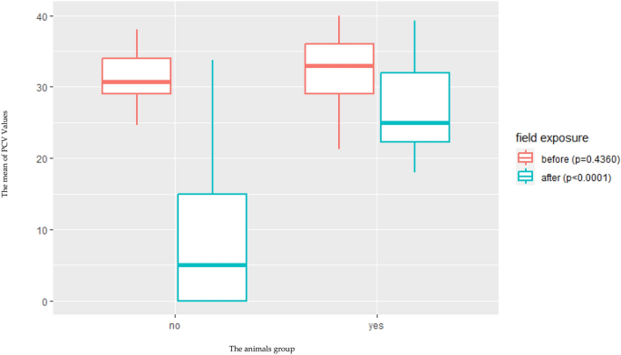
The treatment was applied after animals had persistent fever for two days. The PCV ranged from 26.5 to 40% before challenge in both vaccinated and control groups and dropped after field
challenge to 18–30% in vaccinated animals and 3–34% in the control group (Fig. 1, Tables 1, 2). The TaSp antibodies increased in all animals. The antibodies’ titers before and after
challenge being statistically different in the vaccinated group when compared to the control group (Fig. 2). Animals of control groups became positive by both Tams-1 and 18S rRNA PCR within
ten days post challenge, and this was confirmed by sequencing. The obtained sequences were identical, and we submitted one of them as an example to the GenBank and it is available under
accession number MN704771. Post-mortem examinations revealed that the body cavities of all diseased animals were filled with yellow straw-colored fluid and that petechial hemorrhages were
widely distributed on the serous membranes. The lymph nodes, liver and gall bladder were enlarged, edematous and fragile with petechial hemorrhages. Extensive necrosis and hemorrhages were
seen in the medulla of the affected lymph nodes. Schizont-infected cells that represent the schizont stages of _T. annulata_ were confirmed microscopically in the lymph nodes of animals with
acute theileriosis. Multiple necrotic foci, edema and foci of lymphoid cell reaction were reported in the liver sections. The spleens were enlarged, edematous and fragile with petechial
hemorrhages. Multiple necrotic foci and depletion of lymphoid elements in both white and red pulps were noticed, associated with hemorrhage and hemosiderosis. Both lungs were emphysematous
and dark in color with several areas of hepatization. Also, frothy exudate was found in the bronchi and bronchioles. Microscopic examination revealed alveolar emphysema, lobular interstitial
pneumonia, and foci of lymphoid cell reaction. Myocardial section showed multiple necrotic foci associated with hemorrhage and interstitial lymphoid cell reaction (Fig. 3). CO-DETECTION OF
OTHER TBPS AFTER CHALLENGE RLB assays were performed on blood samples collected from animals before and after vaccination and after challenge. The results revealed that all animals were
negative for tick-borne pathogens at the start of the study. Only vaccinated animals turned positive for _T. annulata_ after inoculation of the attenuated cell line. After field challenge
all animals became positive for _T. annulata_, some of them also tested positive for other tick-borne pathogens (Table 3). All co-infections were considered as subclinical because the blood
smears and 18S rRNA PCR were negative for all pathogens except for _T. annulata_ which confirmed with Tams-1 PCR and sequencing.as All the obtained sequences were identical, we submitted
only one of them as an example to the GenBank and it is available under accession number MN704770. DISCUSSION Tropical theileriosis is a major obstacle to the development of livestock
production in most tropical and subtropical countries, including Egypt. Control of ticks and tick-borne diseases in Egypt depends largely on the use of chemical acaricides. However, the
development of resistance to acaricides is limiting this approach. This situation creates the need for other effective control measures20,21. Vaccination against tropical theileriosis using
an attenuated cell culture vaccine is an interesting opportunity for the control and/or eradication of tropical theileriosis. This approach has been previously reported in several other
North African countries, including Sudan and Tunisia, but not in Egypt11,18,22,23,24. In this study, we described the development of an attenuated cell line of _T. annulata_ (Egyptian
strain). This Egyptian strain was prepared and attenuated according to previously published protocols23,24. The quality of this attenuated cell line and the appropriate dose were also
determined based on previous trials and studies which concluded that the infected cell line at passage 85 was sufficient to be used as a vaccine dose. This passage was close to the
attenuation passage in the Sudanese cell line24 and some Tunisian cell lines18,22. On the other hand, it was far from other Tunisian cell lines, which need more than two hundred passages to
be attenuated and ready to use, and this finding confirmed that each locality should have its own local strain, as described before in many studies11,18,22,23,24. After vaccination and field
challenge, both control and vaccinated animals were infested with ticks. All collected ticks were identified as _Hyalomma excavatum_, which is considered one of the main biological vectors
of _T. annulata_ in Egypt and _Rhipicephalus annulatus_, which may accidentally carry and transmit it mechanically by feeding on infected or carrier animals. This finding was in agreement
with previous finding in Egypt which concluded that both previously mentioned species are the most common species in Egypt and especially in the Egyptian oasis3. The recorded clinical signs
were more severe in the control group compared to the vaccinated group. Control animals gradually became anorexic and died. This finding was in agreement with some previous studies that
specified the same clinical signs for _T. annulata_ vaccination and challenge trials1,9,11,25. PCV results in both vaccinated and control groups did not significantly differ before challenge
(p = 0.4360) using Generalized linear model (GLM) but in contrast, two to three weeks after the challenge the PCV dropped significantly in the control groups when compared with the
vaccinated group (p < 0.0001). This result indicates a heavy infection with _T. annulata_ in the control group (Fig. 1). This finding is supported by previous finding which indicated
severe drop in the PCV in case of _T. annulata_ infection and its co-infection due to the increase in erythrophagocytosis, lysis of parasitized erythrocytes due to multiplication of the
parasite and subsequent removal by the reticuloendothelial system, and the increase in osmotic fragility of parasitized erythrocytes26,27,28. The antibodies’ titers raised in all vaccinated
animals according to the results obtained by using TaSP-ELISA assay when compared with the data collected from control group. This finding indicated that the attenuated cell line can enhance
the production of statistically significant titer of the protective antibodies without significant clinical infection (P > 0.0001)22,24. The protection rate in the vaccinated groups was
100% and it is closely similar to the finding recorded in China which ranged from 99.5 to 99.9%29,30,31,32,33,34,35. Co-infection with other tick-borne pathogens was detected through the RLB
assay which is more sensitive when compared to blood smears, PCR, and nested PCR, suggesting that all co-infection occurred at low levels2,36. The higher co-infections in the unvaccinated
(control) group compared to the vaccinated group could be attributed to the heterologous or non-specific immunological effects (NSE) that can be induced when using live attenuated vaccines.
These NSE have been described in several studies showing that innate immune cells, such as natural killer cells, monocytes, and macrophages, can provide non-specific protection against
certain infections in vaccinated animals receiving live attenuated vaccines. This process referred to as "trained immunity", is regulated by epigenetic reprogramming of innate
immune cells and is responsible for the protective, non-specific effects induced by vaccines37,38,39,40. As all co-infections reported in the control groups were due to intracellular
pathogens such as Babesia and Anaplasma, we suggested that this NSE could play a critical role in preventing these co-infections in vaccinated animals by recognizing infected RBCs by various
receptors of the host innate immune system and activating cytokines and phagocytosis, which play a crucial role in clearing parasitized RBCs as previously mentioned in several studies41,42.
The post-mortem lesions observed in died control animals were similar to the previously mentioned lesions in studies that investigated fatal cases of _T. annulata_ infection11,43. In view
of the data obtained, we could conclude that the use of the live attenuated Egyptian cell line infected with _T. annulata_ has potential efficacy in protecting both exotic and crossbred
cattle against the clinical form of _T. annulata_ even in highly endemic areas and should be recommended as a vaccine against tropical theileriosis in Egypt as in many other countries to
overcome this problem in our Egyptian field. This is also consistent with previous studies that have recommended _T. annulata_ attenuated cell line as a vaccine to control tropical
theileriosis in many countries11,22,23,24. However, before mass production and field application, we recommend further studies to assess its polymorphism and interaction with wild
circulating strains to ensure that vaccinated cattle are effectively immune to challenge with the same parasite strain and protected against a heterologous strain of this parasite as well as
co-infections. In this context, T-cells play a crucial role in both induction and maintenance of the immunity. It has been shown that the generation of cytotoxic T lymphocytes (CTL) is
closely related to the control of the infection—macroschizont-infected cells are killed in an MHC class I restricted manner42,44. Any strain-specificity induced by immunization is likely to
be manifested by CTL45. Besides CTLs, CD4 + T-cells also play an important role in protective immunity to _T. annulata_ infection. They produce macrophage-activating cytokines such as
IFN-gamma which produce mediators such as NO to destroy the intracellular schizonts. Based on the above, the protection observed in the animals after immunization with attenuated schizont in
our study could also be mediated by CTL and CD4 + T cells and it is one of the recommended research points in our future studies to identify this role in detail. Moreover, this
recommendation is supported by a recent study indicating that there is a great heterogeneity of the field _T. annulata_ population in the same geographical area of our study46,47. These
studies also recommended the production of cocktail vaccines including more than one isolate may be required as a further step to overcome the parasite’s heterogeneity in the future. Work is
still ongoing to estimate the duration of immunization and the need for booster doses. CONCLUSION In conclusion, Egyptian _T. annulata_ attenuated cell line is efficient and can protect
both exotic and cross breed cattle reared in endemic areas against Tropical theileriosis. By using this attenuated cell line as a vaccine, the case fatality rates will decrease dramatically.
Although, the obtained results are promising, we still recommend more trials targeting different animals in different areas like Nile Delta and Lower Egypt because these areas may have
different epidemiological status which may be affect the target animals, target age or time of vaccination. PATENT Patent number EG 2019121920A1 ‘Attenuated Tissue Culture Vaccine Against
_Theileria annulate_—Egyptian strain submitted to Patent office, Academy of Scientific research and technology, Ministry of scientific research, Egypt_._ METHODS ISOLATION OF T. ANNULATA
INFECTED CELLS Heparinized blood was collected from a three-year-old cow naturally infected with _T. annulata._ This cow was admitted to the Teaching Veterinary Hospital, Faculty of
Veterinary Medicine, Assiut University from EL-Ghanim district, Assiut Governorate. The animal exhibited typical clinical signs of tropical theileriosis. Its body temperature was 41.5 °C,
with congested conjunctival mucosa and enlarged superficial lymph nodes. An aspirate was taken from the prescapular lymph nodes. Following sampling and diagnosis, the cow was treated with
buparvaquone (MSD Animal health, New Jersey, USA) at 2.5 mg/kg body weight by deep intramuscular injection, followed by a second dose after 72 h. The animal was also treated with
non-steroidal anti-inflammatory drugs (Butafenil, Vetoquinol, France) and antibiotics (Marbocyl 10%, Vetoquinol, France) to control possible secondary bacterial infections. PREPARATION AND
ATTENUATION OF T. ANNULATA INFECTED CELL LINE _T. annulata_ strain Assiut is a single isolation initiated in 2015 from peripheral blood mononuclear cells (PBMC) of an ~ 3-year-old infected
cow from El-Ghanayem, Assiut, Egypt, admitted to the Veterinary Teaching Hospital, Faculty of Veterinary Medicine, Assiut University and confirmed positive for_ T. annulata_ infection.
Preparation of the PBMC from heparinized blood was performed by gradually transferring three mL of blood to a Falcon tube containing an equal amount of Ficoll® 400 (Sigma Aldrich, Germany)
followed by centrifugation at 1800 rpm for 40 min. The PBMC layer was subsequently collected and transferred to a new Falcon tube in which it was washed three times with RPMI 1640 (Cat. No,
BE12-115F, Lonza, Switzerland) with a centrifugation step at 2000 rpm for 10 min between washes. The pellet was subsequently resuspended in 2 mL complete RPMI 1640 media with 10% inactivated
fetal calf serum (LSP, UK), 1% Amphotericin B (Lonza, Switzerland), 1% Gentamicin (Lonza, Switzerland), 2% Streptomycin and Penicillin (Lonza, Switzerland). Lymph node aspirates were washed
three times in RPMI 1640 and resuspended in 2 mL complete media. Both samples were then transferred to two separate filter-cap tissue culture flasks 25 T containing 5 mL complete RPMI 1640
and were incubated with 5% CO2 at 37 °C. The media was changed every 72 h. _T. annulata_ schizont-infected leukocytes grew continuously and were passaged under sterile laboratory conditions
up to 114 serial passages. The obtained cell line was tested to confirm that it was free from any bacterial, mycoplasma or fungal contamination by the regular inoculation of 100 µL from the
cell line on enriched 4% sheep blood agar (BioLab ’BAN20500’) and EcoBio, Sabouraud-Dextrose-Agar 4% PH Euro-USP, (BioLab ’ESDA20500’) and incubate the plates at 37 °C for 48 h and 25 °C for
two week, respectively before inspection48. The DNA extracts from the cultures were tested using 18S rRNA and RLB assays, followed by sequencing3. The viability of the cell line was
evaluated using Trypan blue (Lonza, Switzerland) exclusion counting before storage of each passage in liquid nitrogen and prior to preparation of the vaccine doses Cell viability ranged
between 97 and 99% before storage and vaccination22,23,24. EVALUATION OF THE APPROPRIATE DOSE AND PASSAGE FOR VACCINATION Different groups of exotic male calves, aged between 6 and 9 months,
were used to evaluate the appropriate dose and passage for the vaccination trial. Each group consisted of three animals free of _T. annulata_ infection, its antibodies, and ticks. These
animals were kept in tick-free pens for one month as a pre-inoculation period, then inoculated with the attenuated _T. annulata_ infected cell line and kept for a further three to four weeks
as a post-inoculation period. Each group was injected subcutaneously in the middle third of the neck with a dose of 4 mL containing 500,000, or 1 × 10⁶ cells/ml, using different passages,
including passages 45, 65, and 85, and observed for the development of clinical signs, including fever, respiratory, and/or ocular signs. Blood was routinely examined for infection 48–72 h
after injection. Three blood samples were collected from each animal: one was collected directly from the ear vein for the preparation of Giemsa-stained thin blood smears; the second and
third ones were collected from the jugular vein. The second one was EDTA blood collected for DNA extraction, followed by Tams-1 and 18S rRNA PCR, and the third blood sample was collected at
21 days post-inoculation for serum preparation and tested for the detection of the positive antibody against _T. annulata_49. These animals were then transported and kept in the field for
natural challenges. From the data collected during these trials, we concluded that passage 85 at a dose of 4 ml (1 × 10⁶ cells/ml) was the highly immunogenic dose that could induce the
strongest immune response without any adverse effects. It was able to protect the vaccinated animals during the natural challenge under field conditions, as we didn't observe any
clinical signs other than a transient fever up to the time of slaughter (Table 4). EXPERIMENTAL ANIMALS Twenty-four exotic breed (Friesian) six-month old male calves were purchased from
Assiut’s official governmental farm, which is located approx. 50 km north of Assiut city. Twenty-four crossbred male calves of the same age were purchased from a private farm in Arab
EL-Awamer village, which is located approx. 75 km north to Assiut city. PREPARATION OF ANIMALS FOR VACCINATION TRAIL All calves were kept in tick-free pens for 6–8 weeks prior to
immunization. During this time, they were regularly observed and examined to confirm that they were free from any disease or parasitic infection. Blood samples from each animal were
regularly collected and examined before and during the field challenge. Complete blood counts (CBC) and Giemsa-stained blood smears were made to assess the animal’s health status. PCR assays
targeting the merozoite-piroplasm surface antigen 1 (Tams-1) of _T. annulata_ and the 18S rRNA gene of _Babesia and Theileria_ species were performed to test the animals for the presence of
Babesia and Theileria infections. In addition to this, all animals were screened by an indirect TaSp-ELISA to check for previous _T. annulata_ exposure prior to the start of the vaccination
trial8,50. IMMUNIZATION Both the crossbred and exotic groups were randomly divided into two equal subgroups (vaccination and control groups). Each animal in the vaccination group was
inoculated subcutaneously in the middle third of the neck with 4 mL (1 × 10⁶ cells/ml) of the passage 85 attenuated cell line. Animals in the control group were injected with RPMI 1640
medium only. Both groups were kept in tick-free enclosures for 3–4 weeks under continuous clinical, hematological, and parasitological monitoring, including molecular and serological testing
as described above. Both groups were then transported to EL-Wady EL-Geded governorate, which is in the western desert plateau and occupies approximately 44% of the total area of the Arab
Republic of Egypt51 and is considered an endemic area for _T. annulata_, with a prevalence rate of 63.6% based on molecularly confirmed infection1, and has two main tick species, _Hyalomma_
spp. (_Hyalomma anatolicum and Hyalomma excavatum)_, the main vector of _T. annulata_1,3,11. All animals were kept under natural conditions at this site for three months to be subjected to a
field challenge. FIELD MONITORING Calves were observed daily to assess the tick infestation rate and to monitor the development of clinical signs. Rectal temperature was recorded three
times a day: before sunrise, at noon and after sunset. The mean of the three values was considered as the daily body temperature. Blood samples were collected from the jugular vein three
times a week on ethylenediaminetetraacetic acid (EDTA) and examined for the presence of _T. annulata_ developmental stages. Lymph node aspirates from animals that showed enlarged lymph nodes
were collected and used for the preparation of Giemsa-stained lymph smears, which were subsequently examined microscopically. The parasitemia’s percentage in each animal was calculated
using the following equation (Parasitemia % = (Number of the parasitized Red Blood corpuscles (RBCs)/12,500 RBCs × 100 which represent the total number of RBCs in 25:50 Microscopic Fields).
Serum samples were collected every month for screening using the TaSp ELISA9,25. PCR was used for detection of _T. annulata_ infection in the blood samples collected from animals before and
after immunization and challenge in both groups52,53. Positive PCR products were purified using QIAGEN PCR purification Kit (Cat. No. 28104, Qiagen, UK) according to manufacturer’s
instructions and sequenced in both directions using an ABI 310 Sequencer at the Molecular Biology Research & Studies Institute of Assiut University54. Sequences were subjected to BLAST
similarity searches. A RLB assay for the simultaneous detection of Theileria/Babesia and Anaplasma/Ehrlichia was employed to determine if tick-borne pathogens other than _T. annulata_ were
present in the blood samples2,3. NECROPSY FINDING Post-mortem examinations were performed at the Department of Pathology and Clinical Pathology, Faculty of Veterinary Medicine, Assiut
University, on calves that died during the experiment. Lymph nodes, liver, spleen lung and hearts were collected from the dead animals for further investigation14. TICK COLLECTION AND
IDENTIFICATION Ticks were manually collected from animals using a forceps and identified morphologically according to standard morphological keys55. STATISTICAL ANALYSIS General linear model
analysis was conducted in R. Pairwise analyses were attached by least square means analyses for multiple comparisons under the lsmean package with Tukey adjustment. Significance levels were
interpreted as: P-Value ≤ 0.00*** (Highly significant),0.001** (Moderate significant),0.01* (Mild significant), 0.05 (Non-significant)56,57,58. ETHICAL APPROVAL The study is reported in
accordance with ARRIVE guidelines. the working protocol, procedures, and all the experiments were ethically reviewed and approved by the Scientific Research Committee and Ethics Board of
Assiut University, Egypt, approval number is IRB no: 04-2022-300018. The working protocol, procedures, and all the experiments conformed to recognized standards of Animal research applied by
Assiut University and the animal welfare code in Egypt. Additional ethical approval was obtained from both Assiut governorate (Assiut University), 71526, (616, 24.02.2019) Egypt and New
Valley Governorate (686, 2.3.2019) and the Veterinary authorities in the New Valley Governorate (485, 3.3.2019). DATA AVAILABILITY All data generated or analyzed during this study are
included in this submitted article. REFERENCES * Al-Hosary, A., Ahmed, L., Ahmed, J., Nijhof, A. & Clausen, P.-H. Epidemiological study on tropical theileriosis (_Theileria_ _annulata_
infection) in the Egyptian Oases with special reference to the molecular characterization of Theileria spp. _Ticks Tick-Borne Dis._ 9, 1489–1493 (2018). Article PubMed Google Scholar *
Al-Hosary, A. _et al._ Epidemiology and genotyping of _Anaplasma marginale_ and co‑infection with piroplasms and other Anaplasmataceae in cattle and buffaloes from Egypt. _Parasites Vectors_
13, 4372 (2020). * Al-Hosary, A. _et al._ Tick species identification and molecular detection of tick-borne pathogens in blood and ticks collected from cattle in Egypt. _Ticks Tick-Borne
Dis_. 12, 101676 (2021). * Shaw, M. K. Cell invasion by Theileria sporozoites. _Trends Parasitol._ 19, 2–6 (2003). * Dobbelaere, D. A. E. & Rottenberg, S. Theileria-induced leukocyte
transformation. _Bioinformation_ 6, 377–382 (2003). CAS Google Scholar * von Schubert, C. _et al._ The transforming parasite Theileria co-opts host cell mitotic and central spindles to
persist in continuously dividing cells. _PLoS Biol._ 8, e1000499 (2010). Article Google Scholar * Beniwal R.K. _et al._ Responses in animals vaccinated with the _Theileria annulata_
(Hisar) cell culture vaccine. _Trop. Anim. Health. Prod._ 109S-113S, 109S–113S (1997). * Pipano, E. & Shkap, V. Vaccination against tropical theileriosis. _Ann. N. Y. Acad. Sci._ 916,
484–500 (2000). Article ADS CAS PubMed Google Scholar * Al-Hosary, Ahmed, J., Nordengrahn, A. & Merza, M. Assessment of the first commercial ELISA Kit for the diagnosis of
_Theileria annulata_. _J. Parasitol. Res._ 2015, 1–4 https://doi.org/10.1155/2015/787812 (2015). * Amer, A. A., Mourad, M. I. & Salem, H. A. Theileriosis in Friesian cattle in Upper
Egypt. _Assiut Vet. Med. J._ 36, 148–152 (1987). Google Scholar * Gharbi, M. _et al._ Current status of tropical theileriosis in Northern Africa: A review of recent epidemiological
investigations and implications for control. _Transbound Emerg. Dis._ 67, 8–25 (2020). Article PubMed Google Scholar * Boussaadoun, M. A., Gharbi, M., Sayeh, L., Soudani, M. C. &
Darghouth, M. A. Epidemiological situation of bovine tropical theileriosis (_Theileria_ _annulata_ infection) in the Northwest Tunisia. _J. Adv. Parasitol_ 2, 69–74 (2015). Article Google
Scholar * Coles, E. H. _Veterinary Clinical Pathology_ Vol. 4 (W. B. Saunders Company, 1986). Google Scholar * Jubb, K. V. F., Kennedy, P. C. & Palmer, N. _Textbook of Pathology of
Domestic Animals_ Vol. 3 (Academic Press, 1992). Google Scholar * Al-Hosary, A. A. T. Comparison between conventional and molecular methods for diagnosis of bovine babesiosis (_Babesia_
_bovis_ infection) in tick infested cattle in upper Egypt. _J. Parasit. Dis._ 41, 243–246 (2017). Article PubMed Google Scholar * Lihan, T., Williamson, S., Kirvar, E. & Brown, C. G.
_T__heileria_ _annulata_: Carrier state and immunity. _J. Cell. Sci._ 110, 1441–1451 (1998). Google Scholar * Bakheit, M. A. & Latif, A. A. The innate resistance of Kenana cattle to
tropical theileriosis (_Theileria_ _annulata_ infection) in the Sudan. _Ann. N.Y. Acad. Sci._ 969, 159–163 (2002). Article ADS CAS PubMed Google Scholar * Gharbi, M. G. & Darghouth,
M. A. Control of tropical theileriosis (_Theileria_ _annulata_ infection in cattle) in North Africa. _Asian Pac. J. Trop. Dis._ 5, 505–510 (2015). Article Google Scholar * Zweygarth, E.
_et al._ Serum-free in vitro cultivation of _Theileria_ _annulata_ and _Theileria__ parva_ schizont-infected lymphocytes. _Transbound Emerg. Dis._ 67, 35–39 (2020). Article CAS PubMed
Google Scholar * Aboelhadid, S. M., Arafa, W. M., Mahrous, L. N., Fahmy, M. M. & Kamel, A. A. Molecular detection of _Rhipicephalus (Boophilus) annulatus_ resistance against
deltamethrin in middle Egypt. _Vet. Parasitol. Reg. Stud. Rep._ 13, 198–204 (2018). * El-Ashram, S., Aboelhadid, S. M., Kamel, A. A., Mahrous, L. N. & Fahmy, M. M. First Report of Cattle
Tick _Rhipicephalus (Boophilus) annulatus_ in Egypt Resistant to Ivermectin. _Insects._ 10, 404 (2019). * Darghouth, M. A. Review on the experience with live attenuated vaccines against
tropical theileriosis in Tunisia: Considerations for the present and implications for the future. _Vaccine_ 26S, G4–G10 (2008). Article Google Scholar * Sharieff, O. _et al._ Establishment
of Theileria cell culture system in the Sudan: A Mini-review. _Sudan J. Vet. Sci. Anim. Husb._ 42, 147–158 (2003). Google Scholar * Sharieff, O. E., Hassan, S. M., Salih, D. A. & El
Hussein, A. M. Attenuation of two Field Isolates of _Theileria annulata_ and their Potential as Vaccine Candidates against Tropical Theileriosis in the Sudan. _Int. J. Sci. Res. Sci. Eng.
Technol._ 3, 54–58 (2017). * Mohamed, A. M., Abdel-Rady, A., Ahmed, L. S. & El-Hosary, A. Evaluation of indirect TaSP enzyme-linked immunosorbent assay for diagnosis of tropical
theileriosis in cattle (_Bos indicus_) and water buffaloes (_Bubalus_ _bubalis_) in Egypt. _Vet. Parasitol._ 186, 486–489 (2012). Article CAS PubMed Google Scholar * Sandhu, G. S. _et
al._ Haematological and biochemical studies on experimental _Theileria_ _annulata_ infection in crossbred calves. _Vet. Res. Commun._ 22, 347–354 (1998). Article ADS CAS PubMed Google
Scholar * Alam, T. H. & Nasr, S. M. Haematological and biochemical investigation in bovine babesiosis and theileriosis. _Benha Vet. Med. J._ 22, 118–126 (2011). Google Scholar * Kaur,
R. _et al._ Epidemiology, haematology and molecular characterization of haemoprotozoon and rickettsial organisms causing infections in cattle of Jammu region, North India. _MC Vet. Res._ 17,
219 (2021). CAS Google Scholar * Zhang, Z. A general review on the prevention and treatment of _Theileria_ _annulata_ in China. _Vet. Parasitol._ 70, 77–81 (1997). Article CAS PubMed
Google Scholar * Shen, Y. & Li, X. Control of _Theileria_ _annulata_ infection with a gelatin protected attenuated _Theileria_ _annulata_ schizont vaccin. _Zhongguo Shou Yi Za Zhi_ 19,
20–21 (1993). Google Scholar * Song, S. _et al._ observation on the efficacy of gelatin protected attenuated _Theileria_ _annulata_ schizont vaccine in Shanxi province. _Heilongjiang Anim.
Husbandry Vet. Med._ 9, 26–27 (2000). Google Scholar * Song, S. _et al._ observation on the efficacy of gelatin protected attenuated _Theileria_ _annulata_ schizont vaccine in Buffalos.
_Ningxia Agric. For. Sci._ 3, 19–20 (2000). Google Scholar * Singh, S. _et al._ Impact of field vaccination with a _Theileria annulata_ schizont cell culture vaccine on the epidemiology of
tropical theileriosis. _Vet. Parasitol._ 101, 91–100 (2001). * Yin, H., Luo, J. X. & Lu, W. S. Control of Tropical theileriosis with attenuated schizont vaccine in China. _Vaccine_ 26S,
G11–G13 (2008). Article Google Scholar * Viseras, J., García-Fernández, P. & Adroher, F. Field trial of immunization with an experimental vaccine against Mediterranean theileriosis in
Spain. _Vet. Res._ 28, 397–403 (1997). CAS PubMed Google Scholar * Omar Abdullah, M. _et al._ Identification of piroplasm infection in questing ticks by RLB: A broad range extension of
tick-borne piroplasm in China. _Parasitol Res._ 115, 2035–2044 (2016). Article Google Scholar * Goodridge, H. S. _et al._ Harnessing the beneficial heterologous effects of vaccination.
_Nat. Rev. Immunol._ 16, 392–400 (2016). Article CAS PubMed PubMed Central Google Scholar * Arega, S. M., Knobel, D. L., Toka, F. N. & Conan, A. Non-specific effects of veterinary
vaccines: A systematic review. _Vaccine_ 40, 1655–1664 (2022). * de Bree, L. C. J. _et al._ Non-specific effects of vaccines: Current evidence and potential implications. _Semin. Immunol._
39, 35–43 (2018). Article PubMed Google Scholar * Ziogas, A. & Netea, M. G. Trained immunity-related vaccines: Innate immune memory and heterologous protection against infections.
_Trends Mol. Med._ 28, 497–512 (2022). Article CAS PubMed Google Scholar * Gowda, D. C. & Wu, X. Parasite recognition and signaling mechanisms in innate immune responses to malaria.
_Front. Immunol._ 19, 3006 (2018). Article Google Scholar * Ahmed, J. S., Glass, E. J., Salih, D. A. & Seitzer, U. Innate immunity to tropical theileriosis. _Innate Immunity_ 14, 5–12
(2008). Article CAS PubMed Google Scholar * Branco, S. _et al._ Fatal cases of _Theileria_ _annulata_ infection in calves in Portugal associated with neoplastic-like lymphoid cell
proliferation. _J. Vet. Sci._ 11, 27 (2019). Article Google Scholar * Seitzer, U. _et al._ Schizonts of _Theileria_ _annulata_ interact with the microtubuli network of their host cell via
the membrane protein TaSP. _Parasitol. Res_ 106, 1085–1102 (2010). Article PubMed Google Scholar * Conze, G. _et al._ Evidence for strain specificity in cytotoxic T-lymphocyte-mediated,
major histocompatibility complex class I-dependent killing of _Theileria_ _annulata_-infected cells. _Parasitol. Res._ 84, 593–595 (1998). Article CAS PubMed Google Scholar * Khatri, N.,
Nichani, A., Sharma, R., Khatri, M. & Malhotra, D. Effect of vaccination in the field with the _Theileria_ _annulata_ (Hisar) cell culture vaccine on young calves born during the winter
season. _Vet. Res. Commun._ 25, 179–188 (2001). Article CAS PubMed Google Scholar * Morrison, W. & McKeever, D. Current status of vaccine development against Theileria parasites.
_Parasitology_ 133, 169–187 (2006). * Basu, S. _et al._ Evolution of bacterial and fungal growth media. _Bioinformation_ 11, 182–184 (2015). Article PubMed PubMed Central Google Scholar
* Singh, D. K., Varshney, B. C., Rtcuav, P. R. S. & Tharun, M. Response of cross bred calves to immunization with _Theileria_ _annulata_ schizont infected lymphoid cell cultures. _Ind.
Vet. J._ 70, 605–608 (1993). Google Scholar * Gharbi, M. & Darghouth, M. A. (2015) Control of tropical theileriosis (_Theileria annulata_ infection in cattle) in North Africa. _Asian
Pac. J. Trop. Dis._ 5, 505–510. * Ministry of Agriculture & Land Reclamation. _Agricultural Research & Development Council_ (Ministry of Agriculture & Land Reclamation, 2009).
Google Scholar * Kirvar, E. _et al._ Detection of _Theileria_ _annulata_ in cattle and vector ticks by PCR using the Tams1 gene sequences. _Parasitology_ 120, 245–254 (2000). Article CAS
PubMed Google Scholar * Gubbels, J. M. _et al._ Simultaneous detection of bovine theileria and babesia species by reverse line blot hybridization. _J. Clin. Microbiol._ 37, 1782–1789
(1999). Article CAS PubMed PubMed Central Google Scholar * Kieleczawa, J. & Mazaika, E. Optimization of protocol for sequencing of difficult templates. _J. Biomol. Tech._ 21, 97–102
(2010). PubMed PubMed Central Google Scholar * Estrada-Peña, A., Bouattour, A., Camicas, J. L. & Walker, A. R. _Ticks of Domestic Animals in the Mediterranean Region_. vol. 131
(University of Zaragoza, 2004). * R Core, T. _R: A Language and Environment for Statistical Computing_ (R Foundation for Statistical Computing, 2019). * RStudio, T. RStudio Team. _RStudio:
Integrated Development for R_ 2019 (RStudio Inc, 2019). Google Scholar * Russell, V. Least-squares means: The R Package lsmeans. _J. Stat. Softw._ 69, 1–33 (2016). Google Scholar Download
references ACKNOWLEDGEMENTS DFG project “Molecular epidemiology network for promotion and support of delivery of life vaccines against _Theileria parva_ and _Theileria annulata_ infection in
Eastern and Northern Africa” (DFG SE862/2-1 and DFG CL166/4‐2). FUNDING Open Access funding enabled and organized by Projekt DEAL. The financial support was granted through DFG project
“Molecular epidemiology network for promotion and support of delivery of life vaccines against _Theileria parva_ and _Theileria annulata_ infection in Eastern and Northern Africa” (DFG
SE862/2-1 and DFG CL166/4‐2). AUTHOR INFORMATION AUTHORS AND AFFILIATIONS * Department of Animal Medicine (Infectious Diseases), Faculty of Veterinary Medicine, Assiut University, Assiut,
71526, Egypt Amira AL-Hosary & Laila S. Ahmed * Field Veterinarian, EL-Minia’s Veterinary Directorate, EL-Minia, Egypt Ahmed M. Radwan * Department of Pathology and Clinical Pathology,
Faculty of Veterinary Medicine, Assiut University, Assiut, 71526, Egypt Sary Kh. Abdelghaffar * Department of Pathology and Clinical Pathology, School of Veterinary Medicine, Badr University
in Assiut, Assiut, Egypt Sary Kh. Abdelghaffar * Institute of Infectology, Friedrich-Loeffler-Institut, Südufer 10, Insel Riems, 17943, Greifswald, Germany Susanne Fischer * Institute of
Parasitology and Tropical Veterinary Medicine, Freie Universität Berlin, 14163, Berlin, Germany Ard M. Nijhof, Peter-Henning Clausen & Jabbar S. Ahmed * Veterinary Center for Resistance
Research, Freie Universität Berlin, 14163, Berlin, Germany Ard M. Nijhof Authors * Amira AL-Hosary View author publications You can also search for this author inPubMed Google Scholar *
Ahmed M. Radwan View author publications You can also search for this author inPubMed Google Scholar * Laila S. Ahmed View author publications You can also search for this author inPubMed
Google Scholar * Sary Kh. Abdelghaffar View author publications You can also search for this author inPubMed Google Scholar * Susanne Fischer View author publications You can also search for
this author inPubMed Google Scholar * Ard M. Nijhof View author publications You can also search for this author inPubMed Google Scholar * Peter-Henning Clausen View author publications You
can also search for this author inPubMed Google Scholar * Jabbar S. Ahmed View author publications You can also search for this author inPubMed Google Scholar CONTRIBUTIONS A.A.H., A.M.R.,
L.S. and J.A. planned and coordinated the study. A.A.H. and A.M.R. performed field and laboratory work. S.F. carried out the statistical analysis. All authors analyzed the data. A.A.H.
drafted the manuscript. All the authors critically revised the manuscript. All authors read and approved the final manuscript. CORRESPONDING AUTHORS Correspondence to Amira AL-Hosary or Ard
M. Nijhof. ETHICS DECLARATIONS COMPETING INTERESTS The authors declare no competing interests. ADDITIONAL INFORMATION PUBLISHER'S NOTE Springer Nature remains neutral with regard to
jurisdictional claims in published maps and institutional affiliations. RIGHTS AND PERMISSIONS OPEN ACCESS This article is licensed under a Creative Commons Attribution 4.0 International
License, which permits use, sharing, adaptation, distribution and reproduction in any medium or format, as long as you give appropriate credit to the original author(s) and the source,
provide a link to the Creative Commons licence, and indicate if changes were made. The images or other third party material in this article are included in the article's Creative
Commons licence, unless indicated otherwise in a credit line to the material. If material is not included in the article's Creative Commons licence and your intended use is not
permitted by statutory regulation or exceeds the permitted use, you will need to obtain permission directly from the copyright holder. To view a copy of this licence, visit
http://creativecommons.org/licenses/by/4.0/. Reprints and permissions ABOUT THIS ARTICLE CITE THIS ARTICLE AL-Hosary, A., Radwan, A.M., Ahmed, L.S. _et al._ Isolation and propagation of an
Egyptian _Theileria annulata_ infected cell line and evaluation of its use as a vaccine to protect cattle against field challenge. _Sci Rep_ 14, 8565 (2024).
https://doi.org/10.1038/s41598-024-57325-2 Download citation * Received: 02 April 2023 * Accepted: 18 March 2024 * Published: 12 April 2024 * DOI: https://doi.org/10.1038/s41598-024-57325-2
SHARE THIS ARTICLE Anyone you share the following link with will be able to read this content: Get shareable link Sorry, a shareable link is not currently available for this article. Copy to
clipboard Provided by the Springer Nature SharedIt content-sharing initiative KEYWORDS * _Theileria annulata_ * Cell line * Cattle * Vaccine * Egypt