- Select a language for the TTS:
- UK English Female
- UK English Male
- US English Female
- US English Male
- Australian Female
- Australian Male
- Language selected: (auto detect) - EN
Play all audios:
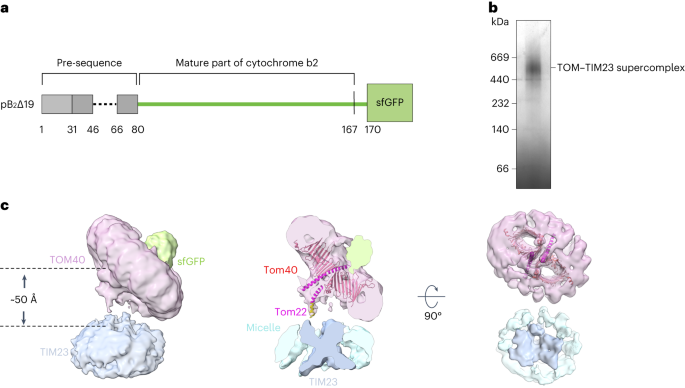
ABSTRACT Over half of mitochondrial proteins are imported from the cytosol via the pre-sequence pathway, controlled by the TOM complex in the outer membrane and the TIM23 complex in the
inner membrane. The mechanisms through which proteins are translocated via the TOM and TIM23 complexes remain unclear. Here we report the assembly of the active TOM–TIM23 supercomplex of
_Saccharomyces cerevisiae_ with translocating polypeptide substrates. Electron cryo-microscopy analyses reveal that the polypeptide substrates pass the TOM complex through the center of a
Tom40 subunit, interacting with a glutamine-rich region. Structural and biochemical analyses show that the TIM23 complex contains a heterotrimer of the subunits Tim23, Tim17 and Mgr2. The
polypeptide substrates are shielded from lipids by Mgr2 and Tim17, which creates a translocation pathway characterized by a negatively charged entrance and a central hydrophobic region.
These findings reveal an unexpected pre-sequence pathway through the TOM–TIM23 supercomplex spanning the double membranes of mitochondria. Access through your institution Buy or subscribe
This is a preview of subscription content, access via your institution ACCESS OPTIONS Access through your institution Access Nature and 54 other Nature Portfolio journals Get Nature+, our
best-value online-access subscription $29.99 / 30 days cancel any time Learn more Subscribe to this journal Receive 12 print issues and online access $209.00 per year only $17.42 per issue
Learn more Buy this article * Purchase on SpringerLink * Instant access to full article PDF Buy now Prices may be subject to local taxes which are calculated during checkout ADDITIONAL
ACCESS OPTIONS: * Log in * Learn about institutional subscriptions * Read our FAQs * Contact customer support SIMILAR CONTENT BEING VIEWED BY OTHERS STRUCTURAL BASIS OF MITOCHONDRIAL PROTEIN
IMPORT BY THE TIM23 COMPLEX Article 21 June 2023 MAPPING PROTEIN INTERACTIONS IN THE ACTIVE TOM-TIM23 SUPERCOMPLEX Article Open access 29 September 2021 CENTRAL ROLE OF TIM17 IN
MITOCHONDRIAL PRESEQUENCE PROTEIN TRANSLOCATION Article Open access 01 August 2023 DATA AVAILABILITY The cryo-EM maps of the supercomplexes with pB2167Δ19–sfGFP and pB2167Δ19–DHFR–sfGFP, and
the substrate-engaged TOM complex have been deposited in the Electron Microscopy Data Bank under accession numbers EMD-34661, EMD-34662 and EMD-34660. The atomic structure coordinates of
the substrate-engaged TOM complex have been deposited in the PDB under the accession number 8HCO. Source data are provided with this paper. REFERENCES * Neupert, W. & Schatz, G. How
proteins are transported into mitochondria. _Trends Biochem. Sci._ 6, 1–4 (1981). Article CAS Google Scholar * Bykov, Y. S., Rapaport, D., Herrmann, J. M. & Schuldiner, M. Cytosolic
events in the biogenesis of mitochondrial proteins. _Trends Biochem. Sci._ 45, 650–667 (2020). Article CAS PubMed Google Scholar * Dudek, J., Rehling, P. & van der Laan, M.
Mitochondrial protein import: common principles and physiological networks. _Biochim. Biophys. Acta_ 1833, 274–285 (2013). Article CAS PubMed Google Scholar * Mokranjac, D. &
Neupert, W. Thirty years of protein translocation into mitochondria: unexpectedly complex and still puzzling. _Biochim. Biophys. Acta_ 1793, 33–41 (2009). Article CAS PubMed Google
Scholar * Wiedemann, N. & Pfanner, N. Mitochondrial machineries for protein import and assembly. _Annu. Rev. Biochem._ 86, 685–714 (2017). Article CAS PubMed Google Scholar *
Hansen, K. G. & Herrmann, J. M. Transport of proteins into mitochondria. _Protein J._ 38, 330–342 (2019). Article CAS PubMed Google Scholar * Vogtle, F. N. et al. Global analysis of
the mitochondrial N-proteome identifies a processing peptidase critical for protein stability. _Cell_ 139, 428–439 (2009). Article PubMed Google Scholar * Callegari, S., Cruz-Zaragoza, L.
D. & Rehling, P. From TOM to the TIM23 complex—handing over of a precursor. _Biol. Chem._ 401, 709–721 (2020). Article CAS PubMed Google Scholar * Mokranjac, D. How to get to the
other side of the mitochondrial inner membrane—the protein import motor. _Biol. Chem._ 401, 723–736 (2020). Article CAS PubMed Google Scholar * Daum, G., Gasser, S. M. & Schatz, G.
Import of proteins into mitochondria—energy-dependent, 2-step processing of the intermembrane space enzyme cytochrome-B2 by isolated yeast mitochondria. _J. Biol. Chem._ 257, 3075–3080
(1982). Google Scholar * Hurt, E. C., Pesoldhurt, B. & Schatz, G. The amino-terminal region of an imported mitochondrial precursor polypeptide can direct cytoplasmic
dihydrofolate-reductase into the mitochondrial matrix. _EMBO J._ 3, 3149–3156 (1984). Article CAS PubMed PubMed Central Google Scholar * Kiebler, M. et al. Identification of a
mitochondrial receptor complex required for recognition and membrane insertion of precursor proteins. _Nature_ 348, 610–616 (1990). Article CAS PubMed Google Scholar * Shiota, T. et al.
Molecular architecture of the active mitochondrial protein gate. _Science_ 349, 1544–1548 (2015). Article CAS PubMed Google Scholar * Araiso, Y. et al. Structure of the mitochondrial
import gate reveals distinct preprotein paths. _Nature_ 575, 395–401 (2019). Article CAS PubMed Google Scholar * Guan, Z. Y. et al. Structural insights into assembly of human
mitochondrial translocase TOM complex. _Cell Discov._ https://doi.org/10.1038/s41421-021-00252-7 (2021). * Tucker, K. & Park, E. Cryo-EM structure of the mitochondrial protein-import
channel TOM complex at near-atomic resolution. _Nat. Struct. Mol. Biol._ 26, 1158–1166 (2019). Article CAS PubMed PubMed Central Google Scholar * Wang, W. et al. Atomic structure of
human TOM core complex. _Cell Discov._ 6, 67 (2020). Article CAS PubMed PubMed Central Google Scholar * Su, J. Y. et al. Structural basis of Tom20 and Tom22 cytosolic domains as the
human TOM complex receptors. _Proc. Natl Acad Sci USA_ https://doi.org/10.1073/pnas.2200158119 (2022). * Hines, V. et al. Protein import into yeast mitochondria is accelerated by the
outer-membrane protein Mas70. _EMBO J._ 9, 3191–3200 (1990). Article CAS PubMed PubMed Central Google Scholar * Ramage, L., Junne, T., Hahne, K., Lithgow, T. & Schatz, G. Functional
cooperation of mitochondrial protein import receptors in yeast. _EMBO J._ 12, 4115–4123 (1993). Article CAS PubMed PubMed Central Google Scholar * Sollner, T., Griffiths, G., Pfaller,
R., Pfanner, N. & Neupert, W. Mom19, an import receptor for mitochondrial precursor proteins. _Cell_ 59, 1061–1070 (1989). Article CAS PubMed Google Scholar * Sollner, T., Pfaller,
R., Griffiths, G., Pfanner, N. & Neupert, W. A mitochondrial import receptor for the Adp/Atp carrier. _Cell_ 62, 107–115 (1990). Article CAS PubMed Google Scholar * Kiebler, M. et
al. The mitochondrial receptor complex—a central role of Mom22 in mediating preprotein transfer from receptors to the general insertion pore. _Cell_ 74, 483–492 (1993). Article CAS PubMed
Google Scholar * Zarsky, V. & Dolezal, P. Evolution of the Tim17 protein family. _Biol. Direct_ 11, 54 (2016). Article PubMed PubMed Central Google Scholar * Bauer, M. F.,
Sirrenberg, C., Neupert, W. & Brunner, M. Role of Tim23 as voltage sensor and presequence receptor in protein import into mitochondria. _Cell_ 87, 33–41 (1996). Article CAS PubMed
Google Scholar * Truscott, K. N. et al. A presequence- and voltage-sensitive channel of the mitochondrial preprotein translocase formed by Tim23. _Nat. Struct. Biol._ 8, 1074–1082 (2001).
Article CAS PubMed Google Scholar * Martinez-Caballero, S., Grigoriev, S. M., Herrmann, J. M., Campo, M. L. & Kinnally, K. W. Tim17p regulates the twin pore structure and voltage
gating of the mitochondrial protein import complex TIM23. _J. Biol. Chem._ 282, 3584–3593 (2007). Article CAS PubMed Google Scholar * Matta, S. K., Pareek, G., Bankapalli, K., Oblesha,
A. & D’Silva, P. Role of Tim17 transmembrane regions in regulating the architecture of presequence translocase and mitochondrial DNA stability. _Mol. Cell. Biol_.
https://doi.org/10.1128/MCB.00491-16 (2017). * Sim, S. I., Chen, Y. & Park, E. Structural basis of mitochondrial protein import by the TIM complex. Preprint at _bioRxiv_
https://doi.org/10.1101/2021.10.10.463828 (2021). * Gebert, M. et al. Mgr2 promotes coupling of the mitochondrial presequence translocase to partner complexes. _J. Cell Biol._ 197, 595–604
(2012). Article CAS PubMed PubMed Central Google Scholar * Ieva, R. et al. Mgr2 functions as lateral gatekeeper for preprotein sorting in the mitochondrial inner membrane. _Mol. Cell_
56, 641–652 (2014). Article CAS PubMed Google Scholar * Lee, S. et al. The Mgr2 subunit of the TIM23 complex regulates membrane insertion of marginal stop-transfer signals in the
mitochondrial inner membrane. _FEBS Lett._ 594, 1081–1087 (2020). Article CAS PubMed Google Scholar * Schleyer, M. & Neupert, W. Transport of proteins into
mitochondria—translocational intermediates spanning contact sites between outer and inner membranes. _Cell_ 43, 339–350 (1985). Article CAS PubMed Google Scholar * Dekker, P. J. T. et
al. The Tim core complex defines the number of mitochondrial translocation contact sites and can hold arrested preproteins in the absence of matrix Hsp70–Tim44. _EMBO J._ 16, 5408–5419
(1997). Article CAS PubMed PubMed Central Google Scholar * Chacinska, A. et al. Mitochondrial translocation contact sites: separation of dynamic and stabilizing elements in formation of
a TOM–TIM–preprotein supercomplex. _EMBO J._ 22, 5370–5381 (2003). Article CAS PubMed PubMed Central Google Scholar * Neupert, W. A perspective on transport of proteins into
mitochondria: a myriad of open questions. _J. Mol. Biol._ 427, 1135–1158 (2015). Article CAS PubMed Google Scholar * Pedelacq, J. D., Cabantous, S., Tran, T., Terwilliger, T. C. &
Waldo, G. S. Engineering and characterization of a superfolder green fluorescent protein. _Nat. Biotechnol._ 24, 79–88 (2006). Article CAS PubMed Google Scholar * Meier, S., Neupert, W.
& Herrmann, J. M. Conserved N-terminal negative charges in the Tim17 subunit of the TIM23 translocase play a critical role in the import of preproteins into mitochondria. _J. Biol.
Chem._ 280, 7777–7785 (2005). Article CAS PubMed Google Scholar * Williamson, M. P. The structure and function of proline-rich regions in proteins. _Biochem. J._ 297, 249–260 (1994).
Article CAS PubMed PubMed Central Google Scholar * Dekker, P. J. T. et al. Identification of Mim23, a putative component of the protein import machinery of the mitochondrial inner
membrane. _FEBS Lett._ 330, 66–70 (1993). Article CAS PubMed Google Scholar * Emtage, J. L. T. & Jensen, R. E. Mas6 encodes an essential inner membrane component of the yeast
mitochondrial protein import pathway. _J. Cell Biol._ 122, 1003–1012 (1993). Article CAS PubMed Google Scholar * Jumper, J. et al. Highly accurate protein structure prediction with
AlphaFold. _Nature_ 596, 583 (2021). Article CAS PubMed PubMed Central Google Scholar * Qi, L. B. et al. Cryo-EM structure of the human mitochondrial translocase TIM22 complex. _Cell
Res_ 31, 369–372 (2021). Article CAS PubMed Google Scholar * Zhang, Y. T. et al. Structure of the mitochondrial TIM22 complex from yeast. _Cell Res_ 31, 366–368 (2021). Article CAS
PubMed Google Scholar * Humphreys, I. R. et al. Computed structures of core eukaryotic protein complexes. _Science_ https://doi.org/10.1126/science.abm4805 (2021). * Gomkale, R. et al.
Mapping protein interactions in the active TOM–TIM23 supercomplex. _Nat. Commun._ https://doi.org/10.1038/s41467-021-26016-1 (2021). * Gold, V. A. M. et al. Visualizing active membrane
protein complexes by electron cryotomography. _Nat. Commun._ https://doi.org/10.1038/ncomms5129 (2014). * Alder, N. N., Jensen, R. E. & Johnson, A. E. Fluorescence mapping of
mitochondrial TM23 complex reveals a water-facing, substrate-interacting helix surface. _Cell_ 134, 439–450 (2008). Article CAS PubMed Google Scholar * Malhotra, K., Sathappa, M.,
Landin, J. S., Johnson, A. E. & Alder, N. N. Structural changes in the mitochondrial Tim23 channel are coupled to the proton-motive force. _Nat. Struct. Mol. Biol._ 20, 965–972 (2013).
Article CAS PubMed Google Scholar * Singha, U. K. et al. Protein translocase of mitochondrial inner membrane in _Trypanosoma brucei_. _J. Biol. Chem._ 287, 14480–14493 (2012). Article
CAS PubMed PubMed Central Google Scholar * Weems, E. et al. Functional complementation analyses reveal that the single PRAT family protein of _Trypanosoma brucei_ is a divergent homolog
of Tim17 in _Saccharomyces cerevisiae_. _Eukaryot. Cell_ 14, 286–296 (2015). Article CAS PubMed PubMed Central Google Scholar * Pyrihova, E. et al. A single Tim translocase in the
mitosomes of _Giardia intestinalis_ illustrates convergence of protein import machines in anaerobic eukaryotes. _Genome Biol. Evol._ 10, 2813–2822 (2018). Article CAS PubMed PubMed
Central Google Scholar * Filipuzzi, I. et al. Stendomycin selectively inhibits TIM23-dependent mitochondrial protein import. _Nat. Chem. Biol._ 13, 1239–1244 (2017). Article CAS PubMed
PubMed Central Google Scholar * Kumazaki, K. et al. Structural basis of Sec-independent membrane protein insertion by YidC. _Nature_ 509, 516–520 (2014). Article CAS PubMed Google
Scholar * Bai, L., You, Q. L., Feng, X., Kovach, A. & Li, H. L. Structure of the ER membrane complex, a transmembrane-domain insertase. _Nature_ 584, 475–478 (2020). Article CAS
PubMed PubMed Central Google Scholar * Pleiner, T. et al. Structural basis for membrane insertion by the human ER membrane protein complex. _Science_
https://doi.org/10.1126/science.abb5008 (2020). * Wu, X. D. et al. Structural basis of ER-associated protein degradation mediated by the Hrd1 ubiquitin ligase complex. _Science_
https://doi.org/10.1126/science.aaz2449 (2020). * McDowell, M. A. et al. Structural basis of tail-anchored membrane protein biogenesis by the GET insertase complex. _Mol. Cell_ 80, 72
(2020). Article CAS PubMed Google Scholar * Anghel, S. A., McGilvray, P. T., Hegde, R. S. & Keenan, R. J. Identification of Oxa1 homologs operating in the eukaryotic endoplasmic
reticulum. _Cell Rep._ 21, 3708–3716 (2017). Article CAS PubMed PubMed Central Google Scholar * Smalinskait, L., Kim, M. K., Lewis, A. J. O., Keenan, R. J. & Hegde, R. S. Mechanism
of an intramembrane chaperone for multipass membrane proteins. _Nature_ 611, 161–166 (2022). Article Google Scholar * Sundaram, A. et al. Substrate-driven assembly of a translocon for
multipass membrane proteins. _Nature_ 611, 167–172 (2022). Article CAS PubMed PubMed Central Google Scholar * Chen, S., Schultz, P. G. & Brock, A. An improved system for the
generation and analysis of mutant proteins containing unnatural amino acids in _Saccharomyces cerevisiae_. _J. Mol. Biol._ 371, 112–122 (2007). Article CAS PubMed Google Scholar * Zheng,
S. Q. et al. MotionCor2: anisotropic correction of beam-induced motion for improved cryo-electron microscopy. _Nat. Methods_ 14, 331–332 (2017). Article CAS PubMed PubMed Central Google
Scholar * Zhang, K. Gctf: real-time CTF determination and correction. _J. Struct. Biol._ 193, 1–12 (2016). Article CAS PubMed PubMed Central Google Scholar * Zivanov, J. et al. New
tools for automated high-resolution cryo-EM structure determination in RELION-3. _eLife_ https://doi.org/10.7554/eLife.42166 (2018). * Emsley, P., Lohkamp, B., Scott, W. G. & Cowtan, K.
Features and development of Coot. _Acta Crystallogr. D_ 66, 486–501 (2010). Article CAS PubMed PubMed Central Google Scholar * Mitochondrial import receptor subunit TOM22. _AlphaFold
Protein Structure Database_ (2022); https://alphafold.ebi.ac.uk/entry/P49334 * Adams, P. D. et al. PHENIX: a comprehensive Python-based system for macromolecular structure solution. _Acta
Crystallogr. D_ 66, 213–221 (2010). Article CAS PubMed PubMed Central Google Scholar * Mirdita, M. et al. ColabFold: making protein folding accessible to all. _Nat. Methods_ 19, 679–682
(2022). Article CAS PubMed PubMed Central Google Scholar Download references ACKNOWLEDGEMENTS We thank N. Gao, X. Zhang and N. Li for helping with cryo-EM calculation, Q. Li for
helping with yeast genetics, and the National Center for Protein Sciences at Peking University for assistance with protein purification. The cryo-EM data were collected at the cryo-EM
platform of Peking University. The computation was supported by the High-Performance Computing Platform of Peking University. This work is supported by National Natural Science Foundation of
China (NSFC) (31870835 and 32271269 to L.L.). AUTHOR INFORMATION Author notes * These authors contributed equally: Xueyin Zhou, Yuqi Yang, Guopeng Wang, Shanshan Wang. AUTHORS AND
AFFILIATIONS * State Key Laboratory of Membrane Biology, Peking-Tsinghua Center for Life Sciences, School of Life Sciences, Peking University, Beijing, China Xueyin Zhou, Yuqi Yang, Shanshan
Wang, Dongjie Sun, Xiaomin Ou, Yuke Lian & Long Li * Academy for Advanced Interdisciplinary Studies, Peking University, Beijing, China Xueyin Zhou * School of Life Sciences, Peking
University, Beijing, China Guopeng Wang Authors * Xueyin Zhou View author publications You can also search for this author inPubMed Google Scholar * Yuqi Yang View author publications You
can also search for this author inPubMed Google Scholar * Guopeng Wang View author publications You can also search for this author inPubMed Google Scholar * Shanshan Wang View author
publications You can also search for this author inPubMed Google Scholar * Dongjie Sun View author publications You can also search for this author inPubMed Google Scholar * Xiaomin Ou View
author publications You can also search for this author inPubMed Google Scholar * Yuke Lian View author publications You can also search for this author inPubMed Google Scholar * Long Li
View author publications You can also search for this author inPubMed Google Scholar CONTRIBUTIONS X.Z. and Y.Y. prepared the protein samples for structural studies. S.W., X.Z. and Y.Y.
performed the crosslinking assays. G.W., X.Z. and Y.Y. collected the cryo-EM data and determined the structures. D.S. explored the sample preparation strategies at the beginning of the
project. X.O. helped with preparation of the proteins and cryo-grids. Y.L. helped with yeast genetics. L.L. supervised the project. L.L., X.Z., Y.Y., G.W. and S.W. prepared the manuscript.
CORRESPONDING AUTHOR Correspondence to Long Li. ETHICS DECLARATIONS COMPETING INTERESTS The authors declare no competing interests. PEER REVIEW PEER REVIEW INFORMATION _Nature Structural
& Molecular Biology_ thanks Agnieszka Chacinska, Friedrich Förster and Robert Keenan for their contribution to the peer review of this work. Primary Handling Editor: Katarzyna Ciazynska,
in collaboration with the _Nature Structural & Molecular Biology_ team. Peer reviewer reports are available. ADDITIONAL INFORMATION PUBLISHER’S NOTE Springer Nature remains neutral with
regard to jurisdictional claims in published maps and institutional affiliations. EXTENDED DATA EXTENDED DATA FIG. 1 PURIFICATION AND STRUCTURE DETERMINATION OF THE TOM-TIM23 SUPERCOMPLEX
WITH PB2167Δ19-SFGFP. A, Coomassie blue staining of the purified supercomplex in SDS-PAGE. The protein bands marked in SDS-PAGE have been confirmed by mass spectrometry. B, Immunoblots of
the substrate and the major translocase subunits in the purified supercomplex. The degradation bands of the polypeptide substrate are marked by *. C, Representative raw image of the
supercomplex collected by a Titan Krios with a K3 detector. D, Representative 2D classes of the supercomplex. E, Flow chart of data processing. The angles between the two discs in different
3D classes are labeled. See the details of data processing in Methods. F, Gold standard Fourier shell correlation (FSC) curve with the estimated resolution of the final map at 0.143. G,
Eulerian angle distribution of the particles used for supercomplex reconstruction. H, A close-up view of the connection between the TOM and TIM23 discs. The structure of the TOM complex
(6UCU) is fitted in the density. The Tom22 segment in the structure (residues 86–135) is colored magenta. The model of the Tom22 C-terminal segment (residues 136–149) is from the AlphaFold
Protein Structure Database and colored yellow. I, Representative 2D classes of the supercomplex particles. The TIM23 disc is placed in the center of the box for better alignment. The
characteristic X shape of the TIM23 TM region can be observed in the side view. Source data EXTENDED DATA FIG. 2 CRYO-EM 3D RECONSTRUCTIONS OF THE SUPERCOMPLEX WITH PB2167Δ19-DHFR-SFGFP. A,
Representative raw image of the super-complex collected by a Titan Krios with a K3 detector. B, Representative 2D classes. C, Flow chart of data processing for the super-complex. D, Eulerian
angle distribution of the particles used for supercomplex reconstruction. E, Flow chart of data processing focusing on the TOM complex. F, Local resolution estimation of the TOM complex. G,
Eulerian angle distribution of the particles used in the local TOM reconstruction. H, FSC curves with the estimated resolution at 0.143 of the supercomplex and the TOM maps. EXTENDED DATA
FIG. 3 EXAMPLES OF THE FIT OF THE MODELS INTO DENSITY MAPS. Density maps (grey mesh) and the models of the selected peptide segments are shown. Residues at the beginning and end of each
segment are indicated. The residue side chains are shown as sticks. EXTENDED DATA FIG. 4 PHOTO-CROSSLINKING OF THE POLYPEPTIDE SUBSTRATES TO THE SUBUNITS IN THE SUPERCOMPLEX. Similar
crosslinking assays as described in Fig. 3, showing the controls without UV exposure. A, Crosslinking between the C-terminal segment of the substrate and Tom40. The same protein samples were
immunoblotted with the anti-Flag antibody to detect the substrates (upper panel) and with the anti-Tom40 antibody (lower panel). The non-specific bands in the immunoblots are marked by *.
B, Crosslinking between the N-terminal segment of the substrate and Tim17. The same protein samples were immunoblotted with the anti-Flag antibody to detect the substrates (upper panel) and
with the anti-Strep antibody to detect Tim17 (lower panel). C, Sequence alignment of Tim17 C-tail. The proline residue is highlighted in green. The multiple-sequence alignment was performed
with ClustalOmega. The sequences are from _Saccharomyces cerevisiae_ (_S. cerevisiae_, Uniprot P39515), _Schizosaccharomyces pombe_ (_S. pombe_, Uniprot P87130), _Neurospora crassa_ (_N.
crassa_, Uniprot P59670), _Homo sapiens_ (_H. sapiens_, Uniprot Q99595), _Mus musculus_ (_M. musculus_, Uniprot Q545U2), _Caenorhabditis elegans_ (_C. elegans_, Uniprot O44477). D,
Crosslinking between the substrate and the Tim17 C-tail deletion mutant (Tim17ΔC) from strains yLS290/pLSB4-X. Protein extraction and crosslinking were performed same as in b. The
non-specific bands in the immunoblots are marked by *. E, Crosslinking between the N-terminal segment of the substrate and Mgr2 in yeast strains yLS240/pLSB4-X. The same protein samples were
immunoblotted with the anti-Flag antibody to detect the substrates (upper panel) and with the anti-HA antibody to detect Mgr2 (lower panel). The crosslinking bands are marked by red arrows.
F, Dependence of supercomplex formation on Mgr2. The supercomplex band is absent in BN-PAGE (upper panel) when Mgr2 is deleted. The two lower strips are from SDS-PAGE to detect Mgr2 and
Tim17. G, Crosslinking between the polypeptide substrate and Tim17 from the Mgr2 knockout yeast strains. The protein samples were immunoblotted with the anti-Strep antibody to detect the
substrates (left panel) and with the anti-HA antibody to detect Tim17 (right panel). The crosslinking bands at the residue positions 139 and 144 are marked by red arrows. Residue 162 of the
substrate is located in Tom40 and shows no crosslinking to Tim17. Source data EXTENDED DATA FIG. 5 DETECTION OF PHOTO-CROSSLINKING BETWEEN THE SUBSTRATE AND TIM23. A, The Bpa was
incorporated into the polypeptide substrate at the indicated residues in the N-terminal segment. The supercomplexes were assembled with these Bpa-incorporated substrates in yeast strains
yLS310/pLSB4-X, and then purified by pulling down Tim23 (Strep-His tagged) and the substrate (Flag tagged) sequentially. The purified supercomplexes were subjected to UV irradiation, and
immunoblotted with the anti-Flag antibody to detect the substrate (upper panel) and with the anti-Strep antibody to detect Tim23 (lower panel). The substrate-crosslinked bands were detected
in the upper panel, similar to the immunoblotting results in Extended Data Fig. 4b. However, immunoblotting of Tim23 did not reveal any crosslinking bands in the lower panel. The
non-specific bands in the immunoblots are marked by *. B, The supercomplexes were assembled as in A, and then mitochondria were subjected to UV irradiation. Tim23 was pulled down using Ni
resin under denaturing conditions. Immunoblotting with the anti-Flag antibody (upper panel) and the anti-Strep antibody (lower panel) did not detect any crosslinking bands between the
substrate and Tim23. C, The supercomplexes with Bpa-incorporated substrates were assembled in yeast strains expressing Strep-His tagged Tim17 (left panels, yeast strains yLS210/pLSB4-X) or
Mgr2 (right panels, yeast strains yLS421/pLSB4-X). After UV irradiation of mitochondria, Tim17 or Mgr2 was pulled down using Ni resin. Immunoblotting with the anti-Flag antibody (upper
panels) and the anti-Strep antibody (lower panels) detected the substrate-crosslinked band of Tim17 or Mgr2, respectively. The non-specific bands in the immunoblots are marked by *. D, Bpa
was incorporated at different positions in Mgr2. The supercomplexes were assembled with HA tagged Mgr2 and Strep-His tagged Tim17 (upper pannel) or Tim23 (lower pannel). Tim17 or Tim23 was
pulled down first and then subjected to UV irradiation. The Ni eluents were then subjected to HA pull-down before immunoblotting. Source data EXTENDED DATA FIG. 6 TIM23-TIM17-MGR2 MODEL AND
MUTANTS. A, AphaFold2 model of the Tim23-Tim17-Mgr2 heterotrimer, displaying the Predicted Aligned Error (PAE) plot (left) and the model (right) colored according to the predicted Local
Distance Difference Test (pLDDT) score. B, Model of the TM segments of Tim23-Tim17-Mgr2, showing the X shape of the heterotrimer. Tim23, Tim17, and Mgr2 are colored Dodger blue, Indian red,
and yellow, respectively. C, Fitting of the Tim17-Tim23-Mgr2 heterotrimer into the cryo-EM density of the TIM23 disc, shown in three views. The TIM23 density is colored sky blue and the
detergent micelle density is colored light blue. D, Top view of the Tim17-Mgr2 dimer, focusing on the residues 121 and 125 of Tim17, shown in three different views. Tim17 is colored Indian
red and Mgr2 is colored yellow. L121 and I125 of Tim17 and the neighboring residues of Mgr2 are shown as blue and yellow sticks, respectively. The channel is indicated by a dashed circle. E,
Disulfide crosslinking between Tim17 and the substrate. The disulfide bond between Tim17(68C) and the substrate (135C or 139C) was detected in yeast strains yLS273/pLSB9-X. The yeast
strains yLS274 and yLS276 with the wild type (WT) substrate were used as control. Tim17 was Strep-His tagged and the substrate was Flag tagged. The crosslinking products between Tim17 and
the substrate were pulled down by using the Ni and Flag resins sequentially after oxidation of mitochondria by CuPh3. Before SDS-PAGE, the Flag eluents were split into two portions, one
treated DTT and one without the DTT treatment. F, Tim17 mutant selection on 5-FOA plates. The yeast strains, yLS270 and yLS280, were transformed with Tim17 mutation plasmid pLSA4-X and
selected on 5-FOA plates. The mutants that could not grow during selection are shown. Source data EXTENDED DATA FIG. 7 SUBSTRATE CONTACT SITES OF TOM40. A, Sequence alignment of Tom40
focusing on the patch2 region. The substrate interacting residues in patch2 are marked. B, Structure superimposition of yeast and human Tom40. The ribbons of yeast and human Tom40 are
colored salmon and cyan, respectively. The substrate interacting residues of yeast and human Tom40 are shown as sticks and colored magenta and gold, respectively. C, Cryo-EM density of the
substrate-engaged Tom40 channel, focusing on the contact site between GFP and L14-15 of Tom40. The tip of L14-15 (residues 283–290) is not modeled as the local density is not well resolved.
However, it is clear from the map that L14-15 is -in direct contact with GFP. D, Sequence alignment of L14-15 from representative fungus species. The residues are colored according to their
hydrophobicity. EXTENDED DATA FIG. 8 ALPHAFOLD2 MODELING OF TIM23 SUBUNITS. Homo- and heter-oligomers of Tim23, Tim17, and Mgr2 are modeled by AlphaFold2. For each prediction, five models
are generated and ranked on the basis of their pLDDT scores. The PAE plots and the models colored according to the pLDDT scores are displayed. SUPPLEMENTARY INFORMATION REPORTING SUMMARY
PEER REVIEW FILE SUPPLEMENTARY TABLES Supplementary tables. SOURCE DATA SOURCE DATA FIG. 1 Unprocessed gels. SOURCE DATA FIG. 2 Unprocessed western blots. SOURCE DATA FIG. 3 Unprocessed
western blots. SOURCE DATA FIG. 3D Quantitative source data. SOURCE DATA FIG. 4 Unprocessed western blots. SOURCE DATA FIG. 5 Unprocessed western blots. SOURCE DATA FIG. 6 Unprocessed
western blots. SOURCE DATA EXTENDED DATA FIG. 1 Unprocessed gels and western blots. SOURCE DATA EXTENDED DATA FIG. 4 Unprocessed western blots. SOURCE DATA EXTENDED DATA FIG. 5 Unprocessed
western blots. SOURCE DATA EXTENDED DATA FIG. 6 Unprocessed western blots. RIGHTS AND PERMISSIONS Springer Nature or its licensor (e.g. a society or other partner) holds exclusive rights to
this article under a publishing agreement with the author(s) or other rightsholder(s); author self-archiving of the accepted manuscript version of this article is solely governed by the
terms of such publishing agreement and applicable law. Reprints and permissions ABOUT THIS ARTICLE CITE THIS ARTICLE Zhou, X., Yang, Y., Wang, G. _et al._ Molecular pathway of mitochondrial
preprotein import through the TOM–TIM23 supercomplex. _Nat Struct Mol Biol_ 30, 1996–2008 (2023). https://doi.org/10.1038/s41594-023-01103-7 Download citation * Received: 23 May 2023 *
Accepted: 18 August 2023 * Published: 11 September 2023 * Issue Date: December 2023 * DOI: https://doi.org/10.1038/s41594-023-01103-7 SHARE THIS ARTICLE Anyone you share the following link
with will be able to read this content: Get shareable link Sorry, a shareable link is not currently available for this article. Copy to clipboard Provided by the Springer Nature SharedIt
content-sharing initiative