- Select a language for the TTS:
- UK English Female
- UK English Male
- US English Female
- US English Male
- Australian Female
- Australian Male
- Language selected: (auto detect) - EN
Play all audios:
ABSTRACT Exfoliation corrosion (EFC) on extruded Mg-4Li-1Ca alloys as bone implant materials has not been reported. This study aimed to elucidate the degradation mechanism of a corrosion
type-exfoliation occurred on an extruded Mg-4Li-1Ca alloy under the interaction of glucose (Glu) and bovine serum albumin (BSA) in Hank’s solution. The alloy microstructure, chemical
composition, texture, stress distribution and Volta potential maps were characterized using metallographic microscopy, scanning electron microscopy and electron backscattered diffraction,
and scanning Kelvin Probe. The alloy corrosion behavior was investigated via electrochemical testing and hydrogen evolution rate methods. X-ray diffraction, Fourier transform infrared
spectrometry, X-ray photoelectron spectrometer, and fluorescence microscopy were used to analyze the corrosion products and the adsorption of the organic molecules. The experimental results
indicated that the anodic second phase (Mg2Ca) were distributed in lamellar and rod-like patterns along the extrusion direction of the alloy, a lower Volta potential for Mg2Ca, texture and
residual stresses also promote EFC. An increase in Glu concentration exacerbated the EFC, whereas BSA inhibited the EFC of the alloy. Higher Glu concentrations in BSA in turn inhibited EFC
since Glu can complex with amino acids in proteins to form Glu-amino acid amides, which promoted protein adsorption and thereby forming a dense protective film on the alloy surface. The
preferential dissolution of the Mg2Ca phase particles led to the complexation and adsorption of Glu and protein. This study enriches the understanding of the corrosion types in biomedical
Mg-4Li-1Ca alloy and paving the way for enhancing the EFC resistance of the alloy. SIMILAR CONTENT BEING VIEWED BY OTHERS TUNING BIODEGRADABILITY, BONE-BONDING CAPACITY, AND WEAR RESISTANCE
OF ZINC-30% MAGNESIUM INTERMETALLIC ALLOY FOR USE IN LOAD-BEARING BONE APPLICATIONS Article Open access 29 January 2024 CORROSION RESISTANCE AND BIOCOMPATIBILITY OF CARBON ION IMPLANTED
AZ31B MAGNESIUM ALLOY Article Open access 25 October 2024 HEAT TREATMENT DEPENDENT CYTOTOXICITY OF SILICALITE-1 FILMS DEPOSITED ON TI-6AL-4V ALLOY EVALUATED BY BONE-DERIVED CELLS Article
Open access 11 June 2020 INTRODUCTION Magnesium (Mg) alloys are widely utilized in the aerospace, automotive, and electronics industries due to their high specific strength, stiffness, and
damping capacity1,2,3. Additionally, their excellent biocompatibility and favorable mechanical properties make them promising candidates for biomedical applications4,5,6,7,8. However, the
electrode potential of Mg is only −2.37 V vs. SHE, which renders them susceptible to corrosion and therefore, limits their practical applications9. Generally, Mg alloys are susceptible to
various types of corrosion, including galvanic corrosion10, pitting corrosion11,12, intergranular corrosion (IGC)13, exfoliation corrosion (EFC)14,15, dealloying corrosion (DAC)16, stress
corrosion cracking17, filiform corrosion18,19, corrosion fatigue20, and crevice corrosion21. However, IGC, EFC, and DAC have been scarcely reported on the corrosion of Mg alloys18,22. IGC is
a form of localized corrosion caused by micro-galvanic corrosion and is closely related to the size and distribution of the intermetallic particles. EFC is a special form of IGC that occurs
on elongated grains and extruded alloys. This form of corrosion has been extensively studied in aluminum alloys23. It was believed that Mg alloys do not exhibit EFC24. Recently, our group14
reported that EFC occur in Mg-1Li-1Ca after a long exposure to 3.5 wt. % NaCl solution. Song et al.25 noted that the observation of EFC in extruded Mg-Li-Ca alloys is a significant advance
in the field of Mg alloy corrosion. However, this study lacked in-depth analysis of microstructural aspects, including characterization of the texture and the influence of stress. In
addition, EFC can lead to a drastic decrease in mechanical properties, making it impossible to ensure the complete support of the material during its service life. Herein, it is of necessity
to study the possibility of EFC before using a particular alloy. The addition of Lithium (Li) in Mg alloys is beneficial not only to reduce their mass but also enhance their strength via
solid-solution strengthening26. The first in vivo study performed by Witte et al.27 demonstrated that the Mg-Li-RE (LAE442) alloy is more corrosion-resistant than the AZ91 alloy. However, RE
is usually toxic. The replacement of calcium (Ca) may be a good choice for RE in Mg-Li alloys. Ca in Mg alloys has grain-refining effect and improve the alloy strength and formability, and
also enhance the biocompatibility of the alloy since Ca is non-toxic and it is the primary component of human bones9,28. In our previous investigation29 shows that Mg-Li-Ca alloys have
corrosion resistance superior to Mg-Ca alloys in Hank’s solutions. Our another study firstly reveals the formation of Li2CO3 on Mg-Li-Ca alloys and the transformation of pitting corrosion to
general corrosion due to the grain refinement30. Long et al.31 investigated that Mg-4Li-1Ca has a larger UTS, a larger EL and a lower corrosion rate than Mg-1Li-1Ca in Hank’s solution. This
scenario strongly supports the hypothesis that Li plays a positive role in decreasing the degradation rate and changing the corrosion type of biomedical Mg-Li-Ca alloys. Furthermore, the
consequence of biocompatibility of Mg-(1, 9, 15)Li-Ca alloys exhibits no toxicity to L-929 cells in 10% concentration of extracts, an acceptable hemolysis ratio for the Mg-1Li-1Ca alloy, and
no sign of thrombogenicity for the Mg-(1, 9)Li-Ca alloys except for the Mg-15Li-1Ca alloy32. Namely, low content of Li in Mg alloys might be more biocompatible. Further in vivo study33
disclosed that Mg-3.5Li-0.5Ca alloys possess the best in vitro performance for the implants into the mice femurs after 0.5 and 2 months, and a markable growth in cortical bone thickness on
the implanted Mg-3.5Li-0.5Ca alloy rods. The extracts of the Mg-3.5Li-0.5Ca alloy are helpful for the osteogenic differentiation of human bone marrow-derived mesenchymal stem cells along the
canonical Wnt/β-catenin pathway. Hence, Mg-4Li-Ca alloy has the greatest potential for use in bone implantation applications34. Nowadays, China is the world’s largest country in terms of
diabetes prevalence, with 141 million reported cases of diabetes in 2021 and an adult prevalence rate of 12.8%, accounting for 26.2% of the total number of reported cases worldwide, which is
expected to continue to grow to 164 million and 174 million in 2030 and 204535. The ageing population and the large number of people suffering from hyperglycemia and diabetes make it a
major public health problem with considerable implications for the country’s economic development, social security and stability36,37. Elevated blood glucose (Glu) levels in humans inhibit
bone calcium absorption and mineralization, leading to osteoporosis and an increased risk of fractures, particularly in hyperglycemic and diabetic patients and the elderly38. Therefore,
investigating the in vitro corrosion behavior of biomedical Mg alloys in Glu-containing environments has important implications for their practical application. Our previous
studies39,40,41,42,43,44 have demonstrated the significant impact of Glu, amino acids and proteins on the corrosion behavior of Mg alloy bone implants. Glu, as a polyhydroxy aldehyde and
monosaccharide is an energy source and intermediate product of metabolism in living cells. The presence of Glu influences the corrosion degradation behavior of pure Mg and its alloys in
simulated body fluids39. Specifically, Glu reduces the pH of the solution, which increases the corrosion rate of pure Mg and its alloys in physiological saline but decreases the corrosion
rate in Hank’s solution39. This is due to the acidification of the Mg surface by Glu, which converts its aldehyde group into a carboxyl group that facilitates interfacial reactions of Mg
alloys. This promotes the formation of Ca-P salts and hydroxyapatite (HA)45. Proteins are organic macromolecules that are essential for human metabolism. Many studies have investigated the
influence of proteins on the degradation rate of Mg alloys in different environments. It is widely reported that the degradation behavior of Mg alloys is primarily affected by protein
adsorption and chelation46,47,48,49. Proteins act as a diffusion barrier between the metal surface and the surrounding environment by adsorbing onto the surface of the Mg alloy47,48. This
process inhibits the adsorption of corrosive ions, and ultimately reducing the corrosion rate of Mg alloys. The reaction between the Mg alloys and proteins is dependent on various factors
such as the type of Mg alloy, reaction time, and environmental conditions4. Protein adsorption occurs in the initial stages of interaction with the Mg alloy surface, and the adsorbed
proteins can alter the physical morphology and chemical properties of the Mg alloy surface, and thereby affecting its degradation behavior. In fact, the interaction between proteins and the
Mg alloys is a more complex process. Some studies have suggested that proteins can chelate Mg2+ ions, and accelerate the degradation rate of Mg alloys. In contrast, another research has
shown that proteins can react with Mg2+ ions (RCH(NH2)COO)2Mg, and inhibit the degradation behavior of Mg alloys50. Furthermore, protein adsorption can affect subsequent cell adhesion,
proliferation, differentiation, signaling, and tissue regeneration on the surface of Mg alloys. Sivaraman et al.51 demonstrated that the conformation of adsorbed proteins is the primary
determinant of platelet adhesion rather than the amount adsorbed. Similarly, Hu et al.52 reported that different bioactive surfaces induce conformational changes in adsorbed proteins, and
different conformations have a significant effect on cellular activity. Despite the potential impact of Glu and proteins on the conformational changes of Mg alloys, only a few studies have
investigated this phenomenon in pure Mg. Our group41 examined the coupling effect of amino acids and Glu on the degradation behavior of pure Mg in physiological saline, and also43
investigated the impact of Glu and protein on the degradation of pure Mg and found that the presence of Glu and protein together significantly inhibited the degradation of Mg, resulting in a
reduced degradation rate. Further understanding of the mechanisms involved in protein adsorption and chelation can provide insights for the design and development of Mg alloys with improved
corrosion resistance for various biomedical applications. This work aims to investigate the effect of Glu and protein on the EFC behavior of Mg-4Li-1Ca in physiological environment. The
influence of factors such as alloy texture, stress, and second phase on EFC is also analyzed in-depth from the microstructure. Based on the detailed analysis of the experimental results, a
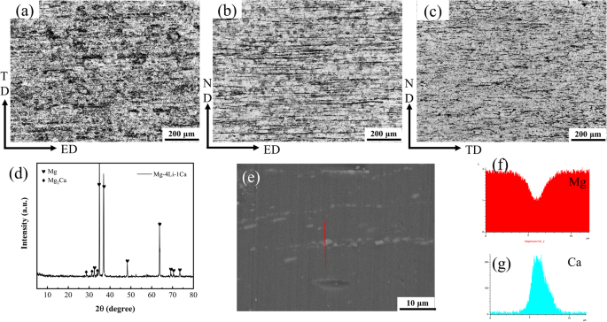
mechanism for EFC on the alloy involving Glu and protein coupling has been proposed. RESULTS Figure 1a–c depicts the metallographic micrographs of the Mg-4Li-1Ca alloy, showing the top
surface, longitudinal, and transverse sections (Supplementary Fig. 1). The intermetallic compounds or second phases (Mg2Ca particles) were observed mainly at the grain boundaries along the
extrusion direction (Fig. 1a). In the LS sample, the Mg2Ca particles were distributed in lines parallel to the extrusion direction; and in the TS sample, the Mg2Ca particles were distributed
in flat, short bars perpendicular to the extrusion direction. The lamellar pattern of Mg2Ca particles distribution in the Mg-4Li-1Ca alloy provides a structural factor for EFC as the Mg2Ca
particles act as the anodic phase relative to the α-Mg matrix and is preferentially corroded during the corrosion process14,31,53. From the electron probe X-ray microanalysis (EPMA) (Fig.
1e–g), it was observed that the intermetallic particles Mg2Ca were linearly distributed in the Mg-4Li-1Ca alloy along the extrusion direction, and the enrichment of Ca element at the
location of the second phase was seen from the line scan X-ray spectroscopy (EDS), which indicated that the presence of the Mg2Ca phase, and the peak of Mg2Ca was visible in the X-ray
diffraction (XRD) plot (Fig. 1d). Scanning Kelvin probe (SKP) testing was employed to detect the Volta potential in a specific region of the sample and characterize the corrosion reaction
activity. Supplementary Fig. 2 illustrates the Volta potential plots for the extruded Mg-4Li-1Ca alloy in SS, LS, and TS samples. The low potential dots and short bars in the Mg-4Li-1Ca
TD-ED plane correspond to the intermetallic Mg2Ca in Fig. 1a, which exhibit the lowest Volta potential. This suggests that the corrosion pits may start first in the planar Mg2Ca particles
during the corrosion process of the extruded Mg-4Li-1Ca alloy. In the LS and TS of the extruded Mg-4Li-1Ca alloy (Supplementary Fig. 2), a gradual increase in potential from the outer to the
inner layer was evident. The lowest Volta in the LS is −488 mV, and in the TS, it was −1027 mV. The highest and lowest Volta potentials in the LS had a difference of 390 mV, and in the TS,
the Volta potential difference was 710 mV. It was thus evident that the outer region had lower reaction activity than the inner region. The microstructural characteristics of the Mg-4Li-1Ca
alloy were investigated using grain orientation analysis, and the results are presented in Fig. 2. The SS plane of the alloy (Fig. 1a) showed a relatively uniform distribution of grain
orientation, dominated by the {0001} base plane. This plane is known for its strong corrosion resistance as compared to the prismatic and conical planes54,55,56. In contrast, the LS and TS
planes (Fig. 1b, c) exhibited a dominant grain orientation of {\(\bar{1}\)2\(\bar{1}\)0} and {01\(\bar{1}\)0}, respectively. The LS plane showed a clear grain-cutting along the extrusion
direction, while the TS plane showed some grain orientation along the same direction. The average grain size was found to be ~5.84 μm for SS, 9.71 μm for LS, and 8.09 μm for TS, with LS and
TS having larger grain sizes than the average size of SS (Supplementary Fig. 3). Furthermore, the grain orientation, polar, and antipodal diagrams indicated that the extruded Mg-4Li-1Ca
alloy had a distinct texture. From Supplementary Fig. 4, it can be observed that the local misorientation on the surface is greater than that on the longitudinal and transverse sections. The
local average misorientation (LAM) analysis also revealed that the surface had the highest local dislocation density. As the local dislocation density was directly proportional to residual
stress, it can be concluded that the surface has the highest residual stress, with all three faces exhibiting residual stress. And the above equation gives \({\rho }^{{GND}}\) of 4.0, 2.24,
and 1.68 × 10¹⁴ m−2 for SS, TS, and LS surfaces, respectively (Supplementary Fig. 4)57,58,59,60. Furthermore, the longitudinal and transverse sections showed orientation-dependent
distribution of residual stress along the extrusion direction, which might provide high-energy corrosion sites for subsequent corrosion studies. Figure 3 illustrates the HER curves of the
Mg-4Li-1Ca alloy in Hank’s solution with different concentrations of Glu content and containing BSA at 37 °C. The HER curve can be used to characterize the change in the degradation rate of
the alloy. The samples displayed higher degradation rate values at the beginning of the immersion test, which may be due to the preferential corrosion of the anodic second phase in the
Mg-4Li-1Ca alloy, releasing H2. The degradation rates of the samples were: 3 g·L−1 Glu > 2 g·L−1 Glu > 1 g·L−1 Glu > 1 g·L−1 Glu + BSA > 2 g·L−1 Glu + BSA > 3 g·L−1 Glu + BSA
solution. It was observed that the corrosion rate of the Mg-4Li-1Ca alloy increased with increasing Glu concentration in the Hank’s solution, while the addition of BSA slowed down the
corrosion of the alloy and the corrosion rate decreased with increasing Glu concentration. This was associated with the properties of Glu in solution, where it underwent acidification and
was converted to gluconic acid. The adsorption of gluconic acid on the surface of the Mg alloy promoted the adsorption of Cl- ions, which accelerated the corrosion39. In contrast, with the
addition of BSA, adsorption of BSA occurred on the surface of the Mg alloy and the synergistic effect of BSA with Glu43 resulted in slowing down the onset of corrosion. Open circuit
potential (OCP), potentiodynamic polarization tests (PDP), and Electrochemical Impedance Spectroscopy (EIS) were employed to investigate the degradation behavior of Mg-4Li-1Ca alloy in six
distinct solutions (Fig. 4 and Supplementary Tables 2, 4). The OCP curves of Mg-4Li-1Ca in all the six solutions exhibited a continuous rise and then stabilized with only small variations
which indicates that Mg dissolves and gradually forms a corrosion product layer at the interface until it reaches a steady state61. The OCP values were found to be the highest in 1 g·L−1
Glu, followed by 2 g·L−1 Glu and 3 g·L−1 Glu, with the degradation of Mg-4Li-1Ca being most prominent in the early stages of degradation. The PDP results indicated that Mg-4Li-1Ca alloy
exhibited the highest _E_corr and lowest _i_corr in 1 g·L−1 Glu, whereas in 3 g·L−1 Glu, the alloy showed the lowest _E_corr and highest _i_corr, implying that Mg-4Li-1Ca alloy degraded the
slowest in 1 g·L−1 Glu solution and the fastest in 3 g·L−1 Glu solution. In contrast, in solutions containing BSA, the degradation rate was found to be slowest in 3 g·L−1 Glu + BSA due to
its highest _E_corr and lowest _i_corr, while the opposite was true in 1 g·L−1 Glu + BSA. As the Glu content decreased, the anodic branch moved towards the noble region, whereas in the
solution containing BSA, the anodic branch moved towards the noble region as the Glu concentration increased. This suggests that the corrosion of the alloy is spontaneous when Glu
concentration increased or when Glu concentration decreased in solutions containing BSA. EIS tests were carried out on three faces of the Mg-4Li-1Ca alloy (SS, LS, TS) and the results are
shown in Supplementary Fig. 5. The circuit fit was carried out and the fitted circuit is shown in the inset of Supplementary Fig. 5a and Supplementary Table 3. LS had the highest radius of
capacitive arc resistance, the largest |Z|, and as can be seen in the circuit fitting data, LS had the greatest charge transfer resistance (_R_ct), indicating that LS surface had the best
corrosion resistance. The results of the Nyquist, Bode and Bode phase plots are consistent, with the LS face having the best corrosion resistance and the TS face having the worst corrosion
resistance. Figure 4e, f present the EIS curves for Mg-4Li-1Ca alloy in different solutions. The Nyquist plots for the Mg-4Li-1Ca alloy in each of the six solutions contain high-frequency
reactance, low-frequency reactance and low-frequency susceptibility reactance. The alloy in Hank’s solutions with different concentrations of Glu exhibited the largest capacitive arc radius
in the 1 g·L–1 Glu solution and the smallest in the 3 g·L−1 Glu solution, while the solutions containing BSA had the largest and smallest capacitive arc radii in the 3 g·L−1 Glu + BSA and 1
g·L−1 Glu + BSA solutions, respectively. This indicated that the elevated Glu content accelerated the corrosion of the Mg-4Li-1Ca alloy, while the elevated Glu content in the solution
containing BSA inhibited the corrosion of the Mg-4Li-1Ca alloy. To gain further insight into the corrosion mechanism of the Mg-4Li-1Ca alloy, a circuit fit was carried out and the fitted
circuit is shown in the inset of Fig. 4e, f and Supplementary Table 4. _L_ and _R_L denote the inductance and resistance, respectively, and are used to describe low-frequency inductive
loops62. The presence of inductive loops indicates the occurrence of corrosion pits, which is mainly associated with the preferential corrosion of the anodic second phase (Mg2Ca) of the
Mg-4Li-1Ca alloy. The _R_ct represents the resistance to the transfer of charges and a higher _R_ct value implies a lower dissolution rate of Mg63. The _R_ct values followed the order: 3
g·L−1 Glu < 2 g·L−1 Glu < 1 g·L−1 Glu, 1 g·L−1 Glu+BSA < 2 g·L−1 Glu + BSA < 3 g·L−1 Glu + BSA. Furthermore, the low-frequency impedance modulus |_Z_| in the Bode diagram is a
key indicator for assessing corrosion resistance, with a higher |_Z_| indicating better corrosion resistance64. It was observed that Z increased in the decreasing order: 3 g·L−1 Glu < 2
g·L−1 Glu < 1 g·L−1 Glu, 1 g·L−1 Glu + BSA < 2 g·L−1 Glu + BSA < 3 g·L−1 Glu + BSA. Figures 5 and 6 depict the EFC behavior of the Mg-4Li-1Ca alloy in Hank’s solutions with varying
Glu concentrations or containing BSA after different immersion periods (30, 60, 90, 120, 150, 180 days) at 37 °C. EFC became more severe as the Glu concentration and immersion time
increased. The surface layer with finer grains exhibited greater corrosion resistance as compared to the inter layer with coarser granules. This phenomenon may explain why EFC occurred more
easily on longitudinal sections with anisotropic microstructures. In other words, the initial corrosion potential differed significantly between the outer surface and the interior, leading
to macroscopic galvanic corrosion at the interface between the outer surface and the interior and slight corrosion of the interior. In addition, the preferential dissolution of Mg2Ca
particles along the grain boundaries of the elongated microstructure might be the initiation of corrosion pits. This finding was consistent with our previous report14, the extruded
Mg-1Li-1Ca exhibited EFC in the form of uncorroded Mg alloy flakes buckling from the substrate after 90 D immersion in a 3.5 wt.% NaCl solution, but the EFC was less severe than in the SBF.
The inclusion of BSA resulted in a more uniform corrosion morphology as compared to the localized corrosion observed in the absence of BSA. This can be attributed to the preferential
adsorption of BSA on the material, creating a protective layer that prevents the aggressive ions (e.g., Cl−) from attacking the material. EFC was observed after 120 days in the
BSA-containing solution, whereas it occurred in just 30 days in the Hank’s solution without BSA. Furthermore, the extent of EFC decreased as the Glu concentration increased, primarily due to
Glu promoting the adsorption of BSA on the Mg alloy surface and inhibiting EFC development43. Figures 7, 8, Supplementary Figs. 6,7 present the Scanning electron microscopy (SEM) images and
EDS data of the Mg-4Li-1Ca alloy after immersion in various solutions. It was observed that the corrosion product films generated by the Mg-4Li-1Ca alloy after immersion in all the
solutions experienced severe rupture. Based on the corrosion morphology of the surfaces immersed in different concentrations of Glu solution for 30 days, it was observed that the higher the
Glu concentration, the more corrosion products on the surface, indicating that the increase in Glu concentration promoted the degradation of the Mg-4Li-1Ca alloy. The EDS data revealed that
the main elements of the corrosion products in the BSA-free solution were C, O, Mg, P, and Ca, while in the BSA-containing solution, the presence of N was also observed, primarily due to the
BSA adsorbed on the surface of the Mg alloy. Figures 9 and 10 display the cross-SEM images and EDS patterns of the Mg-4Li-1Ca alloy. The cross-SEM images reveal that the Mg-4Li-1Ca alloy
experienced significant delamination of Mg when immersed in Hank’s solutions with different Glu concentrations for 30, 60, 90, and 120 days. This was primarily due to the preferential
corrosion of the lamellar anodic second phase Mg2Ca-particles distribution in the Mg-4Li-1Ca alloy, resulting in EFC. However, in the BSA-containing solution, the phenomenon of Mg
delamination due to EFC was not observed until 120 days, and no significant Mg delamination due to EFC was observed in the 3 g·L−1 Glu + BSA solution even after 180 days. This was mainly due
to the presence of Glu, which promoted protein adsorption43 and inhibited the development of EFC. Fourier transform infrared spectrometry (FT-IR) spectroscopy was utilized to investigate
the chemical composition and functional groups of corrosion products on the surface of a sample immersed in different solutions. The FT-IR spectra in Fig. 11 confirm the presence of an
Mg(OH)2 film through the Mg-OH stretching vibrations and O-H group absorption bands. The typical vibration modes of Glu molecules were observed through the C-H bands. The H2PO4− and PO43−
absorption peaks indicated the presence of phosphate in the corrosion products, and the CO32- groups in the FT-IR confirmed the presence of carbonates. Additionally, the presence of C=O or
C=O/C=N bonds suggested the adsorption of Glu or Glu and BSA onto the surface of the Mg alloy. Figure 12 displays the XRD patterns of Mg-4Li-1Ca after various immersion times in different
solutions. The dominating peaks in all spectra were related to the Mg phase65. A small amount of Mg(OH)2 was generated on the surface of Mg-4Li-1Ca in Hank’s solution after immersion, as
shown by the peaks at 18° and 38°. Furthermore, MgCO3 was present on the surface after different immersion times, mostly due to the formation of MgCO3 by CO2 and Mg(OH)2. The phosphate and
Ca2+ in solution generated Ca(H2PO4)2·H2O on the Mg-4Li-1Ca alloy surface. In Fig. 13, the total X-ray photoelectron spectrometer (XPS) spectrum of the Mg-4Li-1Ca alloy surface after 2 h (h)
of immersion, the N1s spectrum in the BSA-containing solution, and the C1s fine spectra of the surface of Mg-4Li-1Ca alloy immersed in different solutions for different times (0.5 h, 2 h,
24 h) are presented. The total spectrum (Fig. 13a) suggests the presence of C, O, Mg, Ca, and P in Hank’s solutions with different concentrations of Glu without BSA, while the presence of N
was detected in the solution containing BSA, indicating protein adsorption, which was consistent with the EDS results. In the N1s spectrum (Fig. 13b–d), the binding energy of N1s
photoelectrons showed three peaks at 399.1 eV, 399.9 eV, and 400.7 eV, corresponding to C=N66, O=C-NH, and N-O/C-N, respectively. This indicates the adsorption of protein on the Mg-4Li-1Ca
alloy surface. From the C1s fine spectra of the sample surface after immersion in the solution without BSA (Fig. 13e–j), three peaks at 284.8 eV, 285.5 eV, and 288.8 eV, corresponding to
C-C/C-H, C-O, and C=O, respectively, can be seen, indicating that Glu underwent acidification and was converted to gluconic acid39. In the fine spectrum of C1s containing BSA (Fig. 13h–j),
284.8 eV, 286.3 eV, 288.2 eV and 289.6 eV correspond to the presence of C-C/C-H, C-O, C=O and N-C=O bonds, respectively. This finding suggested the presence of Glu compounds and protein
compounds on the surface43. The laser confocal microscopy fluorescent image of BSA and Glu adsorption on the Mg alloy surface at various immersion durations is shown in Fig. 14. Fluorescein
isothiocyanate for fluorescent labeling of organic molecules67,68. As can be seen from Fig. 14, when the Mg-4Li-1Ca alloy was immersed in a solution containing Glu and BSA for 0.5 h, small
amounts of Glu and BSA were locally adsorbed on the surface of the sample, and the amount of Glu and BSA adsorbed on the surface of Mg-4Li-1Ca increased with the increase in soaking time.
From the adsorption results on the surface as well as the metallographic images and SKP results, Glu and BSA were preferentially adsorbed on the second phase Mg2Ca particles. And the
adsorption points of Glu and BSA were brighter, which indicated that the pre-existing Glu and BSA were more easily adsorbed on the second phase particles. This was mainly because the second
phase Mg2Ca particles was anodic, which preferentially corroded. The fluorescent features appeared in a striped distribution along the extrusion direction after corrosion, which was
consistent with the second distribution in the OM images (Fig. 1a–c). The Mg2+ and Ca2+ generated after corrosion form complexes and adsorb with carboxyl groups in gluconic acid and carboxyl
and amino groups in proteins, making Glu and proteins preferentially adsorb at the location of the second phase particles. Accordingly, the increase in Glu content accelerated the
adsorption of Glu, while the addition of BSA inhibited the adsorption of both Glu and BSA with the increase in Glu content. DISCUSSION The previous study found that 1, 2, 3 g·L−1 Glu in
Hank’s solution inhibit the corrosion of pure Mg as the Glu content increased39. In contrast, in this study, the increase in Glu concentration in Hank’s solution accelerated the corrosion of
Mg-4Li-1Ca alloy, which can be related to the presence of galvanic coupling corrosion between the large amount of anodic second phase and α-Mg in the alloy. Imani et al.69 found that the
addition of 0.1 g·L−1 of BSA to 2 g·L−1 of Glu in SBF solution inhibited the corrosion of WE43 alloy, which was consistent with the fact that BSA addition inhibited the corrosion of Mg alloy
in this experiment. In comparison to the EFC morphology of Mg-1Li-1Ca in 3.5 wt.% NaCl solution with long-term immersion14, the EFC of the Mg-4Li-1Ca alloy in Hank’s solution was less
seriously due to the presence of phosphates and the protective nature of the Hank’s solution. The macroscopic peeling of Mg-4Li-1Ca alloy in Hank’s solution tends to be single or several
layers warped as compared to the book-bound-like multi-layer peeling observed in Mg-1Li-1Ca in 3.5 wt.% NaCl solution14 and also in Al alloys in the marine atmosphere23. The effect of
organic molecules on the degradation behavior of Mg alloys has been widely reported4,70, but the most studies show the results of short-term degradation. However, for bone implant
applications, the material must maintain their mechanical properties in the human body for at least 12 weeks. Hence, the effect of organic molecules on the long-term degradation of Mg alloys
is critical. Moreover, the effect of organic molecules on the degradation of Mg alloys may change with time. Hereby, the EFC mechanism of Mg-4Li-1Ca alloy during long-term immersion were
investigated under the coupling of Glu and BSA. Mg is a very reactive in salt solutions. Initially, Mg in solution produces large amounts of Mg2+, OH- and H2 molecules. As a result, the pH
of the solution increases. The chemical reactions involved in the dissolution of Mg are listed below: $${\rm{Mg}}({\rm{s}})\to {{\rm{Mg}}}^{2+}({\rm{aq}})+{2{\rm{e}}}^{-}({\rm{Anode\;
reaction}})$$ (1) $${2{\rm{H}}}_{2}{\rm{O}}+{2{\rm{e}}}^{-}\to {{\rm{H}}}_{2}({\rm{g}})+{2{\rm{OH}}}^{-}({\rm{aq}})({\rm{Cathodic\; reaction}})$$ (2)
$${{\rm{Mg}}}^{2+}({\rm{aq}})+{2{\rm{OH}}}^{-}({\rm{aq}})\to {{\rm{Mg}}({\rm{OH}})}_{2}({\rm{s}})({\rm{Product\; generation}})$$ (3) Many of the secondary phase particles (i.e., Mg17Al12,
Al8Mn5, Mg2Si, etc.) in Mg alloys are found to be cathodic in nature due to α-Mg substrate possessing low electrode potential. However, certain second phases have been identified as anodic
(e.g., Mg2Ca, (Mg, Al)2Ca, Mg60Zn32Nd8, Mg35Zn40Nd2571, Mg41Nd516), resulting in differences in the corrosion mechanisms and dissolution rates of Mg alloys9,72. In Mg-Ca alloys, the
secondary phase particles Mg2Ca have been found to be more negative73 and have a lower _i_corr53 than that of the α-Mg substrate. Hence, Mg2Ca particles undergo selective corrosion in Mg-Ca
alloys. Ding et al.14 reported that EFC of extruded Mg-1Li-1Ca alloys must include two conditions: (i) structural factors, and (ii) corrosion factors. For Mg-4Li-1Ca, a pattern of lamellar
distribution of the Mg2Ca anodic second phase particles in the alloy can be observed from metallography, EBSD, and SKP. Furthermore, the Mg2Ca particles being anodic corrodes preferentially
in Hank’s solution, and thus providing an active channel for Mg-4Li-1Ca EFC. PBR is the ratio of the volume _V_com of a compound to the volume _V_M of a metal, as shown in the following
equation14: $${PBR}=\frac{{V}_{{com}}}{{V}_{M}}=\frac{{M}_{{com}}\bullet {\rho }_{M}}{n\bullet A\bullet {\rho }_{{com}}}$$ (4) where _M_com and _ρ_com are the molecular weight and density of
the compound, respectively. A and _ρ_M are the atomic weight and density of the metal, and n is the number of metal atoms in the molecule of the compound. The second phase particles, Mg2Ca,
had a lower electrode potential than the α-Mg phase74 and it acted as an active channel for corrosion reactions, and the resulting corrosion products deposited in the active channel. As the
PBR of these corrosion products was greater than 1, there was a significant wedging effect. The active channels of the corrosion reaction could gradually penetrate deeper into the
substrate, eventually producing corrosion cracks and the corrosion products buckling the lamellar alloy to make it peel off the substrate. The EBSD results indicated that the Mg-4Li-1Ca
alloy grains showed a clear texture along the extrusion direction, which provided a location for the lamellar distribution of the second phase particles. The preferential dissolution of the
intermetallic compounds Mg2Ca resulted in the occurrence of EFC. Moreover, it was also evident from the LAM diagram that residual stresses existed within the alloy, and the high-energy at
the residual stresses accelerated corrosion75,76, while the residual stresses at the LS tended to be distributed along the extrusion direction, accelerating the corrosion cracks along the
extrusion direction. Previous studies had shown that Glu was converted in solution from aldehyde groups to carboxyl groups and thus to gluconic acid, which promoted the enrichment of Cl−
(Eq. 5, 6)39. The Cl− ions attacked the corrosion product Mg(OH)2 (_K_sp = 9.628 × 10⁻⁴) on the alloy surface to produce soluble MgCl2 (Ksp = 54.6), leading to the breakdown of the surface
film and promoting further corrosion of the Mg-4Li-1Ca alloy (Eq. 7). Therefore, the increase in Glu concentration in Hank’s solution accelerated EFC (Fig. 15a).
$${{\rm{CH}}}_{2}{\rm{OH}}{({\rm{CHOH}})}_{4}{\rm{CHO}}\mathop{\to }\limits^{{O}_{2},37{\rm{^\circ }}{\rm{C}}}{\rm{RCOOH}}$$ (5) $$2{\rm{RCOOH}}+{\rm{Mg}}{({\rm{OH}})}_{2}\to
{({\rm{RCOO}})}_{2}{\rm{Mg}}+2{\rm{H}}2{\rm{O}}$$ (6) $${\rm{Mg}}{({\rm{OH}})}_{2}+{{\rm{Cl}}}^{-}\to {{\rm{MgCl}}}_{2}+{2{\rm{OH}}}^{-}$$ (7) Mg-4Li-1Ca alloy did not show any macroscopic
EFC morphology until 120 days in Hank’s solution containing BSA, which suggest that BSA inhibited the occurrence of EFC. However, lowering the Glu content in the solution, EFC became more
severe. The EFC mechanism in Mg-4Li-1Ca alloy is illustrated in Fig. 15b. It has been previously shown that amino acids reacted with Glu to form a new compound containing -C = N-, a Schiff
base, and that amino acids could also form Glu-amino acid amides with amino acids in proteins (Eq. 8)41,43. This allows Glu to promote the adsorption of proteins on the Mg alloy surface.
Consistent results were derived from the fluorescent labeling experiments (Fig. 15b). Due to the preferential corrosion of Mg2Ca, the corrosion produced large amounts of Mg2+ and Ca2+. Glu
and protein complexed with Mg2+ and Ca2+, Ca2+ had stronger complexation with gluconic acid and proteins45 and the increased surface roughness after corrosion allowed Glu and BSA to
preferentially adsorb in the second phase. As the immersion time increased, the proteins produced a dense protective film on the Mg alloy surface, which allowed the Mg alloy to change from
localized corrosion (pitting, preferential corrosion of the second phase particles) to uniform corrosion, inhibiting the occurrence of spalling. As the immersion time increases, the sample
surface layer was corroded away. Previously, our group has found that EFC of Mg-Li-Ca alloys was more likely to occur after the removal of the surface layer. Hence, after corrosion of the
surface layer, the Mg-4Li-1Ca alloy EFC occurs due to a combination of structural and corrosion factors, stresses, hydrogen bubbles, etc.
$${\rm{R}}-{{\rm{NH}}}_{2}+{\rm{R}}{\rm{\mbox{'}}}-{\rm{COH}}\to {\rm{R}}-{\rm{N}}={\rm{CH}}-{\rm{R}}{\rm{\mbox{'}}}+{{\rm{H}}}_{2}{\rm{O}}$$ (8) In summary this study investigated
the effects of different concentrations of Glu and protein in Hank’s solution on the EFC behviour of Mg-4Li-1Ca alloy. The main findings are summarized as follows: * (1) Microstructural
analysis of the Mg-4Li-1Ca alloy using optical metallography and scanning Kelvin probe measurements reveal that the anodic second phase Mg2Ca particles were distributed in a lamellar pattern
along the grain boundaries in the extrusion direction. Mg2Ca had a lower Volta potential and the surface layer of the alloy had a higher Volta potential for greater corrosion resistance.
And from the EBSD, the alloy had significant weaving and residual stresses. This lamellar distribution facilitated the preferential corrosion of Mg2Ca along the grain boundaries, thus
creating the conditions for EFC. Moreover, the texture and the presence of residual stresses of the alloy accelerated the occurrence of EFC. * (2) Mg-4Li-1Ca alloy exhibited EFC after 30
days of immersion in Hank’s solution. Higher Glu content resulted in more severe EFC. However, when BSA was present in Hank’s solution, macroscopic EFC morphology was observed only after 120
days of immersion, indicating that BSA inhibited EFC. But lower Glu content resulted in more severe EFC in this condition. * (3) Fluorescent labeling showed that Glu and protein were mainly
present on the second phase particles, which were preferentially corroding due to their anodic nature. The Mg2+ and Ca2+ generated during corrosion, interact with the carboxyl groups in
gluconic acid and the carboxyl and amino groups in proteins, leading to chelation and electrostatic adsorption. Glu and protein coupled and adsorbed, formed a dense protective film on
Mg-4Li-1Ca surface, which transformed the corrosion from localized corrosion (pitting, preferential corrosion at the second phase) to uniform corrosion, and inhibiting further corrosion. *
(4) Analysis by EDS, FT-IR, XRD, and XPS showed that the corrosion products were mainly MgCO3, Ca(H2PO4)2·H2O, and Mg(OH)2, along with chelation complexes of Glu (or albumin) and metal
cations. Overall, this study provides valuable insights into the microstructural characteristics and corrosion behavior of the Mg-4Li-1Ca alloy, and enriching the literature on the corrosion
types in biomedical Mg-based alloys, and potentially facilitates the improvement in the EFC resistance of the alloys. METHODS MATERIALS PREPARATION In this study, Mg-4Li-1Ca alloy (chemical
composition: Li 3.71 wt.%, Ca 1.15 wt.%, and Mg balance) was used as the test material. The alloy was extruded with a 30:1 extrusion ratio at a rate of 1 m·min−1, with the mold and
extrusion temperatures set at 350 and 300 °C, respectively. The extruded sheets were then cut into rectangular specimens (20 mm × 10 mm × 4.5 mm) in different sections, as illustrated in
Supplementary Fig. 1. Small holes were drilled at one end of the specimens to facilitate suspension using a string for the immersion test. Prior to the immersion, the specimens were ground
with wet SiC abrasive paper (grit sizes ranging from 150 to 1500), and then polished with 1.0 μm diamond paste to obtain a mirror surface. MICROSTRUCTURE OBSERVATION The microstructure and
chemical composition of Mg-4Li-1Ca alloy were examined using optical microscopy (OM, ZEISS Axio Lab A1, Germany) and EPMA (JXA-8230, Japan). Prior to OM observation, specimens were
chemically etched with a solution consisting 100 ml deionized water, 1.5 ml nitric acid (65–68%), 1 g oxalic acid, and 1 ml acetic acid (99%) at room temperature for 30 s. ELECTROCHEMICAL
BEHAVIOR Electrochemical measurements were conducted using a three-electrode cell setup and an electrochemical workstation (Princeton VersaSTAT 4). The cell setup consisted of a standard
three-electrode configuration, with a platinum foil electrode serving as the counter electrode, a saturated calomel electrode (SCE) as the reference electrode, and Mg-4Li-1Ca alloy as the
working electrode. The tests were conducted in physiological environment at room temperature. The OCP was measured for the first 3600 s of immersion. EIS test was carried out in a frequency
range from 100 kHz to 0.1 Hz, and the PDP were carried out from −0.4 V to 0.6 V (vs. OCP) with a scan rate of 1 mV·s−1. The repeatability of each test was ensured in triplicate. Moreover,
_E_corr and _i_corr are estimated by Tafel extrapolation from the anodic and cathodic polarization zones where the overpotential is 60 mV lower or higher than the free corrosion potential.
SCANNING KELVIN PROBE MEASUREMENT Volta potential maps were generated using a SKP (Princeton VERSA SCAN). The measurements were conducted under an air relative humidity of 65%. The probe,
made of tungsten with a diameter of 500 μm, was positioned at a fixed height above the sample surface. A square scanning area of 1000 μm × 1000 μm was used with a scanning step of 50 μm in
both the X and Y directions. IMMERSION TESTS The surface finished samples were suspended in 500 mL beakers using cotton strings. The beakers (with three samples in each beaker) were placed
in an electrothermal thermostat (DHP-420AB, Tianjin Sedeli Experimental Analytical Instruments Manufacturing, China) at 37 ± 0.5 °C. The solution was changed every three days to prevent the
solution from deteriorating and growing bacteria. The samples were removed after 30, 60, 90, and 120 days of immersion for the samples in Hank’s solutions with different concentrations of
Glu (1, 2, 3 g·L−1, since plasma glucose levels range from 4.6 to 6.1 mmol·L−1 in normal subjects, 10 mmol·L−1 after a meal, and 15 mmol·L−1 in hyperglycemic subjects77). The samples were
immersed in Hank’s solution containing BSA (0.1 g·L−1, i.e., the concentration of BSA in human plasma43) with different concentrations of Glu for 90, 120, 150, and 180 days. Three samples
were immersed in each condition to ensure the accuracy of the experiment. The removed samples were rinsed with deionised water, dried using a hairdryer and stored in a sealed container for
subsequent analysis and testing. In this experiment, Hank’s solution was chosen as the corrosion medium to study the influence of different levels of Glu and BSA on the susceptibility of EFC
in Mg-4Li-1Ca. The chemical composition of the solution used for the immersion test is listed in Supplementary Table 1. SURFACE CHARACTERIZATION The morphology and cross-section of the
samples after immersion for different durations were captured using a digital camera. The chemical composition of the samples was identified using field emission scanning electron microscopy
(FE-SEM, Nova Nano SEM450, USA) and EDS (Oxford X-MaxN). Corrosion products were examined using XRD (Rigaku D/MAX 2500PC, Japan), FT-IR (Nicolet 380; Thermo electron, US), and XPS
(ESCALAB250Xi). Fluorescence microscopy (FM, Leica DM2500, Germany) was employed to detect the presence of Glu and albumin. DATA AVAILABILITY The data that support the findings of this study
are available from Prof. Rong-Chang Zeng (Email: [email protected]) upon reasonable request. CODE AVAILABILITY The codes that support the findings of this study are available from the
corresponding author upon reasonable request. REFERENCES * Yang, Y. et al. Research advances in magnesium and magnesium alloys worldwide in 2020. _J. Magnes. Alloy._ 9, 705–747 (2021).
Article CAS Google Scholar * Li, L. Y. et al. Advances in functionalized polymer coatings on biodegradable magnesium alloys—a review. _Acta Biomater._ 79, 23–36 (2018). Article CAS
Google Scholar * Pan, S.-Q., Zhang, F., Wen, C. & Zeng, R.-C. Advances in Mg–Al-layered double hydroxide steam coatings on Mg alloys: a review. _J. Magnes. Alloy._ 11, 1505–1518 (2023).
Article CAS Google Scholar * Cai, L. et al. Advances in bioorganic molecules inspired degradation and surface modifications on Mg and its alloys. _J. Magnes. Alloy._ 10, 670–688 (2022).
Article CAS Google Scholar * Li, D. et al. In vitro and in vivo assessment of the effect of biodegradable magnesium alloys on osteogenesis. _Acta Biomater._ 141, 454–465 (2022). Article
CAS Google Scholar * Costantino, M. D. et al. Inflammatory response to magnesium-based biodegradable implant materials. _Acta Biomater._ 101, 598–608 (2020). Article CAS Google Scholar
* Zhao, Y. et al. pH/NIR-responsive and self-healing coatings with bacteria killing, osteogenesis, and angiogenesis performances on magnesium alloy. _Biomaterials_ 301, 122237 (2023).
Article CAS Google Scholar * Jiao, Z.-J. et al. In vitro degradation and biocompatibility of in-situ fabricated Mg-Al-Ga-LDH/MAO hybrid coating on Mg alloy AZ31. _Surf. Coat. Technol._
472, 129922 (2023). Article CAS Google Scholar * Zeng, R. C., Cui, L. Y. & Ke, W. Biomedical magnesium alloys: composition, microstructure and corrosion. _Acta Metall. Sin._ 54,
1215–1235 (2018). CAS Google Scholar * Jia, J. X., Atrens, A., Song, G. & Muster, T. H. Simulation of galvanic corrosion of magnesium coupled to a steel fastener in NaCl solution.
_Mater. Corros._ 56, 468–474 (2005). Article CAS Google Scholar * Lafront, A. M. et al. Pitting corrosion of AZ91D and AJ62x magnesium alloys in alkaline chloride medium using
electrochemical techniques. _Electrochim. Acta_ 51, 489–501 (2005). Article CAS Google Scholar * Andreatta, F., Apachitei, I., Kodentsov, A. A., Dzwonczyk, J. & Duszczyk, J. Volta
potential of second phase particles in extruded AZ80 magnesium alloy. _Electrochim. Acta_ 51, 3551–3557 (2006). Article CAS Google Scholar * Zeng, R., Kainer, K. U., Blawert, C. &
Dietzel, W. Corrosion of an extruded magnesium alloy ZK60 component—the role of microstructural features. _J. Alloy. Compd._ 509, 4462–4469 (2011). Article CAS Google Scholar * Ding,
Z.-Y. et al. Exfoliation corrosion of extruded Mg-Li-Ca alloy. _J. Mater. Sci. Technol._ 34, 1550–1557 (2018). Article CAS Google Scholar * Morishige, T. H., Goto, T., Nakamura, E. &
Takenaka, T. J. M. T. Exfoliation corrosion behavior of cold-rolled Mg-14 mass% Li-1 mass% Al alloy in NaCl solution. _Mater. Trans._ 54, 1863–1866 (2013). Article CAS Google Scholar *
Gao, G.-J. et al. Dealloying corrosion of anodic and nanometric Mg41Nd5 in solid solution-treated Mg-3Nd-1Li-0.2Zn alloy. _J. Mater. Sci. Technol._ 83, 161–178 (2021). Article CAS Google
Scholar * Unigovski, Y., Eliezer, A., Abramov, E., Snir, Y. & Gutman, E. M. Corrosion fatigue of extruded magnesium alloys. _Mater. Sci. Eng. A_ 360, 132–139 (2003). Article Google
Scholar * Zeng, R., Qi, W., Zhang, F., Cui, H. & Zheng, Y. In vitro corrosion of Mg–1.21Li–1.12Ca–1Y alloy. _Prog. Nat. Sci. Mater. Int._ 24, 492–499 (2014). Article CAS Google
Scholar * Song, Y., Shan, D., Chen, R. & Han, E.-H. Corrosion characterization of Mg–8Li alloy in NaCl solution. _Corros. Sci._ 51, 1087–1094 (2009). Article CAS Google Scholar *
Ding, Z.-Y. et al. In vitro corrosion of micro-arc oxidation coating on Mg-1Li-1Ca alloy—the influence of intermetallic compound Mg2Ca. _J. Alloy. Compd._ 764, 250–260 (2018). Article CAS
Google Scholar * Chen, B. et al. In vitro crevice corrosion of biodegradable magnesium in different solutions. _J. Mater. Sci. Technol._ 52, 83–88 (2020). Article CAS Google Scholar *
Esmaily, M. et al. Fundamentals and advances in magnesium alloy corrosion. _Prog. Mater. Sci._ 89, 92–193 (2017). Article CAS Google Scholar * Sun, S., Fang, Y., Zhang, L., Li, C. &
Hu, S. Effects of aging treatment and peripheral coarse grain on the exfoliation corrosion behaviour of 2024 aluminium alloy using SR-CT. _J. Mater. Res. Technol._ 9, 3219–3229 (2020).
Article CAS Google Scholar * Song, G. L. & Atrens, A. Corrosion mechanisms of magnesium alloys. _Adv. Eng. Mater._ 1, 11–33 (1999). Article CAS Google Scholar * Song, J., She, J.,
Chen, D. & Pan, F. Latest research advances on magnesium and magnesium alloys worldwide. _J. Magnes. Alloy._ 8, 1–41 (2020). Article CAS Google Scholar * Ghorbani, M., Boley, M.,
Nakashima, P. N. H. & Birbilis, N. A machine learning approach for accelerated design of magnesium alloys. _J. Magnes. Alloy_. 11, 3620–3633 (2023). Article CAS Google Scholar *
Witte, F. et al. In vitro and in vivo corrosion measurements of magnesium alloys. _Biomaterials_ 27, 1013–1018 (2006). Article CAS Google Scholar * Yurchenko, N. Y. et al. Effect of
multiaxial deformation on structure, mechanical properties, and corrosion resistance of a Mg-Ca alloy. _J. Magnes. Alloy._ 10, 266–280 (2022). Article CAS Google Scholar * Zeng, R. et al.
Study on corrosion of medical Mg–Ca and Mg–Li–Ca alloys. _Acta Metall. Sin._ 47, 1477–1482 (2011). CAS Google Scholar * Zeng, R.-C., Sun, L., Zheng, Y.-F., Cui, H.-Z. & Han, E.-H.
Corrosion and characterisation of dual phase Mg–Li–Ca alloy in Hank’s solution: the influence of microstructural features. _Corros. Sci._ 79, 69–82 (2014). Article CAS Google Scholar *
Long, L.-X. et al. Comparison of microstructure, mechanical property, and degradation rate of Mg–1Li–1Ca and Mg–4Li–1Ca alloys. _Bioact. Mater._ 26, 279–291 (2023). CAS Google Scholar *
Cui, L., Sun, L., Zeng, R., Zheng, Y. & Li, S. In vitro degradation and biocompatibility of Mg-Li-Ca alloys—the influence of Li content. _Sci. China Mater._ 61, 607–618 (2017). Article
Google Scholar * Xia, D. et al. In vitro and in vivo investigation on biodegradable Mg-Li-Ca alloys for bone implant application. _Sci. China Mater._ 62, 256–272 (2018). Article Google
Scholar * Wang, Y. et al. Corrosion behavior and mechanical property of Mg-4Li-1Ca alloys under micro-compressive stress. _J. Mater. Sci. Technol._ 175, 170–184 (2024). Article Google
Scholar * _IDF Diabetes Atlas_, https://diabetesatlas.org/atlas/tenth-edition/ (2021). * Nanditha, A. et al. Diabetes in Asia and the Pacific: Implications for the Global Epidemic.
_Diabetes Care_ 39, 472–485 (2016). Article CAS Google Scholar * Verma, A. K. et al. A compendium of perspectives on diabetes: a challenge for sustainable health in the modern era.
_Diabetes Metab. Syndr. Obes._ 14, 2775–2787 (2021). Article Google Scholar * Anastasilakis, A. D. et al. The impact of antiosteoporotic drugs on glucose metabolism and fracture risk in
diabetes: good or bad news? _J. Clin. Med_ 10, 996 (2021). Article CAS Google Scholar * Zeng, R. C., Li, X. T., Li, S. Q., Zhang, F. & Han, E. H. In vitro degradation of pure Mg in
response to glucose. _Sci. Rep._ 5, 13026 (2015). Article CAS Google Scholar * Cui, L.-Y. et al. In vitro corrosion of Mg–Ca alloy—the influence of glucose content. _Front. Mater. Sci._
11, 284–295 (2017). Article Google Scholar * Wang, Y. et al. In vitro degradation of pure magnesium—the effects of glucose and/or amino acid. _Materials_ 10, 725 (2017). Article Google
Scholar * Wang, Y. et al. In vitro corrosion of pure Mg in phosphate buffer solution-Influences of isoelectric point and molecular structure of amino acids. _Mater. Sci. Eng. C. Mater.
Biol. Appl_ 105, 110042 (2019). Article CAS Google Scholar * Yan, W. et al. In vitro degradation of pure magnesium-the synergetic influences of glucose and albumin. _Bioact. Mater._ 5,
318–333 (2020). Google Scholar * Li, L. Y. et al. Microbial ingress and in vitro degradation enhanced by glucose on bioabsorbable Mg-Li-Ca alloy. _Bioact. Mater._ 5, 902–916 (2020). Google
Scholar * Li, L.-Y. et al. Corrosion resistance of glucose-induced hydrothermal calcium phosphate coating on pure magnesium. _Appl. Surf. Sci._ 465, 1066–1077 (2019). Article CAS Google
Scholar * Mei, D., Wang, C., Lamaka, S. V. & Zheludkevich, M. L. Clarifying the influence of albumin on the initial stages of magnesium corrosion in Hank’s balanced salt solution. _J.
Magnes. Alloy._ 9, 805–817 (2021). Article CAS Google Scholar * Wang, H. et al. Influence of the second phase on protein adsorption on biodegradable Mg alloys’ surfaces: comparative
experimental and molecular dynamics simulation studies. _Acta Biomater._ 129, 323–332 (2021). Article CAS Google Scholar * Zhang, Z.-Q. et al. Protein conformation and electric attraction
adsorption mechanisms on anodized magnesium alloy by molecular dynamics simulations. _J. Magnes. Alloy._ 10, 3143–3155 (2022). Article CAS Google Scholar * Hou, R.-Q., Scharnagl, N.,
Willumeit-Roemer, R. & Feyerabend, F. Different effects of single protein vs. protein mixtures on magnesium degradation under cell culture conditions. _Acta Biomater._ 98, 256–268
(2019). Article CAS Google Scholar * Zhang, Z. Q. et al. Biodegradation behavior of micro-arc oxidation coating on magnesium alloy-from a protein perspective. _Bioact. Mater._ 5, 398–409
(2020). Google Scholar * Sivaraman, B. & Latour, R. A. The adherence of platelets to adsorbed albumin by receptor-mediated recognition of binding sites exposed by adsorption-induced
unfolding. _Biomaterials_ 31, 1036–1044 (2010). Article CAS Google Scholar * Hu, X. N. & Yang, B. C. Conformation change of bovine serum albumin induced by bioactive titanium metals
and its effects on cell behaviors. _J. Biomed. Mater. Res Part A_ 102, 1053–1062 (2014). Article CAS Google Scholar * Kirkland, N. T. et al. In-vitro dissolution of magnesium-calcium
binary alloys: clarifying the unique role of calcium additions in bioresorbable magnesium implant alloys. _J. Biomed. Mater. Res B Appl Biomater._ 95, 91–100 (2010). Article Google Scholar
* Song, G.-L., Mishra, R. & Xu, Z. Crystallographic orientation and electrochemical activity of AZ31 Mg alloy. _Electrochem. Commun._ 12, 1009–1012 (2010). Article CAS Google Scholar
* Song, G.-L. & Xu, Z. Effect of microstructure evolution on corrosion of different crystal surfaces of AZ31 Mg alloy in a chloride containing solution. _Corros. Sci._ 54, 97–105
(2012). Article CAS Google Scholar * Song, G.-L. & Xu, Z. Crystal orientation and electrochemical corrosion of polycrystalline Mg. _Corros. Sci._ 63, 100–112 (2012). Article CAS
Google Scholar * Calcagnotto, M. E., Ponge, D., Demir, E. & Raabe, D. Orientation gradients and geometrically necessary dislocations in ultrafine grained dual-phase steels studied by 2D
and 3D EBSD. _Mater. Sci. Eng. A_ 527, 2738–2746 (2010). Article Google Scholar * Kubin, L. P. & Mortensen, A. Geometrically necessary dislocations and strain-gradient plasticity: a
few critical issues. _Scr. Mater._ 48, 119–125 (2003). Article CAS Google Scholar * Yan, Z. et al. Deformation behaviors and cyclic strength assessment of AZ31B magnesium alloy based on
steady ratcheting effect. _Mater. Sci. Eng. A_ 723, 212–220 (2018). Article CAS Google Scholar * Gao, H., Huang, Y., Nix, W. D. & Hutchinson, J. W. Mechanism-based strain gradient
plasticity-I. Theory. _J. Mech. Phys. Solids_ 47, 1239–1263 (1999). Article Google Scholar * Song, G., Atrens, A., John, D. S., Wu, X. & Nairn, J. The anodic dissolution of magnesium
in chloride and sulfate solutions. _Corros. Sci._ 39, 1981–2004 (1997). Article CAS Google Scholar * Zhang, C. et al. Ratio of total acidity to pH value of coating bath: a new strategy
towards phosphate conversion coatings with optimized corrosion resistance for magnesium alloys. _Corros. Sci._ 150, 279–295 (2019). Article CAS Google Scholar * Li, C. Q. et al.
Composition and microstructure dependent corrosion behaviour of Mg-Li alloys. _Electrochim. Acta_ 260, 55–64 (2018). Article CAS Google Scholar * Zeng, R.-C. et al. Corrosion resistance
of Zn–Al layered double hydroxide/poly(lactic acid) composite coating on magnesium alloy AZ31. _Front. Mater. Sci._ 9, 355–365 (2015). Article Google Scholar * Mohamad Rodzi, S. N. H.,
Zuhailawati, H. & Dhindaw, B. K. Mechanical and degradation behaviour of biodegradable magnesium–zinc/hydroxyapatite composite with different powder mixing techniques. _J. Magnes.
Alloy._ 7, 566–576 (2019). Article CAS Google Scholar * Xie, T. et al. Selective C-Terminal Conjugation of Protease-Derived Native Peptides for Proteomic Measurements. _Langmuir_ 38,
9119–9128 (2022). Article CAS Google Scholar * Chen, L. et al. Mg-Zn-Y-Nd coated with citric acid and dopamine by layer-by-layer self-assembly to improve surface biocompatibility. _Sci.
China Technol. Sci._ 61, 1228–1237 (2018). Article CAS Google Scholar * Hou, R. et al. Adsorption of proteins on degradable magnesium-which factors are relevant? _ACS Appl. Mater.
Interfaces_ 10, 42175–42185 (2018). Article CAS Google Scholar * Imani, A., Clifford, A. M., Raman, R. K. S. & Asselin, E. Insight into synergetic effects of serum albumin and glucose
on the biodegradation behavior of WE43 alloy in simulated body fluid. _Biomed. Mater_. 18, 015011 (2023). Article Google Scholar * Cai, L. et al. Biodegradation mechanisms of pure Mg in
presence of glucose, vitamin C, and citric acid. _Smart Mater. Manuf._ 1, 100014 (2023). Google Scholar * Cai, C., Song, R., Wang, L. & Li, J. Effect of anodic T phase on surface
micro-galvanic corrosion of biodegradable Mg-Zn-Zr-Nd alloys. _Appl. Surf. Sci._ 462, 243–254 (2018). Article CAS Google Scholar * Liu, J. et al. The special role of anodic second phases
in the micro-galvanic corrosion of EW75 Mg alloy. _Electrochim. Acta_ 189, 190–195 (2016). Article CAS Google Scholar * Seong, J. W. & Kim, W. J. Mg-Ca binary alloy sheets with Ca
contents of ≤1 wt.% with high corrosion resistance and high toughness. _Corros. Sci._ 98, 372–381 (2015). Article CAS Google Scholar * Südholz, A. D., Kirkland, N. T., Buchheit, R. G.
& Birbilis, N. Electrochemical properties of intermetallic phases and common impurity elements in magnesium alloys. _Electrochem. Solid-State Lett._ 14, C5 (2011). Article Google
Scholar * Denkena, B. & Lucas, A. Biocompatible magnesium alloys as absorbable implant materials—adjusted surface and subsurface properties by machining processes. _CIRP Ann._ 56,
113–116 (2007). Article Google Scholar * Xu, J. et al. New insight into the role of microscale residual stresses on initial corrosion behavior of Ti35 alloy. _Corros. Sci._ 206, 110491
(2022). Article CAS Google Scholar * Balint, E., Szabo, P., Marshall, C. F. & Sprague, S. M. Glucose-induced inhibition of in vitro bone mineralization. _Bone_ 28, 21–28 (2001).
Article CAS Google Scholar Download references ACKNOWLEDGEMENTS This work was supported by the National Natural Science Foundation of China (Grant No. 52071191). AUTHOR INFORMATION
AUTHORS AND AFFILIATIONS * Corrosion Laboratory for Light Metals, School of Materials Science and Engineering, Shandong University of Science and Technology, Qingdao, 266590, China Lei Cai,
Mei-Qi Zeng, Meng-Jie Liu, Jing-Yu Bao, Yu-Qian Ren, Lan-Yue Cui, Cheng-Bao Liu, Fen Zhang, Shuo-Qi Li & Rong-Chang Zeng * School of Engineering, University of Newcastle, Callaghan, NSW,
2308, Australia M. Bobby Kannan Authors * Lei Cai View author publications You can also search for this author inPubMed Google Scholar * Mei-Qi Zeng View author publications You can also
search for this author inPubMed Google Scholar * Meng-Jie Liu View author publications You can also search for this author inPubMed Google Scholar * Jing-Yu Bao View author publications You
can also search for this author inPubMed Google Scholar * Yu-Qian Ren View author publications You can also search for this author inPubMed Google Scholar * M. Bobby Kannan View author
publications You can also search for this author inPubMed Google Scholar * Lan-Yue Cui View author publications You can also search for this author inPubMed Google Scholar * Cheng-Bao Liu
View author publications You can also search for this author inPubMed Google Scholar * Fen Zhang View author publications You can also search for this author inPubMed Google Scholar *
Shuo-Qi Li View author publications You can also search for this author inPubMed Google Scholar * Rong-Chang Zeng View author publications You can also search for this author inPubMed Google
Scholar CONTRIBUTIONS M.B.K., L.Y.C., C.B.L., and R.C.Z. conceptualized this work. L.C. conducted the calculation. L.C., M.Q.Z., M.J.L., J.Y.B., and Y.Q.R. performed the experiments. L.C.,
L.Y.C., C.B.L., F.Z., and S.Q.L. investigated the results. M.B.K. and R.C.Z. visualized the results. R.C.Z. supervised this work. All authors discussed the results and contributed to the
writing of the paper. CORRESPONDING AUTHOR Correspondence to Rong-Chang Zeng. ETHICS DECLARATIONS COMPETING INTERESTS The authors declare no competing interests. ADDITIONAL INFORMATION
PUBLISHER’S NOTE Springer Nature remains neutral with regard to jurisdictional claims in published maps and institutional affiliations. SUPPLEMENTARY INFORMATION SUPPLEMENTARY MATERIAL
RIGHTS AND PERMISSIONS OPEN ACCESS This article is licensed under a Creative Commons Attribution 4.0 International License, which permits use, sharing, adaptation, distribution and
reproduction in any medium or format, as long as you give appropriate credit to the original author(s) and the source, provide a link to the Creative Commons license, and indicate if changes
were made. The images or other third party material in this article are included in the article’s Creative Commons license, unless indicated otherwise in a credit line to the material. If
material is not included in the article’s Creative Commons license and your intended use is not permitted by statutory regulation or exceeds the permitted use, you will need to obtain
permission directly from the copyright holder. To view a copy of this license, visit http://creativecommons.org/licenses/by/4.0/. Reprints and permissions ABOUT THIS ARTICLE CITE THIS
ARTICLE Cai, L., Zeng, MQ., Liu, MJ. _et al._ Exfoliation of an extruded Mg-4Li-1Ca alloy in presence of glucose and bovine serum albumin. _npj Mater Degrad_ 8, 8 (2024).
https://doi.org/10.1038/s41529-024-00425-1 Download citation * Received: 27 September 2023 * Accepted: 02 January 2024 * Published: 17 January 2024 * DOI:
https://doi.org/10.1038/s41529-024-00425-1 SHARE THIS ARTICLE Anyone you share the following link with will be able to read this content: Get shareable link Sorry, a shareable link is not
currently available for this article. Copy to clipboard Provided by the Springer Nature SharedIt content-sharing initiative