- Select a language for the TTS:
- UK English Female
- UK English Male
- US English Female
- US English Male
- Australian Female
- Australian Male
- Language selected: (auto detect) - EN
Play all audios:
ABSTRACT Salinity is one of the most severe abiotic stresses that adversely affect plant growth and agricultural productivity. The plant Na+/H+ antiporter Salt Overly Sensitive 1 (SOS1)
located in the plasma membrane extrudes excess Na+ out of cells in response to salt stress and confers salt tolerance. However, the molecular mechanism underlying SOS1 activation remains
largely elusive. Here we elucidate two cryo-electron microscopy structures of rice (_Oryza sativa_) SOS1, a full-length protein in an auto-inhibited state and a truncated version in an
active state. The SOS1 forms a dimeric architecture, with an NhaA-folded transmembrane domain portion in the membrane and an elongated cytosolic portion of multiple regulatory domains in the
cytoplasm. The structural comparison shows that SOS1 adopts an elevator transport mechanism accompanied by a conformational transition of the highly conserved Pro148 in the unwound
transmembrane helix 5 (TM5), switching from an occluded conformation in the auto-inhibited state to a conducting conformation in the active state. These findings allow us to propose an
inhibition–release mechanism for SOS1 activation and elucidate how SOS1 controls Na+ homeostasis in response to salt stress. Access through your institution Buy or subscribe This is a
preview of subscription content, access via your institution ACCESS OPTIONS Access through your institution Access Nature and 54 other Nature Portfolio journals Get Nature+, our best-value
online-access subscription $29.99 / 30 days cancel any time Learn more Subscribe to this journal Receive 12 digital issues and online access to articles $119.00 per year only $9.92 per issue
Learn more Buy this article * Purchase on SpringerLink * Instant access to full article PDF Buy now Prices may be subject to local taxes which are calculated during checkout ADDITIONAL
ACCESS OPTIONS: * Log in * Learn about institutional subscriptions * Read our FAQs * Contact customer support SIMILAR CONTENT BEING VIEWED BY OTHERS ARCHITECTURE AND AUTOINHIBITORY MECHANISM
OF THE PLASMA MEMBRANE NA+/H+ ANTIPORTER SOS1 IN _ARABIDOPSIS_ Article Open access 26 July 2023 STRUCTURAL BASIS FOR THE ACTIVITY REGULATION OF SALT OVERLY SENSITIVE 1 IN _ARABIDOPSIS_ SALT
TOLERANCE Article 26 October 2023 STRUCTURAL INSIGHTS INTO ION SELECTIVITY AND TRANSPORT MECHANISMS OF _ORYZA SATIVA_ HKT2;1 AND HKT2;2/1 TRANSPORTERS Article 03 April 2024 DATA
AVAILABILITY All data generated or analysed in this paper are presented in the main text, figures, extended data figures and supplementary videos or are available from the corresponding
author upon request. The cryo-EM maps of the _Os_SOS1 full-length (_Os_SOS1FL) and truncated versions (_Os_SOS1976) have been deposited in the Electron Microscopy Data Bank with accession
codes EMD-35775 and EMD-35950, respectively, and their structural models have been deposited in the PDB with accession codes 8IWO and 8J2M, respectively (Extended Data Table 1). Source data
are provided with this paper. REFERENCES * Munns, R. & Tester, M. Mechanisms of salinity tolerance. _Annu. Rev. Plant Biol._ 59, 651–681 (2008). CAS PubMed Google Scholar * Yang, Y.
& Guo, Y. Unraveling salt stress signaling in plants. _J. Integr. Plant Biol._ 60, 796–804 (2018). CAS PubMed Google Scholar * Yang, Y. & Guo, Y. Elucidating the molecular
mechanisms mediating plant salt–stress responses. _New Phytol._ 217, 523–539 (2018). CAS PubMed Google Scholar * Zhu, J. K. Salt and drought stress signal transduction in plants. _Annu.
Rev. Plant Biol._ 53, 247–273 (2002). CAS PubMed PubMed Central Google Scholar * Zhu, J. K. Abiotic stress signaling and responses in plants. _Cell_ 167, 313–324 (2016). CAS PubMed
PubMed Central Google Scholar * Gong, Z. et al. Plant abiotic stress response and nutrient use efficiency. _Sci. China Life Sci._ 63, 635–674 (2020). PubMed Google Scholar * Xie, Q.,
Zhou, Y. & Jiang, X. Structure, function, and regulation of the plasma membrane Na(+)/H(+) antiporter salt overly sensitive 1 in plants. _Front. Plant Sci._ 13, 866265 (2022). PubMed
PubMed Central Google Scholar * Wu, S. J., Ding, L. & Zhu, J. K. SOS1, a genetic locus essential for salt tolerance and potassium acquisition. _Plant Cell_ 8, 617–627 (1996). CAS
PubMed PubMed Central Google Scholar * Shi, H., Quintero, F. J., Pardo, J. M. & Zhu, J. K. The putative plasma membrane Na(+)/H(+) antiporter SOS1 controls long-distance Na(+)
transport in plants. _Plant Cell_ 14, 465–477 (2002). CAS PubMed PubMed Central Google Scholar * Shi, H., Ishitani, M., Kim, C. & Zhu, J. K. The _Arabidopsis thaliana_ salt tolerance
gene SOS1 encodes a putative Na+/H+ antiporter. _Proc. Natl Acad. Sci. USA_ 97, 6896–6901 (2000). CAS PubMed PubMed Central Google Scholar * Guo, Y., Halfter, U., Ishitani, M. &
Zhu, J. K. Molecular characterization of functional domains in the protein kinase SOS2 that is required for plant salt tolerance. _Plant Cell_ 13, 1383–1400 (2001). CAS PubMed PubMed
Central Google Scholar * Liu, J., Ishitani, M., Halfter, U., Kim, C. S. & Zhu, J. K. The _Arabidopsis thaliana_ SOS2 gene encodes a protein kinase that is required for salt tolerance.
_Proc. Natl Acad. Sci. USA_ 97, 3730–3734 (2000). CAS PubMed PubMed Central Google Scholar * Ishitani, M. et al. SOS3 function in plant salt tolerance requires N-myristoylation and
calcium binding. _Plant Cell_ 12, 1667–1678 (2000). CAS PubMed PubMed Central Google Scholar * Liu, J. & Zhu, J. K. A calcium sensor homolog required for plant salt tolerance.
_Science_ 280, 1943–1945 (1998). CAS PubMed Google Scholar * Qiu, Q. S., Guo, Y., Dietrich, M. A., Schumaker, K. S. & Zhu, J. K. Regulation of SOS1, a plasma membrane Na+/H+ exchanger
in _Arabidopsis thaliana_, by SOS2 and SOS3. _Proc. Natl Acad. Sci. USA_ 99, 8436–8441 (2002). CAS PubMed PubMed Central Google Scholar * Shi, H., Lee, B. H., Wu, S. J. & Zhu, J. K.
Overexpression of a plasma membrane Na+/H+ antiporter gene improves salt tolerance in _Arabidopsis thaliana_. _Nat. Biotechnol._ 21, 81–85 (2003). CAS PubMed Google Scholar * Yue, Y.,
Zhang, M., Zhang, J., Duan, L. & Li, Z. SOS1 gene overexpression increased salt tolerance in transgenic tobacco by maintaining a higher K(+)/Na(+) ratio. _J. Plant Physiol._ 169, 255–261
(2012). * Xu, F.-C. et al. The Na+/H+ antiporter GbSOS1 interacts with SIP5 and regulates salt tolerance in _Gossypium barbadense_. _Plant Sci._ 330, 111658 (2023). * Brett, C. L.,
Donowitz, M. & Rao, R. Evolutionary origins of eukaryotic sodium/proton exchangers. _Am. J. Physiol. Cell Physiol._ 288, C223–C239 (2005). CAS PubMed Google Scholar * Chanroj, S. et
al. Conserved and diversified gene families of monovalent cation/H(+) antiporters from algae to flowering plants. _Front. Plant Sci._ 3, 25 (2012). CAS PubMed PubMed Central Google
Scholar * Isayenkov, S. V., Dabravolski, S. A., Pan, T. & Shabala, S. Phylogenetic diversity and physiological roles of plant monovalent cation/H(+) antiporters. _Front. Plant Sci._ 11,
573564 (2020). PubMed PubMed Central Google Scholar * Masrati, G. et al. Broad phylogenetic analysis of cation/proton antiporters reveals transport determinants. _Nat. Commun._ 9, 4205
(2018). PubMed PubMed Central Google Scholar * Nunez-Ramirez, R. et al. Structural insights on the plant salt-overly-sensitive 1 (SOS1) Na(+)/H(+) antiporter. _J. Mol. Biol._ 424, 283–294
(2012). CAS PubMed Google Scholar * Guo, Y. et al. Transgenic evaluation of activated mutant alleles of SOS2 reveals a critical requirement for its kinase activity and C-terminal
regulatory domain for salt tolerance in _Arabidopsis thaliana_. _Plant Cell_ 16, 435–449 (2004). CAS PubMed PubMed Central Google Scholar * Martinez-Atienza, J. et al. Conservation of
the salt overly sensitive pathway in rice. _Plant Physiol._ 143, 1001–1012 (2007). CAS PubMed PubMed Central Google Scholar * Hunte, C. et al. Structure of a Na+/H+ antiporter and
insights into mechanism of action and regulation by pH. _Nature_ 435, 1197–1202 (2005). CAS PubMed Google Scholar * Wohlert, D., Kuhlbrandt, W. & Yildiz, O. Structure and substrate
ion binding in the sodium/proton antiporter PaNhaP. _Elife_ 3, e03579 (2014). PubMed PubMed Central Google Scholar * Dong, Y. et al. Structure and mechanism of the human NHE1–CHP1
complex. _Nat. Commun._ 12, 3474 (2021). CAS PubMed PubMed Central Google Scholar * Flynn, G. E., Black, K. D., Islas, L. D., Sankaran, B. & Zagotta, W. N. Structure and
rearrangements in the carboxy-terminal region of SpIH channels. _Structure_ 15, 671–682 (2007). CAS PubMed PubMed Central Google Scholar * Quintero, F. J. et al. Activation of the plasma
membrane Na/H antiporter salt-overly-sensitive 1 (SOS1) by phosphorylation of an auto-inhibitory C-terminal domain. _Proc. Natl Acad. Sci. USA_ 108, 2611–2616 (2011). CAS PubMed PubMed
Central Google Scholar * Lee, C. et al. A two-domain elevator mechanism for sodium/proton antiport. _Nature_ 501, 573–577 (2013). CAS PubMed PubMed Central Google Scholar * Paulino,
C., Wohlert, D., Kapotova, E., Yildiz, O. & Kuhlbrandt, W. Structure and transport mechanism of the sodium/proton antiporter MjNhaP1. _Elife_ 3, e03583 (2014). PubMed PubMed Central
Google Scholar * Winkelmann, I. et al. Crystal structure of the Na(+)/H(+) antiporter NhaA at active pH reveals the mechanistic basis for pH sensing. _Nat. Commun._ 13, 6383 (2022). CAS
PubMed PubMed Central Google Scholar * Dong, Y. et al. Structural basis of autoinhibition of the human NHE3–CHP1 complex. _Sci. Adv._ 8, eabn3925 (2022). CAS PubMed PubMed Central
Google Scholar * Winklemann, I. et al. Structure and elevator mechanism of the mammalian sodium/proton exchanger NHE9. _EMBO J._ 39, e105908 (2020). PubMed PubMed Central Google Scholar
* Matsuoka, R. et al. Structure, mechanism and lipid-mediated remodeling of the mammalian Na(+)/H(+) exchanger NHA2. _Nat. Struct. Mol. Biol._ 29, 108–120 (2022). CAS PubMed PubMed Central
Google Scholar * Ache, P. et al. GORK, a delayed outward rectifier expressed in guard cells of _Arabidopsis thaliana_, is a K(+)-selective, K(+)-sensing ion channel. _FEBS Lett._ 486,
93–98 (2000). CAS PubMed Google Scholar * Xue, J., Han, Y., Zeng, W., Wang, Y. & Jiang, Y. Structural mechanisms of gating and selectivity of human rod CNGA1 channel. _Neuron_ 109,
1302–1313.e4 (2021). CAS PubMed PubMed Central Google Scholar * Lu, Y. et al. Structural basis for the activity regulation of a potassium channel AKT1 from _Arabidopsis_. _Nat. Commun._
13, 5682 (2022). CAS PubMed PubMed Central Google Scholar * Zagotta, W. N. et al. Structural basis for modulation and agonist specificity of HCN pacemaker channels. _Nature_ 425, 200–205
(2003). CAS PubMed Google Scholar * Lee, C. H. & MacKinnon, R. Voltage sensor movements during hyperpolarization in the HCN channel. _Cell_ 179, 1582–1589.e7 (2019). CAS PubMed
PubMed Central Google Scholar * Wang, X. H. et al. Structural basis for activity of TRIC counter-ion channels in calcium release. _Proc. Natl Acad. Sci. USA_ 116, 4238–4243 (2019). CAS
PubMed PubMed Central Google Scholar * Mastronarde, D. N. Automated electron microscope tomography using robust prediction of specimen movements. _J. Struct. Biol._ 152, 36–51 (2005).
PubMed Google Scholar * Wu, C., Huang, X., Cheng, J., Zhu, D. & Zhang, X. High-quality, high-throughput cryo-electron microscopy data collection via beam tilt and astigmatism-free
beam-image shift. _J. Struct. Biol._ 208, 107396 (2019). CAS PubMed Google Scholar * Punjani, A., Rubinstein, J. L., Fleet, D. J. & Brubaker, M. A. cryoSPARC: algorithms for rapid
unsupervised cryo-EM structure determination. _Nat. Methods_ 14, 290–296 (2017). CAS PubMed Google Scholar * Zivanov, J. et al. New tools for automated high-resolution cryo-EM structure
determination in RELION-3. _eLife_, https://doi.org/10.7554/eLife.42166 (2018). Article PubMed PubMed Central Google Scholar * asarnow/pyem: UCSF pyem v0.5. _Zenodo_. (2019);
https://github.com/asarnow/pyem * Pettersen, E. F. et al. UCSF Chimera—a visualization system for exploratory research and analysis. _J. Comput. Chem._ 25, 1605–1612 (2004). CAS PubMed
Google Scholar * Jumper, J. et al. Highly accurate protein structure prediction with AlphaFold. _Nature_ 596, 583–589 (2021). CAS PubMed PubMed Central Google Scholar * Emsley, P.,
Lohkamp, B., Scott, W. G. & Cowtan, K. Features and development of Coot. _Acta Crystallogr. D_ 66, 486–501 (2010). CAS PubMed PubMed Central Google Scholar * Liebschner, D. et al.
Macromolecular structure determination using X-rays, neutrons and electrons: recent developments in Phenix. _Acta Crystallogr. D_ 75, 861–877 (2019). CAS Google Scholar * Barad, B. A. et
al. EMRinger: side chain-directed model and map validation for 3D cryo-electron microscopy. _Nat. Methods_ 12, 943–946 (2015). CAS PubMed PubMed Central Google Scholar * Pettersen, E. F.
et al. UCSF ChimeraX: structure visualization for researchers, educators, and developers. _Protein Sci._ 30, 70–82 (2021). CAS PubMed Google Scholar Download references ACKNOWLEDGEMENTS
This project is financially supported by the Strategic Priority Research Program of the Chinese Academy of Sciences (XDA24020305 to Y.C.) and the National Key Research and Development
Program of China (2020YFA0509903 to Y.C. and 2021YFA1300702 to M.S.). AUTHOR INFORMATION Author notes * These authors contributed equally: Xiang-yun Zhang, Ling-hui Tang, Jia-wei Nie,
Chun-rui Zhang, Xiaonan Han. AUTHORS AND AFFILIATIONS * State Key Laboratory of Molecular Developmental Biology, Institute of Genetics and Developmental Biology, Chinese Academy of Sciences,
Beijing, China Xiang-yun Zhang, Ling-hui Tang, Jia-wei Nie, Chun-rui Zhang, Qi-yu Li, Li Qin, Mei-hua Wang, Xiahe Huang, Min Su, Yingchun Wang & Yu-hang Chen * University of Chinese
Academy of Sciences, Beijing, China Xiang-yun Zhang, Ling-hui Tang, Jia-wei Nie, Chun-rui Zhang, Qi-yu Li, Li Qin, Mei-hua Wang, Yingchun Wang, Rui-ming Xu, Qi Xie & Yu-hang Chen *
National Laboratory of Biomacromolecules, CAS Center for Excellence in Biomacromolecules, Institute of Biophysics, Chinese Academy of Sciences, Beijing, China Xiaonan Han & Rui-ming Xu *
College of Grassland Science and Technology, China Agricultural University, Beijing, China Feifei Yu * State Key Laboratory of Plant Environmental Resilience, College of Biological
Sciences, China Agricultural University, Beijing, China Yan Guo * State Key Laboratory of Plant Genomics, Institute of Genetics and Developmental Biology, The Innovative Academy of Seed
Design, Chinese Academy of Sciences, Beijing, China Qi Xie * National Center of Technology Innovation for Maize, State Key Laboratory of Crop Germplasm Innovation and Molecular Breeding,
Syngenta Group China, Beijing, China Qi Xie Authors * Xiang-yun Zhang View author publications You can also search for this author inPubMed Google Scholar * Ling-hui Tang View author
publications You can also search for this author inPubMed Google Scholar * Jia-wei Nie View author publications You can also search for this author inPubMed Google Scholar * Chun-rui Zhang
View author publications You can also search for this author inPubMed Google Scholar * Xiaonan Han View author publications You can also search for this author inPubMed Google Scholar *
Qi-yu Li View author publications You can also search for this author inPubMed Google Scholar * Li Qin View author publications You can also search for this author inPubMed Google Scholar *
Mei-hua Wang View author publications You can also search for this author inPubMed Google Scholar * Xiahe Huang View author publications You can also search for this author inPubMed Google
Scholar * Feifei Yu View author publications You can also search for this author inPubMed Google Scholar * Min Su View author publications You can also search for this author inPubMed Google
Scholar * Yingchun Wang View author publications You can also search for this author inPubMed Google Scholar * Rui-ming Xu View author publications You can also search for this author
inPubMed Google Scholar * Yan Guo View author publications You can also search for this author inPubMed Google Scholar * Qi Xie View author publications You can also search for this author
inPubMed Google Scholar * Yu-hang Chen View author publications You can also search for this author inPubMed Google Scholar CONTRIBUTIONS X.Z. performed protein purification, cryo-EM data
collection, yeast experiments and data analysis; L.T. and X. Han performed cryo-EM data collection, structural determination and structural analysis; C.Z. performed model building,
structural analysis and sequence analysis; J.N., X. Han, Q.L., X. Huang, L.Q. and M.W. performed experiments; F.Y., M.S., R.X., Y.W., Y.G. and Q.X. analysed data; Y.C. initiated the project,
planned and analysed experiments, supervised the research and wrote the manuscript with input from all authors. CORRESPONDING AUTHOR Correspondence to Yu-hang Chen. ETHICS DECLARATIONS
COMPETING INTERESTS The authors declare no competing interests. PEER REVIEW PEER REVIEW INFORMATION _Nature Plants_ thanks Jose M. Pardo, Huazhong Shi and the other, anonymous, reviewer(s)
for their contribution to the peer review of this work. ADDITIONAL INFORMATION PUBLISHER’S NOTE Springer Nature remains neutral with regard to jurisdictional claims in published maps and
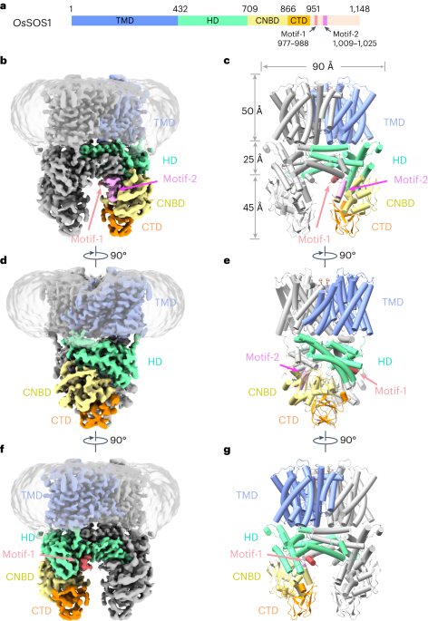
institutional affiliations. EXTENDED DATA EXTENDED DATA FIG. 1 STRUCTURE-BASED SEQUENCE ALIGNMENTS FOR THE REPRESENTATIVE SOS1S FROM BOTH DICOTS AND MONOCOTS. The cryo-EM structure of OsSOS1
is used to restrict sequence gaps to inter-helical segments, with the superior coils and arrows defining extents of the secondary elements. The sequences include representative members from
dicots and monocots: Oryza sativa Japonica Group (XP_015619351.1), Zea mays (XP_008645741.2), Sorghum bicolor (XP_002443674.1), Aegilops tauschii (XP_020190641.1), Brachypodium distachyon
(XP_003576505.1), Dendrobium officinale (XP_020699162.1), Asparagus officinalis (XP_020256864.1), Musa acuminata subsp. malaccensis (XP_018686090.1), Phoenix dactylifera (XP_008798100.1),
Amborella trichopoda (XP_006849492.1), Arabidopsis thaliana (NP_178307.2), Brassica rapa (XP_009114217.1), Eutrema salsugineum (XP_006395824.1), Beta vulgaris subsp. vulgaris
(XP_010680103.2), Glycine max (NP_001244939.1), Daucus carota (XP_017226136.1), Cucumis melo (XP_008466844.1), Vitis vinifera (NP_001268140.1), Juglans regia (XP_018828660.1), Eucalyptus
grandis (XP_010066529.2). The highly conserved inhibitory motifs (motif-1 and motif-2) are shown in boxes. Some critical residues are highlighted as following: 1. The putative
phosphorylation residues are highlighted in cyan (S1113 and S1135 in OsSOS1). 2. The mutated residues are highlighted in yellow. EXTENDED DATA FIG. 2 STRUCTURAL DETERMINATION OF THE
_OS_SOS1FL. (A) The workflow for image processing of the _Os_SOS1FL. 2D classification of the _Os_SOS1FL particles from the cryo-EM sample are shown. (B) Fourier shell correlation (FSC)
curve suggests an overall resolution at 3.1 Å, as estimated using the 0.143 cut-off criterion (Left). The FSC curves for cross-validation between the model and the unmasked (Black) or masked
(Pink) cryo-EM maps of _Os_SOS1 (Right). (C) Representative cryo-EM density map for the TM segments of TMD, helical elements of HD, CNBD, CTD and auto-inhibitory motif segments. EXTENDED
DATA FIG. 3 STRUCTURAL DETERMINATION OF THE _OS_SOS1976. (A) The workflow for image processing of the _Os_SOS1976. 2D classification ns of the _Os_SOS1976 particles from the cryo-EM sample
are shown. (B) Fourier shell correlation (FSC) curve suggests an overall resolution at 3.4 Å, as estimated using the 0.143 cut-off criterion (Left). The FSC curves for cross-validation
between the model and the unmasked (Black) or masked (Pink) cryo-EM maps of _Os_SOS1976 (Right). (C) Representative cryo-EM density map for the TM segments of the TMD. EXTENDED DATA FIG. 4
DOMAIN ORGANIZATION OF _OS_SOS1 AND SALT TOLERANCE ASSAY. (A) Ribbons drawing of the _Os_SOS1 protomer is shown, orientated as in Fig. 1a. (B) Salt tolerance assay of the _Os_SOS1 and its
C-terminal truncated mutants in the Na+-extrusion defective yeast cells (_S. cerevisiae_ AXT3K). EXTENDED DATA FIG. 5 STRUCTURAL ANALYSIS OF THE TMD OF THE _OS_SOS1. (A) Superimposition of
the TMD (_Os_SOS1, as colored in Fig. 2a) with archaeal Na+/H+ exchanger NhaP from _Pyrococcus abyssii_ (pink), with an r.m.s.d. of 2.6 Å/386 superimposed Ca and 20% sequence identity. (B)
Superimposition of the TMD (_Os_SOS1, as colored in Fig. 2a) with human Na+/H+ exchanger _Hs_NHE1 from (light purple), with an r. m. s. d. of 2.9 Å/389 superimposed Ca and 26% sequence
identity. (C) Cavities within the TMD. Zoom view are shown, as indicated. Two endogenously bound lipid molecules at the dimeric interface are shown as sticks. (D) A conserved inter-helices
salt bridge (R341 _vs_ E171) and a kink (~18°) at the middle of TM4 in the TMD. EXTENDED DATA FIG. 6 STRUCTURAL ANALYSIS OF THE HD OF THE _OS_SOS1. (A) Ribbons drawing and domain topology of
the _Os_SOS1 HD, with one protomer in green and the other in grey. (B) Electrostatic potential at helical domain surface. Electronegative and electropositive potential are colored in
degrees of red and blue saturation, respectively. One protomer is drawn as ribbon diagram, and colored in green. Highly conserved charged residues at the dimeric HD interface and
ion-conducting pathway are shown in stick. Two conserved glycine-mediate kinks, H6 (~35°) and H7 (~34°), are indicated. EXTENDED DATA FIG. 7 STRUCTURE-BASED SEQUENCE ALIGNMENT OF CNBDS. The
structures for both CNBDs (_Os_SOS1 and HCN) have been superimposed and used to restrict sequence gaps to inter-secondary structural elements. Superior coils and arrows define extents of the
secondary structural elements in _Os_SOS1 CNBD (top) and HCN CNBD (bottom). Two loss-of-function mutations in the b-roll structure of the CNBD, including G777D (_sos1-8_) and G784E
(_sos1-9_), led to impaired transport function in _Arabidopsis_ are highlighted in magenta. The residues critical for cAMP binding in the HCN CNBD are highlighted in cyan. EXTENDED DATA FIG.
8 STRUCTURAL ANALYSIS OF THE DOMAIN INTERFACES. (A) The TMD-HD domain interface. The dimeric TMD is shown in ribbon drawing, with one protomer in light blue and the other one in grey. The
dimeric HD is shown as surface, colored with electrostatic potential. Electronegative and electropositive potential are shown in degrees of red and blue saturation, respectively. (B) Domain
organization of TMD (light bule) and HD (salmon), with a dihedral angle of ~26°. (C) The HD-CNBD domain interface. The dimeric HD is shown in ribbon drawing, with one protomer in green and
the other one in grey. The CNBD-CTD is shown as surface, colored with electrostatic potential. Electronegative and electropositive potential are shown in degrees of red and blue saturation,
respectively. EXTENDED DATA FIG. 9 COMPARISON OF THE TRUNCATED _OS_SOS1976 WITH THE AUTO-INHIBITED _OS_SOS1FL. (A, B) The elution profile of the truncated SOS1976 (magenta) and the
full-length _Os_SOS1FL (sky-blue) on size-exclusion column Superose 6 (Increase 10/300 GL). The truncated SOS1976 elutes at 13.8 ml, whereas the full-length _Os_SOS1FL elutes at 14.3 ml. The
high-molecular weight aggregates elute at avoid volume are indicated with *. The eluted fraction were analyzed by SDS-PAGE. (C) The superimposition of the auto-inhibited _Os_SOS1FL
structure and the truncated SOS1976 structure, based on their dimeric TMD portions (with an r.m.s.d. of 2.38 Å/848 superimposed Ca), reveals distinctive structural differences occurred in
the cytoplasmic regions. The structure of the truncated SOS1976 is represented as a density map, with the TMD portion depicted in purple and the unmodeled cytoplasmic portion in gray. The
cytosolic portion of the _Os_SOS1FL is represented in cartoon format (light blue). This comparison highlights a substantial conformational change upon SOS1 activation. The masks of the
detergent boundary are illustrated in white. SUPPLEMENTARY INFORMATION REPORTING SUMMARY SUPPLEMENTARY VIDEO 1 Comparison of plant SOS1, SOS1FL (PDB ID: 8IWO, auto-inhibited state) versus
SOS1976(PDB ID: 8J2M, active). SUPPLEMENTARY VIDEO 2 Comparison of human _Hs_NHE1, occluded conformation (PDB ID: 7dsx) versus conducting conformation (PDB ID: 7dsv). SUPPLEMENTARY DATA 1
Validation report for the full-length SOS1FL (auto-inhibited state). SUPPLEMENTARY DATA 2 Validation report for the truncated SOS1976 (constitutively active state). SOURCE DATA SOURCE DATA
FIG. 1 Unprocessed size-exclusion profile and SDS–PAGE gels for ED-9a/b. RIGHTS AND PERMISSIONS Springer Nature or its licensor (e.g. a society or other partner) holds exclusive rights to
this article under a publishing agreement with the author(s) or other rightsholder(s); author self-archiving of the accepted manuscript version of this article is solely governed by the
terms of such publishing agreement and applicable law. Reprints and permissions ABOUT THIS ARTICLE CITE THIS ARTICLE Zhang, Xy., Tang, Lh., Nie, Jw. _et al._ Structure and activation
mechanism of the rice Salt Overly Sensitive 1 (SOS1) Na+/H+ antiporter. _Nat. Plants_ 9, 1924–1936 (2023). https://doi.org/10.1038/s41477-023-01551-5 Download citation * Received: 15 May
2023 * Accepted: 27 September 2023 * Published: 26 October 2023 * Issue Date: November 2023 * DOI: https://doi.org/10.1038/s41477-023-01551-5 SHARE THIS ARTICLE Anyone you share the
following link with will be able to read this content: Get shareable link Sorry, a shareable link is not currently available for this article. Copy to clipboard Provided by the Springer
Nature SharedIt content-sharing initiative