- Select a language for the TTS:
- UK English Female
- UK English Male
- US English Female
- US English Male
- Australian Female
- Australian Male
- Language selected: (auto detect) - EN
Play all audios:
ABSTRACT In _Streptococcus pyogenes_, the type II fatty acid (FA) synthesis pathway FASII is feedback-controlled by the FabT repressor bound to an acyl-Acyl carrier protein. Although FabT
defects confer reduced virulence in animal models, spontaneous _fabT_ mutants arise in vivo. We resolved this paradox by characterizing the conditions and mechanisms requiring FabT activity,
and those promoting _fabT_ mutant emergence. The _fabT_ defect leads to energy dissipation, limiting mutant growth on human tissue products, which explains the FabT requirement during
infection. Conversely, emerging _fabT_ mutants show superior growth in biotopes rich in saturated FAs, where continued FASII activity limits their incorporation. We propose that membrane
alterations and continued FASII synthesis are the primary causes for increased _fabT_ mutant mortality in nutrient‐limited biotopes, by failing to stop metabolic consumption. Our findings
elucidate the rationale for emerging _fabT_ mutants that improve bacterial survival in lipid-rich biotopes, but lead to a genetic impasse for infection. SIMILAR CONTENT BEING VIEWED BY
OTHERS METABOLISM OF L-ARABINOSE CONVERGES WITH VIRULENCE REGULATION TO PROMOTE ENTERIC PATHOGEN FITNESS Article Open access 25 May 2024 CONTEXT-DEPENDENT CHANGE IN THE FITNESS EFFECT OF
(IN)ORGANIC PHOSPHATE ANTIPORTER _GLPT_ DURING _SALMONELLA_ TYPHIMURIUM INFECTION Article Open access 24 February 2025 PROPIONATE PREVENTS LOSS OF THE PDIM VIRULENCE LIPID IN _MYCOBACTERIUM
TUBERCULOSIS_ Article 13 May 2024 INTRODUCTION Bacterial membranes form a mutable permeable barrier that facilitates adaptation to a changing environment. They usually comprise a
phospholipid bilayer composed of a polar head and apolar fatty acid (FA) chains. The FA synthesis pathway (FASII), which is widespread among bacteria, synthesizes saturated and/or
unsaturated FAs joined to an acyl carrier protein (ACP; forming acyl-ACP) (Supplementary Fig. 1a). Unsaturated FAs are produced via a FASII shunt catalyzed by an acyl-ACP-isomerase named
FabM in streptococci. While FASII is conserved (with some enzyme variation), its regulators differ among Firmicutes. In streptococcaceae and enterococcaccae, FASII gene expression is
controlled by a unique MarR-family feedback-type regulator named FabT encoded in the main FASII locus (Supplementary Fig. 1b, c). FabT uses acyl-ACP as corepressor [1,2, for review3]. The
affinity of FabT-(acyl-ACP) binding to a specific DNA palindromic sequence increases with the length of the acyl carbon chain and the presence of an unsaturation4. FabT regulons were
characterized in various streptococcaceae species and in conditions that affect membrane FA composition, including growth temperature, pH or growth phase5,6,7,8,9. FabT exerts greater
repression of genes encoding elongation steps, mainly the _trans_−2-enoyl-ACP reductase II FabK, and less repression of _fabM_. In _Streptococcus pneumoniae fabM_ expression is not repressed
by FabT6. Accordingly, an _S. pneumoniae_ strain lacking a functional FabT produced longer and more saturated FAs6. FabT regulons reportedly also comprise non-FASII genes involved in
transport, DNA and carbohydrate metabolism, protein, purine, and pyrimidine synthesis; however, their identities vary according to reports and the species under study3. To date, FabT
regulons were not analyzed in the presence of exogeneous FAs (eFAs), which enhance FabT transcriptional repression4. This missing information is particularly relevant as numerous host
infection sites are FA-rich. _Streptococcus pyogenes_, also known as Group A _Streptococcus_, GAS, is a major human pathogen causing mainly extracellular infections, which is responsible for
a large variety of clinical manifestations ranging from superficial infections to life-threatening invasive infections10. GAS infections rank among the top ten causes of death due to
bacterial infections worldwide11. GAS isolates mutated in _fabT_ were recovered in non-human primates at the point of intramuscular inoculation, raising the possibility of such populations
forming in the human host5. In a murine model, strains harboring _fabT_ point mutations display smaller size lesions, no loss of body weight, and a lower mortality than their wild-type
counterparts12. In a non-human primate model, a _fabT_ deleted strain shows decreased colonization and dissemination capacities compared to the parental strain5. Its survival is also
decreased in human blood or in the presence of human polymorphonuclear leukocytes5. These reported properties indicate that the _fabT_ mutant variants are poorly adapted for infection, which
led us to question the rationale for their emergence. We solve this question here by performing in-depth analyses of the features of WT and a representative _fabT_ mutant in different
conditions relevant to host infection. We report that the _fabT_ mutant is metabolically wasteful, by consuming sugars and amino acids, yet failing to grow, which accounts for its failure to
cause infection. Conversely, saturated FA-rich environments impose a counter-selective pressure against WT bacteria expressing active FabT. We show that _fabT_ mutant growth is stimulated
around lipid-rich muscle sources in a FASII-dependent manner, while WT growth is inhibited. These findings solve the apparent contradiction between the in vivo emergence of attenuated _fabT_
variants and the need for an active FabT repressor during infection. RESULTS WT AND _FABT_ MUTANT GROWTH PROPERTIES Multiple independent FabT point mutants were harvested from the infection
site of nonhuman primates, some of which mapped to His1055. We chose the FabTH105Y point mutation as being representative of mutations that arose in vivo, which was established in the
_emm28_ reference strain M28PF1 (respectively, mFabT and WT)13,14 (Supplementary Fig. 1b, c)15. A Δ_fabT_ deletion strain was constructed for phenotypic comparisons (Supplementary Fig. 2).
mFabT and WT strains grow similarly in laboratory medium (THY), and in THY supplemented with 0.1% Tween 80 (THY-Tween, a C18:1Δ9 [oleic acid] source) (Supplementary Fig. 2a, b, left; also
see ref. 2). Survival of WT and mFabT strains in mid-exponential phase was also comparable, as assessed by live/dead staining (Supplementary Fig. 2a, b, right). Unlike mFabT, the Δ_fabT_
mutant grew slowly in both THY and THY-Tween media (Supplementary Fig. 2c, d; also see ref. 12). As _fabT_ deletions were not reported to arise in vivo, we chose to study the mFabT mutant
strain as being representative of in vivo mutations. FABTH105Y IMPACTS MEMBRANE LIPID PRODUCTION AND SPECIES The GAS mFabT strain produced greater proportions of longer-length saturated FA
(C18:0) than the WT strain (26.1% _versus_ 6.8%, respectively; Supplementary Table 1), as first reported in _S. pneumoniae_6. In THY-Tween, which supplies C18:1Δ9, the native FabT represses
FASII and contains 1.7 times more C18:1Δ9 than does mFabT. In this condition, the proportion of C18:0 remained higher in mFabT than in the WT (12.4% and 0.2%, respectively; Supplementary
Table 1)2. For comparison, the Δ_fabT_ mutant showed a higher C18:0 to C16:0 ratio than mFabT in both THY and THY-Tween, likely reflecting complete FASII derepression in the deletion strain
(Supplementary Table 1). The more extreme membrane FA differences may account for poor Δ_fabT_ growth in laboratory media. The effects of the FabT mutation on membrane FA composition should
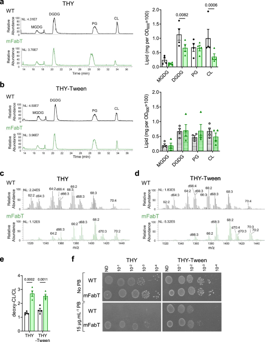
also alter phospholipid metabolism. Lipid analyses of WT and mFabT strains cultured in THY and THY-Tween media identified notable differences in membrane lipid features: i, the primary
differences between WT and mFabT FA composition were reflected in the identifiable lipid species (Supplementary Fig. 3, Supplementary Table 2); ii, THY grown mFabT has ~60% overall lipid
yield compared to that of the WT, extracted from equivalent bacterial OD600 (_N_ = 4); these differences were narrowed to ~5% in cultures grown in THY-Tween (Supplementary Table 3). Among
the detected lipids, diglucosyldiacylglycerol (DGDG) and cardiolipin (CL) amounts were proportionately lower in mFabT compared to WT in THY medium. In THY Tween, levels of both CL and DGDG
decreased for WT, but not for mFabT (Fig. 1a, b). iii, CL species in mFabT appear to be twofold enriched in deoxidized cardiolipin (deoxy-CL) species compared to CL in the WT strain,
regardless of the growth medium (Fig. 1c–e). CL but not deoxy-CL reportedly facilitates oxidative phosphorylation activity by binding protons16. Understanding the reasons for the observed
differences in yield and CL species between WT and mFabT strains will require further study. In conclusion, the greater proportions of saturated and longer FAs in the _fabT_ mutant appear to
cause a reduced overall membrane lipid content and alterations in lipid distribution and composition as compared to the WT. _fabT_ mutants are reportedly more resistant to the cationic
cyclic peptide polymyxin B5,17. Polymyxin B binds negatively charged lipids such as CL18. Putting together these reports and the CL changes noted above, we examined polymyxin B resistance of
mFabT and WT strains in conditions where CL amounts varied (Fig. 1f). Features that decreased CL pools, i.e., _fabT_ mutation or FA availability during WT growth, correlated with greater
polymyxin B resistance. These data support the idea that greater polymyxin B resistance in _fabT_ mutants is due to lower CL pools, and show that availability of environmental FAs can lead
to polymyxin B resistance. They give a rationale for _fabT_ mutant emergence upon polymyxin B selection17. However, they do not explain the in vivo emergence of mFabT mutants at an early
stage of GAS infection. EFFECTS OF _FABT_ ON FASII AND VIRULENCE FACTOR EXPRESSION We first examined the basis for the shift towards greater amounts of C18:0 in the mFabT strain when grown
in THY medium (Supplementary Table 1). In THY-Tween, FASII genes are repressed in WT, but not detectably in mFabT2. In FA-free THY medium however, the only difference in _S. pyogenes_ FASII
gene expression was a greater expression of _fabK_ in mFabT than in the WT strain, leading to an increased _fabK_: _fabM_ ratio in the mFabT mutant, as reported for a _fabT_ mutant in _S.
pneumoniae_ (2,6; Supplementary Fig. 4a). FabK and FabM compete for the same substrate to respectively synthesize saturated and unsaturated FAs (Supplementary Fig. 1a); thus, higher _fabK_
expression relative to _fabM_ is consistent with the higher proportion of saturated FAs in _fabT_ mutants2,6. The underlying reason for a higher _fabK_:_fabM_ ratio in the mFabT mutant was
revealed by analyzing FASII locus organization by qRT-PCR (Supplementary Fig. 4b). All genes from _fabM_ to _accD_ were cotranscribed. In addition, transcription start sites are present
within this operon (Fig. 2a)19. All transcriptional start sites except that of _fabH_ are preceded by FabT consensus DNA binding motifs (5′-ANTTTGATTATCAAATT). Notably, two FabT binding
sites are predicted upstream of _fabK_, but not of the other FASII transcriptional units (starting positions 400693 and 400931 on the _emm28_ M28PF1 genome [CP011535.2]13)3. We suggest that
the double FabT binding sites upstream of _fabK_, and basal levels of free FAs in THY are responsible for differential _fabK fabM_ regulation and hence increased saturated FA synthesis in
the mFabT mutant. We then used RNAseq and qRT-PCR to identify how membrane FA changes might impact and explain the virulence defect during host infection. WT and mFabT expression was
compared in THY, and in THY-Tween (as C18:1Δ9 source) to activate WT FabT repression (3 for Review; Supplementary Table 4, Fig. 2b–e, Supplementary Fig. 4c). Several differences, some
corresponding to multi-gene operons, distinguished the two strains (Supplementary Table 4). Notably, purine synthesis operon genes (M28_Spy0022 to M28_Spy0026) were upregulated in mFabT,
suggestive of increased metabolic activity. An operon encoding adhesins (M28_Spy0107 to M28_Spy0111) was also upregulated; however, expression of virulence genes of the Mga regulon, i.e., C5
peptidase ScpA, and other adhesins, M protein, Sof and SfbX, were downregulated (Supplementary Table 4). Decreased expression of adhesins and other virulence factors such as SLO (_slo_) and
NADase (_nga_) could contribute to the reported poor virulence of _fabT_ mutants in infection20. Addition of C18:1Δ9 activates FabT-mediated repression, which, as reported, turns off FASII
gene expression in the WT strain (Fig. 2c, Supplementary Table 4)4. In contrast, FASII genes (see above), and a non-FASII gene within the FabT regulon (M28_Spy1638, encoding a putative fatty
acid kinase binding protein14;), remain transcriptionally active in the mFabT mutant, showing that FabTH105Y is defective for FASII repression (Fig. 2e, Supplementary Table 4). Despite the
more pronounced difference in FASII gene expression between WT and mFabT in THY-Tween, only few differences were observed in virulence gene expression. We noted that virulence genes lack
FabT binding motifs, suggesting an indirect effect of FabT on their expression. Of note, in THY medium, the mFabT mutation led to longer chain lengths than in WT (determined as C18 to C16
ratios of respectively, 2.3 and 1; Supplementary Table 1). In THY-Tween, these ratios are comparable; the WT C18:C16 ratio increased to 2. We speculate that the higher C18:C16 ratio of
membrane FAs may provoke lower virulence gene expression, as seen for mFabT in THY, and the WT in THY-Tween (Supplementary Table 4). Thus, the greater proportion of longer FAs in the
membrane might be a cause for the decreased expression of virulence genes that lack FabT binding motifs. THE WT STRAIN HAS A FITNESS ADVANTAGE OVER MFABT DURING GROWTH ON HUMAN DECIDUA To
understand the role of _fabT_ and emergence of _fabT_ mutants, we designed ex vivo assays to study infection with WT and _fabT_ variants. mFabT strain capacity to colonize human tissue ex
vivo was assessed by measuring its growth on human tissue. As _emm28_ strains are associated to puerperal fever21,22, we compared ex vivo growth of WT and mFabT on human decidua23. In
control experiments, both strains failed to grow in RPMI, as assessed by growth ratios between cfus after 8 h incubation and the inocula (Supplementary Fig. 2e). In static conditions, growth
of mFabT was 63% lower than that of the WT strain (Fig. 3a). Growth kinetics of WT_eryR_-i_gfp_ and mFabT_eryR_-i_gfp_ GFP-marked strains were then followed in flow conditions at the tissue
surface by time-lapse microscopy (Fig. 3b, c). The surface colonized by the WT strain increased throughout the 4 h growth period (Fig. 3b). In contrast, mFabT strain growth only increased
during the first half-hour of image acquisition. The average thickness of bacterial microcolonies increased for the WT, but decreased for the mFabT strain (Fig. 3c). Altogether, the WT
strain grew with a doubling time of roughly 200 min whereas the mFabT strain did not grow. Thus, in contrast to normal growth in THY medium, the mFabT strain has a major growth defect in the
presence of human decidua. These results provide insight into the nature of the colonization defect and virulence attenuation of _fabT_ mutant strains5,12. IMPAIRED MFABT FITNESS IS DUE TO
DEFECTIVE ADHESION AND POOR GROWTH ON HUMAN CELLS AND IN CELL SUPERNATANTS Bacterial colonization of host tissue comprises an initial adhesion step, followed by bacterial multiplication23.
As GAS has tropism for endometrial and skin tissues, we compared WT and mFabT adhesion capacities on human endometrial cells, and on differentiated (as present in upper skin layers), and
undifferentiated skin keratinocytes (Fig. 4a). The mFabT mutant displayed an adhesion defect on endometrial cells as reported2 and undifferentiated keratinocytes compared to the WT. In
contrast, the strains adhered similarly on differentiated keratinocytes. The adhesion defect, as also suggested by transcriptome analyses (Supplementary Table 4, Fig. 4a), may contribute to
the virulence defect observed in the non-human primate animal models5. GAS replicates mainly extracellularly during infection of endometrium and skin24. Growth comparisons of WT and mFabT
strains on these biotopes, namely endometrial cells, undifferentiated keratinocytes, and differentiated keratinocytes, showed that the mFabT strain was decreased (90%, 53%, and 56%,
respectively) compared to the WT strain (Fig. 4b). The FabTH105Y mutation thus leads to impaired growth in the presence of human cells. To determine whether adhesion is required for the
growth differences between WT and _fabT_ strains, we assayed bacterial growth in uninfected cell supernatant, termed “conditioned supernatant” (Fig. 4c). The mFabT strain displayed a similar
growth defect in endometrial, undifferentiated, and differentiated keratinocyte conditioned supernatants (72%, 50% and 50%, respectively) compared to the WT. This indicates that
cell-secreted products differentially affect growth of WT and mFabT strains. Adhesins differentially produced in WT (Supplementary Table 4, Fig. 4a) could also contribute to promoting higher
bacterial densities during infection. The differences in WT and mFabT growth were visible when grown on endometrial cells or their conditioned supernatants. It is therefore likely that
secreted endometrial cell compounds affecting GAS growth are produced independently of infection. Higher bacterial growth densities on differentiated keratinocytes than on conditioned
supernatants (compare Fig. 4b, c) may be due to greater nutrient availability after infection. Altogether, these data implicate both adhesion and growth defects in poor survival of mFabT in
cell infection environments. Poor mFabT growth suggests that mFabT makes inefficient use of nutrients secreted by eukaryotic cells and/or is more susceptible than WT to secreted bactericidal
molecules. We investigated growth kinetics of WT and mFabT strains in endometrial cell conditioned supernatant in time course experiments (Fig. 4d left). Cfu ratios were similar for both
strains at 4 h, indicating no difference in the lag time. However, from 8 h onwards, cfu ratios were higher for the WT strain (Fig. 4c, d left). The use of a live-cell analysis (IncuCyte®
Systems, Sartorius, Germany), in which both living and dead bacteria are visualized, revealed that mFabT growth did not peak before the WT and that both strains grew similarly until ~3 h;
after 3 h mFabT net growth is visibly slower than that of the WT strain (Supplementary Fig. 2f). mFabT mortality was ~1.5-fold greater than WT at 8 h post inoculation in conditioned
supernatant (Fig. 4d right); these differences are amplified at 16 h and 24 h (Fig. 4d left). We noted that GAS dies rapidly when growth stops, as seen in RPMI (Supplementary Fig. 2e). We
conclude that mFabT grows more slowly and dies more rapidly than the WT strain, and lead us to suggest that mFabT death is triggered by poor growth on nutrient-limited medium. THE MFABT
MUTANT EXHIBITS GREATER METABOLIC TURNOVER IN INFECTION CONDITIONS We investigated a possible metabolic basis for the mFabT growth defect, which could reflect an incapacity to use
cell-secreted products for growth, and/or higher mortality (Fig. 4d). We used a metabolomics approach to assess metabolites that are differentially consumed by mFabT compared to the WT
strain, as performed on conditioned supernatants. Hexoses and amino acids, Asn, Gly, Ile, and Lys were the main metabolites overproduced by uninfected cells (Supplementary Fig. 5,
Supplementary Table 5). The mFabT strain consumed more hexoses and amino acids Asn, Ile, Lys, and Ser, as seen at 16 h (Fig. 5, Supplementary Table 5). This overconsumption is not linked to
a higher bacterial yield, but rather the opposite. It is also not due to faster growth of the mutant relative to WT, but rather to a lower bacterial yield resulting from accrued mortality of
this strain (Fig. 4d, Supplementary Fig. 2f). Two characteristics of mFabT may underlie this futile cycle: 1- The high C18:C16 ratio in mFabT compared to WT (respectively, 2.3 to 1;
Supplementary Table 4) may reduce bacterial fitness by altering activities of membrane components, leading to greater mortality in nutrient-limited medium. 2- Lipid synthesis and FA
availability reportedly play a role in regulating metabolic functions25. Continued FASII expression in mFabT may desynchronize coordination between membrane synthesis and growth, such that
metabolite import remains active even if other growth factors are unavailable. Consistently, increased mFabT metabolite consumption and greater mortality occurred after 8 h incubation, when
growth presumably slows (Fig. 4d, Fig. 5). We propose that membrane alterations and continued FASII synthesis are the primary causes for increased mortality in nutrient‐limited biotopes, by
failing to stop metabolic consumption after 8 h growth. These data give evidence that the mFabT mutant has greater metabolic consumption at the GAS site of infection, but without growth
benefits, which might account for the diminished capacity of _fabT_ mutants to cause infection5. CONTINUED FASII ACTIVITY IN THE MFABT MUTANT LEADS TO LOWER EFA INCORPORATION C18:1Δ9
incorporation is reduced in the mFabT strain (Supplementary Table 1). In _S. pneumoniae_, a _fabT_ mutant reportedly incorporated only traces of C16:16; in an _Enterococcus faecalis fabT_
deletion mutant, incorporation of unsaturated FAs, and to a lesser extent, saturated FAs, was defective4. We evaluated FA incorporation by GAS in medium supplemented with C17:1, which is not
synthesized by GAS; incorporation of this FA generates a discrete peak by gas chromatography (Fig. 6 left). The proportion of C17:1 was threefold higher in the WT than in mFabT (52%:17%).
Continued FASII synthesis in the mFabT mutant might create competition between endogenously synthesized FAs and eFAs for incorporation into membrane phospholipids. To test this, we performed
the same experiments in the presence of platensimycin (Fig. 6 right), a FabF inhibitor that blocks FASII synthesis independently of FabT26. WT and mFabT strains grew similarly in the
presence of C17:1 and platensimycin (Supplementary Table 6), and C17:1 was the major membrane FA in both strains (Fig. 6). In conclusion, the FabTH105Y mutant is less responsive to
environmental FAs than the WT strain. Poor eFA incorporation in mFabT phospholipids thus correlates with continued expression of FASII genes. DE NOVO EMERGENCE OF _FABT_ MUTANTS IN SATURATED
FA ENVIRONMENTS We hypothesized that the defect in eFA incorporation could actually confer a growth advantage to the mFabT strain in toxic lipid environments, and thereby point to
conditions of _fabT_ mutant emergence. Indeed, FA incorporation can negatively affect bacterial integrity, and free FAs are considered part of the first line of host defense against skin
infections27. Notably, increased C18:0 levels in the mFabT mutant compared to WT (2; Supplementary Table 1) led us to examine saturated FAs as potential selective pressure for _fabT_
emergence. WT and mFabT growth and eFA incorporation were then compared in the presence of C14:0 and C16:0, both of which are found among host lipids28. Both FAs inhibited WT growth, and
were nonetheless efficiently incorporated in WT membranes; in striking contrast, mFabT failed to incorporate C14:0, and incorporated >twofold less C16:0, while strain growth was robust
(Fig. 7a, b). This growth advantage incited us to consider that spontaneous _fabT_ mutants could emerge in the presence of saturated FAs. As proof of concept, we grew the WT strain in THY
liquid medium without or with C14:0, and then streaked cultures on solid medium containing C14:0. Single colonies appeared on this medium, and _fabT_ genes were sequenced. Mutations in
_fabT_ were obtained in both selection procedures and encoded FabT variants FabTT65M and FabTG99S (Supplementary Fig. 1b, c). Both these variants were also identified in a primate infection
study5. We compared growth and FA-incorporation phenotypes of the isolated FabTT65M and FabTG99S mutants, as well as Δ_fabT_ mutant as reference, to those of WT and mFabT strains
(Supplementary Fig. 6). Growth at 4 h of the newly isolated mutants, mFabT and Δ_fabT_, was essentially unaffected by C14:0. Moreover, the mutant expressing FabTT65M, mFabT and Δ_fabT_
strains incorporated ≤2% C14:0 compared to ~53% ± 6 in the WT. The mutant expressing FabTG99S incorporated 20% ± 3, well below that in the WT strain. These results provide a rationale for
emergence of _fabT_ mutants, by generating a growth advantage in lipid-containing biotopes as may be present at the infection locus. EVIDENCE THAT EFA EXCLUSION BY MFABT CONFERS A GROWTH
ADVANTAGE IN A SIMULATED MUSCLE BIOTOPE We hypothesized that _fabT_ mutations could provide a transient advantage in lipid-rich biotopes, which would explain their emergence in muscle5. An
ex vivo model was devised to assess effects of muscle on GAS growth. For this, commercial organic 15% fat meat, i.e., muscle from cattle, was placed on lawns of WT_eryR_-i_gfp_ and
mFabT_eryR_-i_gfp_. This is within the percent fat range estimated for human muscle (8–30%; in contrast, uterine fluid lipid concentrations are >1000-fold lower29,30). Remarkably, WT
strains developed a weak inhibitory halo surrounding the muscle samples. In strong contrast, the mFabT strain grew directly and vigorously around muscle sources (Fig. 7c). This result is
consistent with growth inhibition of WT but not mFabT GAS, notably by saturated eFAs (Fig. 7a). To confirm that vigorous growth of mFabT around muscle relates to its lower incorporation of
FAs from muscle lipids, we tested the effects of adding the FASII inhibitor platensimycin to growth medium and plates. All media contained C17:1, which is incorporated and allows mFabT
growth in the presence of the FASII inhibitor (Fig. 6, Supplementary Table 6). As in Fig. 7c, mFabT growth was robust around muscle sources in the absence of FASII inhibitor (Fig. 7d upper).
In contrast, growth was strongly inhibited around the muscle source when platensimycin was present (Fig. 7d lower). The marked inhibitory effect of the muscle sample on the _fabT_ mutant
when FASII is blocked gives strong evidence that incorporation of muscle lipids negatively affects GAS growth. We note that indirect effects, consequent to FASII inhibition by platensimycin
and/or by FabT repression, may contribute to growth inhibition. We conclude that continued FASII activity in mFabT mutants reduces eFA incorporation and thus protects bacteria from toxicity
of host lipids as present in muscle biotopes. DISCUSSION Our work establishes the causal origin for _fabT_ mutation emergence and provides an explanation for its disappearance during
invasion (Fig. 8): GAS is genetically designed to incorporate eFA from lipid-containing environments, and repress FASII. We showed that WT GAS growth is inhibited by saturated FAs in such
environments. Counter-selection can lead to emergence and outgrowth of _fabT_ mutants, which explains their detection at a non-invasive step of infection5. At this step, the _fabT_ mutation
would confer a transient advantage over non-mutant strains. We first showed the _fabT_ growth advantage, and mutant emergence, using C14:0 selection. We then demonstrated that the lipid-rich
inter-muscular environment allows _fabT_ mutant growth in a FASII-dependent manner, while inhibiting the WT strain. While the overall muscle and uterine fluids environments where GAS
multiply are very different, it is notable that their lipid concentrations, differ by ~1000-fold (150 mg/g muscle, compared to 20–200 µg/ml in uterine fluid29,30). We suggest that exposure
to high inter-muscular lipid availability would contribute to WT mortality. However, the _fabT_ mutation has a cost for virulence: it results in a multiplication defect and higher mortality
in the presence of human cells, which is confirmed on human decidua tissue and its fluids. Continued FASII activity in _fabT_ mutant strains provokes a state of futile bacterial metabolism
where increased metabolite uptake does not lead to improved growth. Our findings indicate that futile FASII synthesis by mFabT is detrimental for GAS virulence. We showed previously that
blocking FASII with antibiotic inhibitors, mutation, or deletion did not prevent infection by _Streptococcus agalactiae_, nor by other Firmicute pathogens31,32,33. FASII inhibition and eFA
incorporation in phospholipids corresponds to the natural feedback inhibition in response to eFAs3,31. The contrary, i.e., making FASII synthesis constitutive by inhibiting FabT, is
detrimental for in vivo infection. FabT is thus a promising target for therapeutic design against specific Gram-positive pathogens including GAS, _S. agalactiae_, _S. pneumoniae_, and _E.
faecalis_. METHODS ETHICS STATEMENT The study of the human maternal-fetal membranes was approved by the local ethics committee (Comité de Protection des Personnes Ile de France III, no.
Am5724-1-COL2991, 05/02/2013). Participants were all female. All participants provided written informed consent prior to inclusion in the study at the Department of Obstetrics, Port Royal
Maternity, Cochin University Hospital, Paris, France. BACTERIAL STRAINS AND CULTURE CONDITIONS The strains used in this study are described in Supplementary Table 7. GAS strains were grown
under static condition at 37 °C in Todd Hewitt broth supplemented with 0.2% Yeast Extract (THY) or on THY agar (THYA) plates, or in brain heart infusion (BHI) liquid or agar medium when
specified. Medium was supplemented with 0.1% Tween 80 (THY-Tween; Sigma-Aldrich, Ref. P1754) as indicated, as a source of C18:1Δ9. THY was also supplemented with the saturated FAs, C14:0 and
C16:0, or the unsaturated FA C17:1 (Larodan, Sweden) in the presence of FA-free bovine serum albumin (0.025%, Sigma-Aldrich, Ref. A6003) The FASII inhibitor platensimycin (Tebubio, France)
was added at (0.5 to 1 µg/ml) as indicated. For WT_eryR_-i_gfp_ and mFabT_eryR_-i_gfp_ (Supplementary Table 7) strains, medium was supplemented with 5 to 10 μg/ml of erythromycin (Ery).
Strains were prepared as follows, unless specified otherwise: overnight cultures were diluted to an OD600 = 0.05 and grown in THY to the exponential phase (OD600 comprised between 0.4 and
0.5). For GFP expression, exponential-phase bacteria were further diluted to OD600 = 0.1 in THY supplemented with 10 μg/ml erythromycin, and 20 ng/ml anhydrotetracycline to induce GFP
expression, grown for 90 min at 37 °C, and diluted in RPMI as indicated below. For growth in saturated FAs, WT and mFabT THY precultures were diluted in THY or THY-C14:0 or THY-C16:0 to
OD600 = 0.05, transferred to 50 mL falcon tubes, and incubated at 37 °C. Growth was determined by OD600 readings at designated time points. STRAIN CONSTRUCTIONS Primers used for cloning and
strain verification are described in Supplementary Table 8. The Δ_fabT_ strain corresponds to a _fabT_ deleted mutant. It was obtained by homologous recombination of the plasmid pG1-∆_fabT_
following a published protocol34,35. The DNA fragments encompassing _fabT_ were cloned in BamHI – EcoRI digested pG1 using the In Fusion cloning kit® (Clonetech). This led to the deletion of
_fabT_ from nucleotides 49 to 389, as confirmed by PCR. The mFabT_eryR_-i_gfp_ strain harboring an integrated inducible _gfp_ gene was constructed as described for the WT_eryR_-i_gfp_
strain23. Whole genome sequencing was performed on the mFabT and mFabT_eryR_-i_gfp_ constructed strains, and no surreptitious mutations were found (Bioproject PRJNA926803, accession number
SAMN34247893 for mFabT, SAMN34247911 for mFabT_eryR_-i_gfp_). FABT MODELLING Overall structure of FabT dimer (Supplementary Fig. 1) was predicted by Alphafold36. We used UCSF ChimeraX37 for
molecular graphics and further analyses. ChimeraX was developed by the Resource for Biocomputing, Visualization, and Informatics at the University of California, San Francisco, with support
from NIH R01-GM129325 and the Office of Cyber Infrastructure and Computational Biology, NIAID. GROWTH CURVES GAS stationary precultures were diluted in THY or THY-Tween to an OD600 = 0.05,
and transferred to 96-well plates, which were incubated at 37 °C in a Thermo Scientific Multiskan GO (ThermoFisher Scientific). Growth was determined by shaking plates immediately before
measuring absorbance at OD600 every 10 min. LIVE/DEAD ANALYSIS After bacterial growth in the HEC-1-A conditioned supernatant during 8 h, bacterial mortality was determined using the
LIVE/DEAD® BacLightTM Bacterial Viability Kit (ThermoFisher Scientific, Ref. L7012) as described for flow cytometry utilization using an ACCURI C6 cytometer (BD Biosciences, Le pont de
Claix, France) from the CYBIO Core Facility. Bacteria were grown in HEC-1-A conditioned supernatant for 8 h for testing. Results of three independent experiments were analyzed using the BD
Accuri C6 software. REAL-TIME BACTERIAL GROWTH Test strains were diluted to 103 bacteria per ml in endometrial conditioned supernatant and incubated at 37 °C with 5% CO2. Bacterial growth
was followed by imaging with 20X magnification every 10 min using IncuCyte® Live-Cell Analysis Systems (Sartorius). The bacteria-covered surface was determined using built-in Incucyte
software. Experiments were performed in independent triplicates, and visualizations were routinely done on nine positions. FATTY ACID ANALYSIS Strains were grown until OD600 = 0.4–0.5. Fatty
acids were extracted and analyzed as described2,14,31,32,33. Briefly, analyses were performed in a split-splitless injection mode on an AutoSystem XL Gas Chromatograph (Perkin-Elmer)
equipped with a ZB-Wax capillary column (30 m × 0.25 mm × 0.25 mm; Phenomenex, France). Data were recorded and analyzed by TotalChrom Workstation (Perkin-Elmer). FA peaks were detected
between 12 and 40 min of elution, and identified by comparing to retention times of purified esterified FA standards (Mixture ME100, Larodan, Sweden). Results are shown as percent of the
specific FA compared to total peak areas (TotalChrom Workstation; Perkin Elmer). LIPID ANALYSIS Strains were grown as 200 ml cultures in THY or THY-Tween until OD600 = 0.4–0.5. Lipid
extractions and identifications were performed as described32,38,39,40. Lipid separation was realized by normal phase HPLC (U3000 ThermoFisher Scientific) using a Inertsil Si 5 µm column
(150 × 2.1 mm I.D.) from GL Sciences Inc (Tokyo, Japan). Lipids were quantified using a Corona-CAD Ultra and identified by mass-spectrometry negative ionization and MS2/MS3 fragmentations
(LTQ-Orbitrap Velos Pro). The adducts observed for molecular ions were: CH3COO- for MGDG, CH3COO- and H- for DGDG, H- for PG and CL. The concentration of each lipid class was determined as
described using as standards (from Avanti, Germany) DGDG, 840524P-5MG; MGDG, 840523P-5MG; CL (heart CA), 840012P-25MG; PG (egg), 841138P-25MG41. Lipid spectra were analyzed on XcaliburTM
software (ThermoFisher Scientific, version 4.2.47). Lipid concentrations are presented as milligrams per OD600 = 100 for all samples. The Lipid Maps Structure Search program
https://www.lipidmaps.org/resources/tools/bulk-structure-search/create?database=COMP_DB was used to identify FA moieties on identified lipid species. Primary lipid data are available at “
https://doi.org/10.21228/M88C05 ”42. POLYMYXIN B ASSAY Polymyxin B sensitivity was assayed as described5. Bacteria were grown to OD600 = 0.4–0.5 in THY or THY-Tween. Serial dilutions were
prepared in PBS, and 2.5 µl of each dilution was inoculated onto THY or THY-Tween plates containing or not 20 µg ml–1 polymyxin B (Sigma-Aldrich, Ref. 81271). Plates were incubated at 37 °C
for ~24 h and photographed. Experiments were done in biological triplicates. IN SILICO ANALYSIS Geneious prime Biomatters development, www.geneious.com was used to identify
5′-ANTTTGATTATCAAATT-3′, the putative FabT binding sequence, on the M28PF1 genome, accepting up to 2 mismatches. RNA ISOLATION AND ILLUMINA RNA-SEQ SEQUENCING GAS strains were cultured at 37
°C in THY or THY-Tween, and cells were harvested during exponential growth (OD600 between 0.4 and 0.5). Independent triplicate cultures were prepared for each condition. For RNA
preparation, 2 volumes of RNA protect* (Qiagen) was added to cultures prior centrifugation (10 min 12,000 × _g_) and total RNA was extracted after lysing bacteria by a 30 min 15 mg ml–1
lysozyme, 300 μ ml–1 mutanolysin treatment at 20 °C followed by two cycles of Fast-prep (power 6, 30 s) at 4 °C. RNA extraction (Macherey-Nagel RNA extraction kit; Germany) was done
according to supplier instructions. RNA integrity was analyzed using an Agilent Bioanalyzer (Agilent Biotechnologies, Ca., USA). 23S and 16S rRNA were depleted from the samples using the
MICROBExpress Bacterial mRNA enrichment kit (Invitrogen, France); depletion was controlled on Agilent Bioanalyzer (Agilent Biotechnologies). Libraries were prepared using an Illumina TS kit.
Libraries were sequenced generating 10,000,000 to 20,000,000 75-bp-long reads per sample. RNA-SEQ DATA ANALYSIS The MGAS6180 strain sequence (NCBI), which is nearly identical to
M28PF113,21, was used as a reference sequence to map sequencing reads using the STAR software (2.5.2b) BIOCONDA (Anaconda Inc). RNA-seq data were analyzed using the _hclust_ function and a
principal component analysis in R 3.5.1 (version 2018-07-02). For differential expression analysis, normalization and statistical analyses were performed using the SARTools package and
DESeq243,44 _p_-values were calculated and adjusted for multiple testing using the false discovery rate controlling procedure45. We used UpsetR to visualize set intersections in a matrix
layout comprising the mFabT _versus_ the WT strain grown in THY and in THY-Tween, and growth in THY-Tween _versus_ THY for each strain46,47. Transcriptome data generated in this study are
provided in the Source data file in Tabs labelled Fig. 2b–e, and deposited in the arrayexpress (https://www.ebi.ac.uk/biostudies/arrayexpress) repository with accession #E-MTAB-14449.
FIRST-STRAND CDNA SYNTHESIS, QUANTITATIVE PCR (QRTPCR) AND PCR First-strand cDNA used for quantitative PCR and PCR experiments was done as follows: 500 nanograms of total RNA was treated
with SuperScriptTM II reverse transcriptase and random primers according to manufacturer’s instructions (Invitrogen, Life Technologies, France). Quantitative PCR was carried out to determine
FASII and non-FASII gene expression with SYBR Green PCR kits (Applied Biosystems, Life Technologies, France) as per manufacturer’s instructions (Supplementary Table 8). _gyrA_ and _rpoB_
were used as housekeeping reference genes. Relative quantification of specific gene expression was calculated with the 2 −ΔΔCt method using _gyrA_ as the reference gene and expressed in
log2-fold change. Each assay was performed in triplicate on each sample as described48. PCR was carried out for FASII operon mapping using six pairs of primer pairs flanking neighboring
FASII genes (Supplementary Table 8) as described4. PCR amplicons were examined on a 1% agarose gel. GAS GROWTH CAPACITY ANALYSIS IN RPMI GAS bacteria were grown in THY to an OD600 = 0.4 to
0.5. Cultures were washed twice in PBS and diluted in RPMI medium without glutamine (Gibco, Ref. 32404-014) to a final concentration of 103 bacteria per ml, and then incubated at 37 °C + 5%
CO2 for 8 h. Serial dilutions were plated on THYA solid medium. Cfus were determined after 24 h growth at 37 °C and normalized to the inoculum for each experiment. EX VIVO GAS GROWTH
CAPACITY ANALYSIS Human placentas were recovered from healthy female patients with uncomplicated singleton pregnancy, no recent antibiotic treatment and who had just given birth. Placentas
with attached maternal-fetal membranes were collected and processed as described23 with the following modifications. Tissues were obtained after vaginal delivery, and the samples used were
from regions located far from the cervix, known as zone of intact morphology49. Human decidual explants were infected within hours of their reception. Ex vivo bacterial growth capacity in
the presence of decidua human tissue was done as follows: exponentially growing bacteria were washed twice in PBS and diluted in RPMI at a final concentration of 104 bacteria per ml. Decidua
tissue were washed twice in PBS. One ml of bacteria were then added to tissue, followed by incubation at 37 °C + 5% CO2. After 8 h, the tissues were shredded using Precellys Evolution
(Bertin Technologies) (6 × 20 s at 5500 rpm with a 20 s pause between shaking). Serial dilutions of shredded material were plated on THYA plates. The number of cfus was determined after 24 h
of growth at 37 °C. Live bacterial multiplication on human tissue: infection of maternal-fetal explants, image acquisition and treatments were realized as described23. CELL CULTURE HEC-1-A
(ATCC_ HTB-112TM) endometrial epithelial cells were cultured as described50, in McCoy’s 5A medium (Gibco, Ref. 26600080) supplemented with 10% fetal bovine serum at 37 °C, 5% of CO2. HaCaT
(Addex-Bio T0020001) keratinocytes were cultivated as recommended, in DMEM high glucose medium (Gibco, Ref. 31966) supplemented with 10% fetal bovine serum at 37 °C, 5% of CO2. HaCaT cells
were maintained in the undifferentiated state by cultivation in poor DMEM medium (Gibco, Ref. 21068-028) supplemented with 1X glutaMax (Gibco, Ref. 1X) sodium pyruvate (Gibco, Ref. 2%) fetal
bovine serum and 8% chelated fetal bovine serum using Chelex® 100 Resin (BioRad, Ref. 142–1253). To differentiate HaCaT cells, 2.8 mM CaCl2 (Sigma-Aldrich, Ref. 21115) was added to the
medium. Cells were differentiated after seven days as described50. All cell lines were routinely evaluated for cellular morphology and growth characteristics by microscopy. BACTERIAL
ADHESION CAPACITY GAS adhesion capacity was evaluated as described after growing bacteria in THY to an OD600 of 0.4 to 0.534. Values were normalized to the inoculum for each experiment.
BACTERIAL GROWTH CAPACITY IN THE PRESENCE OF EUKARYOTIC CELLS OR CULTURE SUPERNATANTS GAS were cultured in THY to OD600 = 0.4–0.5. Bacteria were washed twice in PBS and diluted in RPMI
medium without glutamine (Gibco, Ref. 32404–014) to infect cell cultures or inoculate filtered cell culture supernatants (conditioned supernatants) at a final concentration of 103 and 104
bacteria per ml, respectively. Confluent cells in 24-well plates were starved 24 h before the experiment, i.e., incubated in RPMI medium without glutamine, and washed twice in PBS. Cells
were infected with 1 ml of bacteria and incubated at 37 °C + 5% CO2. After 8 h, supernatants were recovered and cells were lysed with 1 ml distilled water. The fractions were pooled and
serial dilutions plated on THYA plates. Conditioned supernatants were prepared by incubating cells in 1 ml RPMI at 37 °C + 5% CO2 for 8 h. The conditioned supernatant was recovered,
inoculated with 103 bacteria, and incubated for another 4, 8, 16, or 24 h. Serial dilutions were plated on THYA plates. The number of cfus was determined after 24 h of growth at 37 °C and
normalized to the inoculum for each experiment. METABOLOMIC ANALYSIS HEC-1-A conditioned supernatants were inoculated or not with WT or mFabT strains during 8 or 16 h and prepared as
described above (see “Bacterial growth capacity in the presence of eukaryotic cells or culture supernatants”). The metabolite composition of these supernatants was analyzed by Proteigene
(https://proteigene.com) using MxP® Quant 500 kit (Biocrates) by two analytical methods, LC-MS/MS for small molecules and FIA-MS/MS for lipids. This analysis was repeated on 3 independent
series of supernatants and on RPMI. These analyses have a defined detection threshold (LOD) for each family of metabolite. Metabolomic data can be consulted at “
https://doi.org/10.21228/M88C05 ”42. SPONTANEOUS _FABT_ MUTANT ISOLATION WT strain overnight precultures were diluted in THY and grown at 37 °C. When the THY culture reached mid-exponential
phase, (OD600 = 0.4–0.5), bacteria were either diluted 1/10 and transferred to THY-C14:0 supplemented with BSA 0.025%, or 1.5 × 10⁷ cfu were streaked on THYA supplemented with C14:0
(THYA-C14:0). The THY-C14:0 liquid culture and plates were incubated 60 h at 37 °C. THY-C14:0 liquid cultures were subsequently streaked on THYA-C14:0 plates. Thirteen and six colonies were
isolated, respectively from the THY-C14:0 liquid then THYA-C14:0 plates, and THYA-C14:0 plates. Isolated colonies were subsequently grown on THYA. Eight colonies issued from the liquid
selection, and all six clones from the solid medium were used for _fabT_ sequencing; PCR was performed directly on patched colonies. The oligonucleotides used were FabT-222, and FabTavComp,
(Supplementary Table 8) binding 221 bp 5′ from the T of the TTG translation start site and 565 bp downstream of it, respectively. The _fabT_ gene and surrounding sequences were amplified by
PCR using the Green Taq DNA Polymerase, GenScript, according to the manufacturer’s instruction, with 30 cycles with a hybridizing temperature of 50 °C and an elongation time of 1 min. Sanger
sequencing was carried out by Eurofin Genomics (https://eurofinsgenomics.eu/en/custom-dna-sequencing/portfolio-overview/) on PCR products. These analyses identifed FabTT65M in all eight
sequenced clones from liquid selection, and FabTG99S in one of six clones on solid medium; the other clones displayed a WT FabT. EX VIVO ASSESSMENT OF GAS WT AND MFABT GROWTH ON MUSCLE
(MEAT) LIPIDS WT_eryR_-i_gfp_ and mFabT_eryR_-i_gfp_ strains (Supplementary Table 7)23 were grown overnight in BHI (Ery 5), and then diluted to OD600 = 0.05 in FA-free bovine serum albumin
(referred to as BSA; 0.025%) plus C17:1 100 µM in 1 ml, without or with platensimycin 1 µg/ml, with Ery 5 for selection. After 4 h growth, culture densities were adjusted to OD600 = 1 in
BHI, and 35 µl were spread on 5 cm diameter plates containing 5 ml BHI agar plus BSA 0.025% and C17:1 100 µM, without or with platensimycin 0.5 µg/ml, with Ery 5 for selection. The muscle
samples used were from organic cattle meat bought frozen and pre-ground, containing 15% fat (as declared by supplier; Picard, France). Absence of contaminants was checked by plating without
bacteria. Samples were placed directly on the lawns, and plates were incubated 48 h at 37 °C, and photographed. _N_ = 4 for experiments without platensimycin, and _N_ = 2 for those with
platensimycin. STATISTICAL ANALYSIS Data were analyzed with GraphPad Prism version 9.4.1. The tests used are indicated in figure legends. Statistical significance is indicated by: ns (not
significant, _p_ > 0.05); *, _p_ < 0.05; **, _p_ < 0.01; ***, _p_ < 0.001; ****, _p_ < 0.0001. In addition, the Shapiro–Wilk normality test was used for all _t_-tests.
REPORTING SUMMARY Further information on research design is available in the Nature Portfolio Reporting Summary linked to this article. DATA AVAILABILITY All relevant data are within the
manuscript, supporting information files, source data files, and repositories. Accession codes or references for strains used are: WT strain https://www.ncbi.nlm.nih.gov/nuccore/CP011535;
mFabT, https://www.ncbi.nlm.nih.gov/nuccore/?term=SAMN34247893; mFabT_eryR_-i_gfp_, https://www.ncbi.nlm.nih.gov/nuccore/?term=SAMN34247911; WT_eryR_-i_gfp_, see ref. 23. Transcriptome data
(accession #E-MTAB-14449) is deposited in arrayexpress (https://www.ebi.ac.uk/biostudies/arrayexpress); also see Source data file (Tabs labelled Fig. 2b–e). The lipidomic data generated in
this study (ID ST003401; datatrackID:5074) is deposited in the Metabolomics Workbench repository42 and assigned a digital object identifier (DOI) of “https://doi.org/10.21228/M88C05”. The
metabolomic data generated in this study (ID ST003403; datatrackID:5086) is deposited in the Metabolomics Workbench repository42 and assigned a digital object identifier (DOI) of “. All data
are publicly available as of publication. Any additional information required to reanalyze the data reported in this paper is available from the authors upon request. Source data are
provided with this paper. REFERENCES * Jerga, A. & Rock, C. O. Acyl-Acyl carrier protein regulates transcription of fatty acid biosynthetic genes via the FabT repressor in _Streptococcus
pneumoniae_. _J. Biol. Chem._ 284, 15364–15368 (2009). Article CAS PubMed PubMed Central Google Scholar * Lambert, C. et al. Acyl-AcpB, a FabT corepressor in _Streptococcus pyogenes_.
_J. Bacteriol._ 205, e0027423 (2023). Article PubMed Google Scholar * Lambert, C., Poyart, C., Gruss, A. & Fouet, A. FabT, a bacterial transcriptional repressor that limits futile
fatty acid biosynthesis. _Microbiol. Mol. Biol. Rev._ 86, e0002922 (2022). * Zou, Q., Zhu, L. & Cronan, J. E. The _Enterococcus faecalis_ FabT transcription factor regulates fatty acid
synthesis in response to exogenous fatty acids. _Front. Microbiol._ 13, 877582 (2022). Article PubMed PubMed Central Google Scholar * Eraso, J. M. et al. Genomic landscape of intrahost
variation in group A _Streptococcus_: repeated and abundant mutational inactivation of the _fabT_ gene encoding a regulator of fatty acid synthesis. _Infect. Immun._ 84, 3268–3281 (2016).
Article CAS PubMed PubMed Central Google Scholar * Lu, Y. J. & Rock, C. O. Transcriptional regulation of fatty acid biosynthesis in _Streptococcus pneumoniae_. _Mol. Microbiol._ 59,
551–566 (2006). Article CAS PubMed Google Scholar * Eckhardt, T. H., Skotnicka, D., Kok, J. & Kuipers, O. P. Transcriptional regulation of fatty acid biosynthesis in _Lactococcus
lactis_. _J. Bacteriol._ 195, 1081–1089 (2013). Article CAS PubMed PubMed Central Google Scholar * Faustoferri, R. C. et al. Regulation of fatty acid biosynthesis by the global
regulator CcpA and the local regulator FabT in _Streptococcus mutans_. _Mol. Oral. Microbiol_ 30, 128–146 (2015). Article CAS PubMed Google Scholar * Zhang, J. et al. Inactivation of
transcriptional regulator FabT influences colony phase variation of _Streptococcus pneumoniae_. _mBio_ 12, e0130421 (2021). Article PubMed Google Scholar * Barnett, T. Indraratna, A.
& Sanderson-Smith M. in _Streptococcus pyogenes: Basic Biology to Clinical Manifestations_ (eds Ferretti, J. J. Stevens, D. L. & Fischetti, V. A.) Ch. 13 (University of Oklahoma
Health Sciences Center Library, 2022). * Carapetis, J. R., Steer, A. C., Mulholland, E. K. & Weber, M. The global burden of group A streptococcal diseases. _Lancet Infect. Dis._ 5,
685–694 (2005). Article PubMed Google Scholar * Tatsuno, I. et al. Relevance of spontaneous _fabT_ mutations to a streptococcal toxic shock syndrome to non-streptococcal toxic shock
syndrome transition in the novel-type _Streptococcus pyogenes_ isolates that lost a _salRK_. _APMIS_ 124, 414–424 (2016). Article CAS PubMed Google Scholar * Longo, M. et al. Complete
genome sequence of _Streptococcus pyogenes emm28_ clinical isolate M28PF1, responsible for a puerperal fever. _Genome Announc._ 3, https://doi.org/10.1128/genomeA.00750-15 (2015). * Lambert,
C. et al. A _Streptococcus pyogenes_ DegV protein regulates the membrane lipid content and limits the formation of extracellular vesicles. _PLoS ONE_ 18, e0284402 (2023). Article CAS
PubMed PubMed Central Google Scholar * Zuo, G. et al. Structural insights into repression of the Pneumococcal fatty acid synthesis pathway by repressor FabT and co-repressor acyl-ACP.
_FEBS Lett._ 593, 2730–2741 (2019). Article CAS PubMed Google Scholar * Haines, T. H. & Dencher, N. A. Cardiolipin: a proton trap for oxidative phosphorylation. _FEBS Lett._ 528,
35–39 (2002). Article ADS CAS PubMed Google Scholar * Port, G. C., Vega, L. A., Nylander, A. B. & Caparon, M. G. _Streptococcus pyogenes_ polymyxin B-resistant mutants display
enhanced ExPortal integrity. _J. Bacteriol._ 196, 2563–2577 (2014). Article PubMed PubMed Central Google Scholar * Teuber, M. & Miller, I. R. Selective binding of polymyxin B to
negatively charged lipid monolayers. _Biochim Biophys. Acta_ 467, 280–289 (1977). Article CAS PubMed Google Scholar * Rosinski-Chupin, I., Sauvage, E., Fouet, A., Poyart, C. &
Glaser, P. Conserved and specific features of _Streptococcus pyogenes_ and _Streptococcus agalactiae_ transcriptional landscapes. _BMC Genomics_ 20, 236 (2019). Article PubMed PubMed
Central Google Scholar * Zhu, L. et al. Contribution of Secreted NADase and Streptolysin O to the Pathogenesis of Epidemic Serotype M1 _Streptococcus pyogenes_ Infections. _Am. J. Pathol._
187, 605–613 (2017). Article CAS PubMed PubMed Central Google Scholar * Green, N. M. et al. Genome sequence of a serotype M28 strain of group a streptococcus: potential new insights
into puerperal sepsis and bacterial disease specificity. _J. Infect. Dis._ 192, 760–770 (2005). Article CAS PubMed Google Scholar * Plainvert, C. et al. Invasive group A streptococcal
infections in adults, France (2006–2010). _Clin. Microbiol Infect._ 18, 702–710 (2012). Article CAS PubMed Google Scholar * Weckel, A. et al. _Streptococcus pyogenes_ infects human
endometrium by limiting the innate immune response. _J. Clin. Invest._ 131, https://doi.org/10.1172/JCI130746 (2021). * Pancholi, V. & Caparon, M. in _Streptococcus pyogenes: Basic
Biology to Clinical Manifestations_ (eds Ferretti, J. J. Stevens, D. L. & Fischetti, V. A.) (University of Oklahoma Health Sciences Center Library, 2016). * Vadia, S. et al. Fatty acid
availability sets cell envelope capacity and dictates microbial cell size. _Curr. Biol._ 27, 1757–1767 e1755 (2017). Article CAS PubMed PubMed Central Google Scholar * Wang, J. et al.
Platensimycin is a selective FabF inhibitor with potent antibiotic properties. _Nature_ 441, 358–361 (2006). Article ADS CAS PubMed Google Scholar * Thormar, H. & Hilmarsson, H. The
role of microbicidal lipids in host defense against pathogens and their potential as therapeutic agents. _Chem. Phys. Lipids_ 150, 1–11 (2007). Article CAS PubMed Google Scholar * Ni
Raghallaigh, S., Bender, K., Lacey, N., Brennan, L. & Powell, F. C. The fatty acid profile of the skin surface lipid layer in papulopustular rosacea. _Br. J. Dermatol._ 166, 279–287
(2012). Article CAS PubMed Google Scholar * Tsujii, H., Matsuoka, Y., Obata, R., Hossain, M. S. & Takagi, Y. Fatty acid composition of lipids in day 7-13 blastocysts, serum and
uterine fluid of rabbits. _Reprod. Med. Biol._ 8, 107–112 (2009). Article CAS PubMed PubMed Central Google Scholar * Young, H. J., Jenkins, N. T., Zhao, Q. & McCully, K. K.
Measurement of intramuscular fat by muscle echo intensity. _Muscle Nerve_ 52, 963–971 (2015). Article PubMed PubMed Central Google Scholar * Brinster, S. et al. Type II fatty acid
synthesis is not a suitable antibiotic target for Gram-positive pathogens. _Nature_ 458, 83–86 (2009). Article ADS CAS PubMed Google Scholar * Kenanian, G. et al. Permissive fatty acid
incorporation promotes Staphylococcal adaptation to FASII antibiotics in host environments. _Cell Rep._ 29, 3974–3982 e3974 (2019). Article CAS PubMed Google Scholar * Hays, C. et al.
Type II fatty acid synthesis pathway and cyclopropane ring formation are dispensable during Enterococcus faecalis systemic infection. _J. Bacteriol._ 203, e0022121 (2021). Article PubMed
Google Scholar * Weckel, A. et al. The N-terminal domain of the R28 protein promotes _emm28_ group A _Streptococcus_ adhesion to host cells via direct binding to three integrins. _J. Biol.
Chem._ 293, 16006–16018 (2018). Article CAS PubMed PubMed Central Google Scholar * Six, A. et al. Srr2, a multifaceted adhesin expressed by ST-17 hypervirulent Group B _Streptococcus_
involved in binding to both fibrinogen and plasminogen. _Mol. Microbiol_ 97, 1209–1222 (2015). Article CAS PubMed Google Scholar * Jumper, J. et al. Highly accurate protein structure
prediction with AlphaFold. _Nature_ 596, 583–589 (2021). Article ADS CAS PubMed Google Scholar * Pettersen, E. F. et al. UCSF ChimeraX: structure visualization for researchers,
educators, and developers. _Protein Sci._ 30, 70–82 (2021). Article CAS PubMed Google Scholar * Abreu, S., Solgadi, A. & Chaminade, P. Optimization of normal phase chromatographic
conditions for lipid analysis and comparison of associated detection techniques. _J. Chromatogr. A_ 1514, 54–71 (2017). Article CAS PubMed Google Scholar * Bligh, E. G. & Dyer, W. J.
A rapid method of total lipid extraction and purification. _Can. J. Biochem. Physiol._ 37, 911–917 (1959). Article CAS PubMed Google Scholar * Thedieck, K. et al. The MprF protein is
required for lysinylation of phospholipids in listerial membranes and confers resistance to cationic antimicrobial peptides (CAMPs) on Listeria monocytogenes. _Mol. Microbiol._ 62, 1325–1339
(2006). Article CAS PubMed Google Scholar * Moulin, M. et al. Sex-specific cardiac cardiolipin remodelling after doxorubicin treatment. _Biol. Sex. Differ._ 6, 20 (2015). Article
PubMed PubMed Central Google Scholar * Sud, M. et al. Metabolomics Workbench: an international repository for metabolomics data and metadata, metabolite standards, protocols, tutorials
and training, and analysis tools. _Nucleic Acids Res._ 44, D463–470 (2016). Article CAS PubMed Google Scholar * Anders, S. et al. Count-based differential expression analysis of RNA
sequencing data using R and Bioconductor. _Nat. Protoc._ 8, 1765–1786 (2013). Article PubMed Google Scholar * Varet, H., Brillet-Gueguen, L., Coppee, J. Y. & Dillies, M. A. SARTools:
A DESeq2- and EdgeR-Based R pipeline for comprehensive differential analysis of RNA-Seq data. _PLoS ONE_ 11, e0157022 (2016). Article PubMed PubMed Central Google Scholar * Benjamini, Y.
& Hochberg, Y. Controlling the false discovery rate: a practical and powerful approach to multiple testing. _J. R. Stat. Soc. Ser. B_ 57, 289–300 (1995). Article MathSciNet Google
Scholar * Lex, A., Gehlenborg, N., Strobelt, H., Vuillemot, R. & Pfister, H. UpSet: visualization of intersecting sets. _IEEE Trans. Vis. Comput Graph_ 20, 1983–1992 (2014). Article
PubMed PubMed Central Google Scholar * Conway, J. R., Lex, A. & Gehlenborg, N. UpSetR: an R package for the visualization of intersecting sets and their properties. _Bioinformatics_
33, 2938–2940 (2017). Article CAS PubMed PubMed Central Google Scholar * Plainvert, C. et al. A novel CovS variant harbored by a colonization strain reduces _Streptococcus pyogenes_
virulence. _J. Bacteriol._ e0003923, https://doi.org/10.1128/jb.00039-23 (2023). * Marcellin, L. et al. Immune modifications in fetal membranes overlying the cervix precede parturition in
humans. _J. Immunol._ 198, 1345–1356 (2017). Article CAS PubMed Google Scholar * Malerba, M. et al. Epidermal hepcidin is required for neutrophil response to bacterial infection. _J.
Clin. Invest._ 130, 329–334 (2020). Article CAS PubMed Google Scholar Download references ACKNOWLEDGEMENTS Expert assistance of Benjamin Saint-Pierre (Genom’IC facility of the Institut
Cochin) with transcriptomic experiments is gratefully acknowledged. We thank Alice d’Orfani, Margaux Dubon, Lauryn Moali, Iman Nasr and Laure Detheve, undergraduates in the Institut Cochin
laboratory for technical help, the Imag’IC and Cybio core facilities of the Institut Cochin, and Cédric Broussard, Virginie Salnot and François Guillonneau for helpful discussions. We are
grateful to Jamila Anba-Mondoloni (Micalis Institute) for valuable comments and suggestions on this work. We thank the personnel of the CIC Mère-Enfant Cochin-Necker for the human decidua,
and the MELISA platform (doi.org/10.17180/VQKB-4646) for supporting this work. We acknowledge the use of Alphafold and ChimeraX for FabT in silico design. CL was supported by Université
Paris Cité (BioSPC, n° 51809666), FRM (FDT202106012831) and FEMS (FEMS Congress Attendance Grant for poster n° 7875 in 2021 and FEMS grant - LISSSD 2022 n° LISS-213065). This work was
supported by DIM One Health (RPH17043DJA; AF), Agence Nationale de la Recherche (ANR-16-CE15-0013; AG), under the umbrella of the Joint Programming Initiative on Antimicrobial Resistance
(JPIAMR) ANR funding (ANR-22-AAMR-0007; AG), and the Fondation pour la Recherche Medicale (DBF20161136769; AG). IMAG’IC core facility is supported by the National Infrastructure France
BioImaging (grant ANR-10-INBS-04). AUTHOR INFORMATION Author notes * Clara Lambert Present address: Molecular Microbiology and Structural Biochemistry, CNRS, UMR5086, Université de Lyon,
Lyon, France AUTHORS AND AFFILIATIONS * Université Paris Cité, Institut Cochin, INSERM, U1016, CNRS, UMR8104, Paris, France Clara Lambert, Marine Gaillard, Caroline Bachmann, Antoine
Hautcoeur, Thomas Guilbert, Celine Méhats, Muriel Andrieu, Claire Poyart & Agnes Fouet * Micalis Institute, INRAE, AgroParisTech, Université Paris-Saclay, Jouy en Josas, France Paprapach
Wongdontree, Karine Gloux & Alexandra Gruss * UMS-IPSIT - Plateforme SAMM, Université Paris-Saclay, Orsay, France Bastien Prost & Audrey Solgadi * Lipides: Systèmes Analytiques et
Biologiques, Université Paris-Saclay, Orsay, France Sonia Abreu * AP-HP Centre–Université Paris Cité, Paris, France Claire Poyart Authors * Clara Lambert View author publications You can
also search for this author inPubMed Google Scholar * Marine Gaillard View author publications You can also search for this author inPubMed Google Scholar * Paprapach Wongdontree View author
publications You can also search for this author inPubMed Google Scholar * Caroline Bachmann View author publications You can also search for this author inPubMed Google Scholar * Antoine
Hautcoeur View author publications You can also search for this author inPubMed Google Scholar * Karine Gloux View author publications You can also search for this author inPubMed Google
Scholar * Thomas Guilbert View author publications You can also search for this author inPubMed Google Scholar * Celine Méhats View author publications You can also search for this author
inPubMed Google Scholar * Bastien Prost View author publications You can also search for this author inPubMed Google Scholar * Audrey Solgadi View author publications You can also search for
this author inPubMed Google Scholar * Sonia Abreu View author publications You can also search for this author inPubMed Google Scholar * Muriel Andrieu View author publications You can also
search for this author inPubMed Google Scholar * Claire Poyart View author publications You can also search for this author inPubMed Google Scholar * Alexandra Gruss View author
publications You can also search for this author inPubMed Google Scholar * Agnes Fouet View author publications You can also search for this author inPubMed Google Scholar CONTRIBUTIONS
C.L., M.G., P.W., C.B., A.H., K.G., C.M., B.P., A.S., S.A., M.A., A.G., and A.F. performed and interpreted experiments. C.L., P.W., T.G., B.P., A.S., C.P., A.G., and A.F. analyzed and
interpreted data. C.L., A.G., and A.F. conceived the project, designed research, and wrote the paper. CORRESPONDING AUTHORS Correspondence to Alexandra Gruss or Agnes Fouet. ETHICS
DECLARATIONS COMPETING INTERESTS The authors declare no competing interests. PEER REVIEW PEER REVIEW INFORMATION _Nature Communications_ thanks the anonymous reviewer(s) for their
contribution to the peer review of this work. A peer review file is available. ADDITIONAL INFORMATION PUBLISHER’S NOTE Springer Nature remains neutral with regard to jurisdictional claims in
published maps and institutional affiliations. SUPPLEMENTARY INFORMATION SUPPLEMENTARY INFORMATION PEER REVIEW FILE REPORTING SUMMARY SOURCE DATA SOURCE DATA RIGHTS AND PERMISSIONS OPEN
ACCESS This article is licensed under a Creative Commons Attribution-NonCommercial-NoDerivatives 4.0 International License, which permits any non-commercial use, sharing, distribution and
reproduction in any medium or format, as long as you give appropriate credit to the original author(s) and the source, provide a link to the Creative Commons licence, and indicate if you
modified the licensed material. You do not have permission under this licence to share adapted material derived from this article or parts of it. The images or other third party material in
this article are included in the article’s Creative Commons licence, unless indicated otherwise in a credit line to the material. If material is not included in the article’s Creative
Commons licence and your intended use is not permitted by statutory regulation or exceeds the permitted use, you will need to obtain permission directly from the copyright holder. To view a
copy of this licence, visit http://creativecommons.org/licenses/by-nc-nd/4.0/. Reprints and permissions ABOUT THIS ARTICLE CITE THIS ARTICLE Lambert, C., Gaillard, M., Wongdontree, P. _et
al._ The double-edged role of FASII regulator FabT in _Streptococcus pyogenes_ infection. _Nat Commun_ 15, 8593 (2024). https://doi.org/10.1038/s41467-024-52637-3 Download citation *
Received: 14 February 2024 * Accepted: 16 September 2024 * Published: 04 October 2024 * DOI: https://doi.org/10.1038/s41467-024-52637-3 SHARE THIS ARTICLE Anyone you share the following link
with will be able to read this content: Get shareable link Sorry, a shareable link is not currently available for this article. Copy to clipboard Provided by the Springer Nature SharedIt
content-sharing initiative