- Select a language for the TTS:
- UK English Female
- UK English Male
- US English Female
- US English Male
- Australian Female
- Australian Male
- Language selected: (auto detect) - EN
Play all audios:
ABSTRACT Traditionally, molecular assembly pathways for viruses are inferred from high resolution structures of purified stable intermediates, low resolution images of cell sections and
genetic approaches. Here, we directly visualise an unsuspected ‘single shelled’ intermediate for a mammalian orthoreovirus in cryo-preserved infected cells, by cryo-electron tomography of
cellular lamellae. Particle classification and averaging yields structures to 5.6 Å resolution, sufficient to identify secondary structural elements and produce an atomic model of the
intermediate, comprising 120 copies each of protein λ1 and σ2. This λ1 shell is ‘collapsed’ compared to the mature virions, with molecules pushed inwards at the icosahedral fivefolds by ~100
Å, reminiscent of the first assembly intermediate of certain prokaryotic dsRNA viruses. This supports the supposition that these viruses share a common ancestor, and suggests mechanisms for
the assembly of viruses of the _Reoviridae_. Such methodology holds promise for dissecting the replication cycle of many viruses. SIMILAR CONTENT BEING VIEWED BY OTHERS MOLECULAR SOCIOLOGY
OF VIRUS-INDUCED CELLULAR CONDENSATES SUPPORTING REOVIRUS ASSEMBLY AND REPLICATION Article Open access 06 December 2024 THE PALISADE LAYER OF THE POXVIRUS CORE IS COMPOSED OF FLEXIBLE A10
TRIMERS Article Open access 05 February 2024 VISUALIZING MOLECULAR INTERACTIONS THAT DETERMINE ASSEMBLY OF A BULLET-SHAPED VESICULAR STOMATITIS VIRUS PARTICLE Article Open access 15 August
2022 INTRODUCTION Orthoreoviruses belong to a large family of viruses, the _Reoviridae_, which infect vertebrate, invertebrate and plant hosts and are responsible for economically and
medically important diseases1. The mature virus particles are ~850 Å in diameter and are approximately spherical, with icosahedral symmetry. The particles harbour 10-separate genome segments
and comprise two principle protein layers, the innermost composed of 120 copies of protein λ1 clamped together by 150 copies of σ2 (viral polymerases, λ3, are attached inside adjacent to
the icosahedral fivefold axes). The outer layer consists of 600 copies each of μ1 and σ3. The array of μ1 and σ3 is interrupted at the fivefold axes by pentameric λ2 turrets which
enzymatically cap the mRNA transcripts produced by the polymerase on egress from the particle. Finally, trimers of the fibrous cell adhesion protein σ1 emerge from the centres of the
turrets. Unlike most RNA viruses these particles do not completely uncoat during cell entry, instead μ1, σ3 and σ1 are stripped off, maintaining in the cytoplasm a protein shell (known as
the core) secluding the dsRNA genome from cellular pathogen recognition receptors. These particles are transcription-competent and, in the ribonucleoside triphosphate rich environment of the
cytoplasm, they synthesise and extrude capped mRNAs, derived from the ten genome segments. These are translated to make the viral proteins which not only build new particles but also
re-organise the volumes of the cytoplasm devoted to the manufacture of new virus (virus factories). These early stages of infection are relatively more understood than the later stages of
assembly and egress2. Single-stranded gene segments are thought to interact with each other, facilitating the encapsidation of the complete viral genome3, but how the multi-layered particles
assemble, how the RNAs and replication enzymes are incorporated, what the signal for production of double stranded gene segments is, how assembly is completed by attachment of the final
proteins and how particles leave the cell remain largely open questions. Here we demonstrate that vitrification of infected cells followed by cryo-focussed ion beam (cryo-FIB) milling and
cryo-electron tomography (cryo-ET) allows high resolution reconstruction of assembly intermediates of a mammalian reovirus, allowing atomic models to be constructed which throw light on some
of these fundamental questions. RESULTS IN CELL TOMOGRAPHY OF REOVIRUS USING FIB MILLING The first atomic structures for particles from the _Reoviridae_ family were determined by X-ray
crystallography4,5,6,7, and advances in single particle cryo-electron microscopy (cryo-EM) have recently yielded structures at comparable or higher resolution for several members of the
family8,9,10,11. Structural information is available for two types of purified orthoreovirus particles: the intact virus (we term similar particles that we see inside infected cells
virion-like particles) and partially disassembled transcriptionally competent cores7,12. Cryo-EM has also allowed lower symmetry structures within the particle to be deconvoluted from the
icosahedral structure of the protein shell, yielding insight into the spatial organisation of the polymerase and genome segments9,13,14. To link such atomic descriptions of stable purified
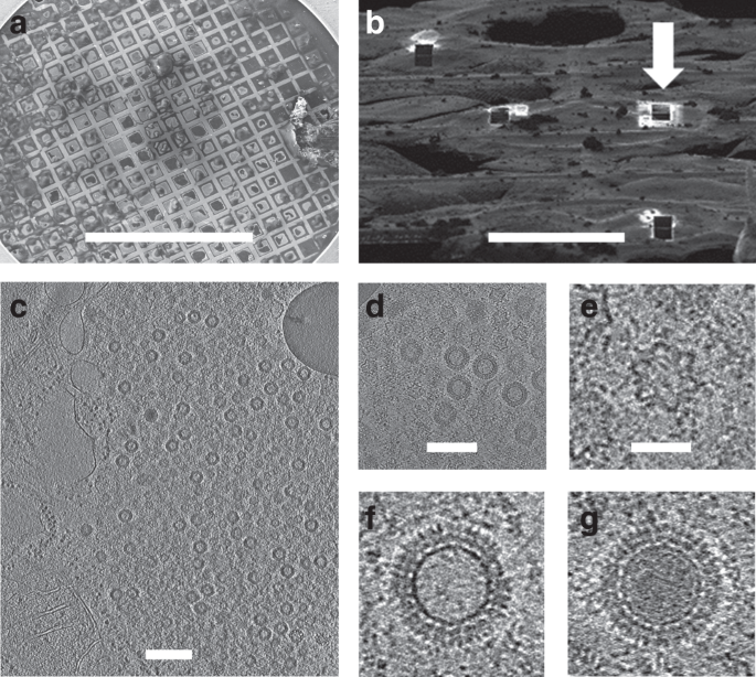
particles to the sequential processes of virus assembly occurring inside infected cells, we grew MA104 cells on gold EM grids (Methods) and infected them with a mammalian orthoreovirus.
Twelve-hours post infection the grids were plunged into liquid ethane and inspected by scanning electron microscopy (Methods and Fig. 1a). Cells were identified and regions chosen for
cutting ~150–200 nm thick lamellae using cryo-FIB milling (Methods and Fig. 1b), a process that preserves high resolution information15. Milled lamellae were then analysed by cryo-ET and the
tilt series analysed to yield three-dimensional reconstructions (Methods, Fig. 1c, d and Supplementary Movie 1). In total eight tomograms were collected, with particles from the five best
used to generate the final structures. Overall, the attrition rate for the plunge–mill–collect workflow was high, with few lamellae proving useful. At 12-h post infection much of the
cytoplasm contained viral factories crowded with virus particles enabling effective data collection even though the milling of lamellae was performed blind. A few enveloped particles were
observed in some tomograms, consistent with co-infection with rotavirus, but the particles in virus factories analysed here are exclusively reovirus (see below). Unexpectedly, numerous star
shaped particles were observed in the virus factories in addition to particles consistent with the expected virion architecture (Fig. 1e). In addition virion-like particles of two types,
‘empty’ and ‘full’, were observed (Fig. 1f, g). The ‘full’ particles presumably contain dsRNA. SUB-TOMOGRAM AVERAGING OF PARTICLES ACHIEVES HIGH RESOLUTION Particles completely contained
within the lamella were picked from five tomograms and classified into categories: 242 stars, 10 full and 65 empty virion-like particles (Methods). These classes were then subjected to
sub-tomogram alignment and averaging, applying icosahedral symmetry, initially using PEET and subsequently emClarity16 (Methods, Supplementary Table 1). Maps for the three categories are
shown in Fig. 2a–c, Supplementary Fig. 1 and Supplementary Movies 2 and 3. The two virion-like particles are extremely similar, with the exception that the density level is much higher in
the interior of the full particles (Fig. 2b, c and Supplementary Fig. 2). An accumulation of empty particles consistent with our observations has been reported for certain reovirus
strains17. Higher resolution analysis, allowing for deviations from exact icosahedral symmetry, was then performed (Supplementary Table 1). For stars, analysis focused on the fivefold
regions using emClarity16 (Methods) yielded a reconstruction at 6.6 Å. For the empty virion-like particles, reconstructions focused on the fivefold regions and pseudo sixfold regions yielded
reconstructions at 6.5 and 5.6 Å respectively (Methods, Supplementary Figs. 3 and 4a). VIRION-LIKE STRUCTURES The virion-like structures can be directly compared to known structures of
purified virions7,12, which fit the density for the in situ structures extremely well (CCs for main chain 0.76 and 0.78 for the fivefold and pseudo sixfold regions of the empty particle,
example fits to various maps are shown in Fig. 2d, f–g, Supplementary Fig. 4b, c and analysed in Supplementary Table 2). Thus purified virus particles provide a good model for the in-cell
empty virion-like particle indicating that the internal RNA does not alter the fundamental organisation of the structure, thus at the fivefold axis on the exterior of the particle, there is
evidence for the σ1 spike being attached (Fig. 2d). Although there is essentially no RNA in these empty particles there is evidence that the λ3 polymerase is attached at the expected
position to the interior of λ1 close to the fivefold axis (Fig. 2d and Supplementary Fig. 1). However there is no evidence for the lattice-work organisation of the N-terminal 181 residues of
λ1 B subunits on the inside of the particle which has been assumed to play a role in organising their assembly around the threefold axes10, indeed there is no evidence for ordered structure
for the first 259 residues of the B subunit (in the A subunit there is density for residue 241 onwards). It is possible that these termini play a role in interactions with, and possibly
organising, the genome in the mature virus. STAR SHAPED PARTICLES COMPRISING INNER SHELL PROTEINS The star shaped icosahedral particles comprise 120 major proteins (Fig. 2h, i) with
additional density features on the outside. Since the inner shell of the virion-like particle is composed of 120 copies of λ1 we modelled these molecules into the density, initially fitting
them as rigid bodies and then using Namdinator for molecular dynamics flexible fitting18 (Methods). The quality of the fit was convincing, with principle secondary structural elements
occupying strong density features (Fig. 2i) and the correlation between model (main chain) and experimental density increasing from 0.50 (single rigid body per molecule) to 0.79 (the quality
of the agreement can be judged from Supplementary Fig. 4d and Supplementary Table 2). Unique features confirmed that these were reovirus particles (Supplementary Fig. 5). Broad features of
the organisation of the molecules parallel the arrangement seen in the virus—five copies of λ1 (termed A molecules) cluster around each fivefold axis and five further copies of λ1 (termed
B), orientated in a similar fashion to the A molecules, are located 35 Å further from the fivefold (Fig. 2i). Since the 60 copies each of the A and B molecules account for the large majority
of the density for these particles we henceforth term them single-layered particles (SLPs). The maximum diameter of the SLPs is very similar to the corresponding layer of virion-like
particles (Fig. 2j), however whilst the latter are roughly spherical the SLP is collapsed inwards at the icosahedral fivefolds by 100 Å, to give the characteristic star shaped cross-section
(Supplementary Movie 4). Superposition of equivalent λ1 molecules between the virion-like particles and SLPs shows that the majority of the movement is caused by molecules A and B tipping
inwards by ~35° (Fig. 3a, b). There are also significant conformational changes within the molecules, the largest being a hinge-like flexing of two parts of the subunit (the same hinge is
used to adjust between the A and B conformations in the virus particles4). There is a 10° flex inwards of the fivefold adjacent part of molecule A and a similar 15° flex in molecule B (Fig.
3a, b). There are also significant conformational rearrangements throughout the A and B molecules (Supplementary Fig. 6) however, at the resolution of this analysis it would be premature to
make a detailed interpretation. CONFORMATIONAL SWITCHING BETWEEN THE TWO PARTICLE TYPES Due to the radically different configurations of the SLP and virion-like particle the contacts between
the molecules differ markedly between the two particles. This is required to accommodate the proteins without compression towards the fivefold axes which are squeezed inwards in the SLP.
The molecules rotate by some 35° and slide partially over each other like the blades of a camera iris. The two principle interfaces between the blades are between molecules, BA′ and AB, and
there are a number of smaller interfaces, AB″, BB′, AA′ and AA″ (Fig. 3c). The largest interfaces (BA′ and AB) have contact areas of roughly 4400 and 4000 Å2 compared to 5900 and 5500 Å2 in
the virion-like particle, respectively. Interface areas are given in Supplementary Table 3; due to the low resolution of the analysis the values for the SLP are very approximate (for
methodology see “Methods”). Overall the contacts in the SLP are significantly weaker, with different residues involved. For the BA′ interface the iris blades are twisted such that at the tip
there is a 30° rotation which allows the A′ molecule to dip inside the B molecule. In this region the interface is formed between what in the virion-like particle is part of the outer
surface of A′ and the region of the B molecule which in that particle forms the inner surface. These contacts use completely different areas to those used in the expanded virion. For the AB
interface the rotation of the iris blades again radically alters the interface towards the fivefold axes, in this case through an unusual ratchet mechanism. If the A molecule is considered
fixed then the change corresponds to a 45° rotation around a pivot helix in B, with the effect that a bundle of helices surrounding the pivot helix rotate by a register of one helix (~12 Å),
analogous to gears slipping by one tooth (Fig. 3d). In most cases interfaces are weakened in the SLP. However the >90° rotation leaves the AB″ and AA″ interfaces almost unchanged
(2100–2200 and 1200–1000 Å2 respectively. This is achieved by tucking the fivefold distal end of A under the edge of the B″ subunit. Overall the λ1 protein structure allows it to occupy two
radically different configurations in the SLP/virion-like particle, but is likely not compatible with stable intermediate structures. As for the empty virion-like particles we see no trace
of density for the first ~300 residues of either λ1 subunit, suggesting that if these residues do play a role in organising the assembly of the λ1 shell, as has been proposed10, then it is
via a loose association consistent with both the expanded and collapsed configurations that becomes ordered when the dsRNA packs against the interior of the core. Σ2 ONLY PARTIALLY CLAMPS
THE SLPS The 120 copies of λ1 are decorated with additional density at 120 sites (Fig. 3e). The density is poorly defined and only the region fairly close to the λ1 shell is seen, so it is
not possible to unambiguously assign it to a particular protein however the overlap between the positions of these features and the attachment points of σ2 in the virus suggests that they
are σ27. The relative positions of σ2 in the SLP and virus are shown in Supplementary Fig. 7. In the virus there are 150 copies per particle, which attach at three distinct points within the
icosahedral asymmetric unit. Two of these are in general positions, the other lies on the icosahedral twofolds. We name these three sites, twofold, threefold adjacent and A-hinge. In the
SLP the threefold adjacent site remains occupied whilst the large rotation at the twofold separates the two half binding sites allowing two σ2 molecules to bind where only one can be
accommodated in the expanded virion (Fig. 3f, g). In contrast, the A-hinge binding site is not occupied due to the iris like movement causing the adjacent B molecule to partially occlude the
site. It is known that in the virion σ2 binding on top of the λ1 layer is required for the stability of the expanded λ1 shell and it has been referred to as a clamp7. In the SLP it appears
that σ2 only partially clamps the structure prior to expansion. SLPS CONTAIN DO NOT CONTAIN RNA OR VISIBLE POLYMERASE There are no significant sites of unexplained density in the interior of
the λ1 layer of the SLP that might be attributed to the λ3 polymerase. The λ3 polymerase could be disordered (the point of attachment to λ1 in the virion-like particle is a point of gross
conformational change between the SLP and virion-like particle and so attachment would necessarily be different), or not found at every vertex, or be displaced away from the fivefold, which
would lead to it being washed out due to fivefold averaging, or it could be simply not present at all. The interior volume of the SLP is 59% of that of the virion-like particle, thus the
dsRNA genome segments cannot fit inside (the dsRNA in the core of the _Reoviridae_ is already at a concentration of ~410 mg/ml5). In addition the density level in the tomogram is far lower
than that in the full virion-like particles (Fig. 2a, c). It is believed that the genome is encapsidated in a single-stranded form. If single-stranded genome segments were present inside the
SLPs then given the reduction in volume the density of RNA would still be 85% of that seen in the virus and the observed density levels do not support this (Fig. 2a, c). DISCUSSION The
structures found here can be directly compared with assembly intermediates for other viruses. The _Cystoviridae_ are a family of bacterial dsRNA viruses and although they are somewhat
smaller and simpler than the _Reoviridae_ (they possess only three genome segments) it has been proposed that they share a common ancestor19. The _Cystoviridae_ have acted as a model system
for understanding assembly and RNA packaging in dsRNA viruses20,21,22 and the first stage in the assembly of these viruses is the formation of a highly indented SLP23. There is a striking
similarity between the indented SLPs of phage Phi8 (representative of the _Cystoviridae_) and reovirus (Fig. 4). In both cases the structures are collapsed at the fivefold axes via
hinge-like motions. Whilst for Phi8 the SLP clearly harbours copies of the viral polymerase, which is not seen in reovirus, in both cases the SLPs contain no genomic RNA, representing a
pre-packaging state in assembly. The _Cystoviridae_ package ssRNA genome segments into preformed SLPs, in a well-defined order which is regulated by specific interactions of the genome
segments with structural features on the outside of the particle which alter as the genome segments enter and inflate the particle22,24. The observation of an analogous pre-packaging SLP
structure for reovirus suggests that incorporation of ssRNA might trigger expansion and addition of the outer protein components. The mechanism for ensuring the correct set of 10-genome
segments is encapsidated for the _Reoviridae_ is however very different from the _Cystoviridae_20. The far larger number of segments and the recent studies of genome segment selection in the
_Reoviridae_ indicate that the mechanism used to ensure encapsidation of a complete set of 10-genome segments includes RNA-RNA recognition between segments, possibly to form a concatamer of
RNA which is then recognized for packaging3,25,26,27. If there is indeed some similarity in the assembly process, with collapsed particles being transformed into an expanded state by the
recruitment of the RNA genome then the empty virion-like particles we observe would be dead-end products that have failed to package the genome. Indeed the proportion of empty virion-like
particles observed is consistent with the known ~50% failure rate in packaging observed in strains which do not organize their virus factories in association with cellular microtubules17.
The observation of only the SLP and completely assembled virion-like particles suggests that the conversion from the SLP to a virus-type structure occurs through the relatively rapid
addition of all the remaining structural components of the virus. This is consistent with the description of an assembly intermediate that accumulates only when there is a temperature
sensitive lesion in the λ2 turret protein delaying assembly events following the addition of λ228—this expanded intermediate contained λ1, σ2 and also the λ3 polymerase protein, and lacked
the complete dsRNA genome. The expanded shape and the additional presence of the λ3 polymerase protein indicate that this particle represents a step in the orthoreovirus assembly pathway
between the abundant collapsed SLPs and completed virions observed in this study. Reovirus and rotavirus build their shells from a different set of proteins, and even those that are
homologous are markedly different in sequence and structure. It seems unlikely therefore that a spatially separated contamination with rotavirus would have any effect on the reovirus
structures we observe, however we cannot rule out that the co-infection perturbs cellular processes and hence the reovirus assembly pathway that we observe. We believe that cryo-ET of
suitably prepared infected cells will be a general vehicle for understanding virus replication-cycles in atomic and molecular detail, with our study already providing a reconstruction at 5.6
Å resolution from only 65 icosahedral particles. It is clear that further unexpected structures will be revealed which are simply not stable enough to be purified. Technical advances will
optimise experimental procedures and improve resolution, and correlation with live-cell imaging and the use of mutant viruses will define the molecular context in the cell and illuminate the
dynamics of the virus replication cycle. METHODS CELL LINES AND VIRUS MA104 cells were cultured in Dulbecco’s modified Eagle’s medium supplemented with 5% (v/v) foetal bovine serum at 37 °C
in 5% CO2. Mammalian orthoreovirus serotype 3 was used (sequence deposited for L1, S1 and S2). CELL PREPARATION FOR CRYO-EM Glow-discharged carbon-coated gold TEM grids (Quantifoil R2/1)
were seeded with 33,000 MA104 cells and allowed to adhere for 4 h at 37 °C. Cells were infected at a multiplicity of infection of 10 and incubated at 37 °C. At 12-h post-infection the grids
were washed three times in PBS, blotted from the back side for 6–8 s and flash frozen in liquid ethane using a manual plunger. FOCUSED ION BEAM MILLING The method was based on a previous
report15. Grids were clipped into autogrid rims (Thermo Scientific) and loaded into a Scios DualBeam system (Thermo Scientific) using a Quorum cryo transfer station (PP3010T) and a custom
built cryo stage cooled to −165 °C. Prior to milling, grids were coated with a ~2 μm layer of platinum using the gas injection system. Milling was performed in a step-wise fashion, using a
30-kV Ga ion beam, and beam currents of 300, 100, 49, and 30 pA to generate a lamella with a final thickness of ~150–200 nm. Milling progress was monitored with the scanning electron
microscope using 3–5 kV beam and 25 pA beam current. CRYO-ET DATA ACQUISITION Tilt series were collected on several positions from three different lamellae using a Thermo Scientific Krios
electron microscope (eBIC Krios III) operating at 300 keV and equipped with a Gatan post-column energy filter (selecting a 20 eV window) on a K2 Summit direct electron detector (Gatan). Data
acquisition was performed covering an angular range from −40° to +40° with 2° angular increments recorded automatically using the dose-symmetric tilting scheme29 under low-dose conditions
using SerialEM software30. Each tilt series was collected with a nominal defocus value between 4.3 and 5.9 µm. Each tilt was acquired as movies (containing 5 frames) in counting mode using a
dose of 2 e−/Å2 per tilt. The total cumulative dose for each tilt series was 82 e−/Å2, with calibrated pixel size of 1.8 Å. TOMOGRAM RECONSTRUCTION AND SUB-TOMOGRAM AVERAGING A total of
eight tilt series were recorded from three lamellae, subjected to movie-frame alignment using Unblur31, but without dose weighting since this is implemented in emClarity16 at the final
sub-tomogram averaging step. Initial analysis used IMOD/PEET32,33, and these preliminary results are shown in Fig. 2a–c. For final analysis the frame-aligned tilt series were aligned and
tomograms reconstructed in the framework of Appion34 using Protomo35. The five tilt series of best quality (thinner lamella, minimum ice contamination) were selected and subjected to
sub-tomogram alignment and averaging using emClarity. Tomograms were also reconstructed at binning 4 using SIRT for visualization. A total of 242 SLPs were picked from five tomograms and
were subsequently aligned and averaged. The 12 fivefold vertices were identified in IMOD isosurface view from the averaged particle map allowing icosahedral symmetry to be applied to the
whole SLP, resulting in an averaged map of SLP at 7.8 Å resolution. The fivefolds were then extracted from the symmetrized particle map and used as a reference for emClarity sub-tomogram
particle picking by template matching. For emClarity processing, a total of 2683 SLP fivefolds were extracted. The 3D alignment procedures of SLP fivefolds were carried out gradually with
binning of 4, 3, 2 and 1, each with 3 iterations. No manual mask was applied during the refinement. Dose-weighted filtering was applied at the final step. The FSC was calculated by the
gold-standard method from even and odd data sets. The resolution of the SLP fivefolds is 6.6 Å at the 0.143 FSC cut off. The same process was carried out for a total of 65 empty virion-like
particles, from those a total of 625 fivefolds and 3039 pseudo sixfolds were extracted, iteratively aligned and averaged. The resolution of these fivefold and pseudo sixfold reconstructions
at the 0.143 FSC cut-off is 6.5 Å and 5.6 Å, respectively. A summary of cryoET data acquisition and data processing are presented in Supplementary Table 1. MODEL BUILDING, ANALYSIS AND
VALIDATION Models were derived from PDB 1EJ6, 2CSE, 1JMU and 3S6X. Initial fitting was performed by placing by hand into the density followed by Chimera36 rigid body fitting. For the
virion-like particle this was followed by molecular dynamics flexible fitting implemented in Namdinator18,37. For the SLP individual proteins (A and B) were fitted in Chimera and then each
λ1 divided into two portions which were further rigid body fitted. Finally the fitting was optimised using Namdinator. Representative FSC curves for the fit of the models to the map are
shown in Supplementary Fig. 4. Validation metrics (including correlation coefficients and FSCs) were determined using Phenix38,39,40 and are presented in Supplementary Table 2. Contact areas
between protein subunits were calculated using the CCP441 program Areaimol42,43. Since contact surface calculations are very sensitive to coordinate error we enlarged the probe radius to
5.0 Å to reduce sensitivity and allow values to be compared with greater confidence. REPORTING SUMMARY Further information on research design is available in the Nature Research Reporting
Summary linked to this article. DATA AVAILABILITY Reovirus sequence data have been deposited at the NCBI, accession codes MT614295-7. Cryo-EM density maps of SLP fivefold and virion-like
particle fivefold and pseudo sixfold have been deposited in EMDB under the accession codes EMD-22165, EMD-22166 and EMD-22164. The resulting atomic models have been deposited in the Protein
Data Bank under the accession codes PDB-6ZTS, PDB-6ZTZ and PDB-6ZTY. REFERENCES * Schiff, L. A., Nibert, M. L. & Tyler, K. L. In _Fifth ed. Fields Virology_ Vol. 2 (eds Knipe, D. M.
& Howley, P. M.) 1853–1915 (Lippincott, Williams and Wilkins, 2007). * Coombs, K. M. Reovirus structure and morphogenesis. _Curr. Top. Microbiol. Immunol._ 309, 117–167 (2006). CAS
PubMed Google Scholar * Borodavka, A., Dykeman, E. C., Schrimpf, W. & Lamb, D. C. Protein-mediated R. N. A. folding governs sequence-specific interactions between rotavirus genome
segments. _Elife_ https://doi.org/10.7554/eLife.27453 (2017). * Grimes, J. M. et al. The atomic structure of the bluetongue virus core. _Nature_ 395, 470–478 (1998). Article ADS CAS
Google Scholar * Gouet, P. et al. The highly ordered double-stranded RNA genome of bluetongue virus revealed by crystallography. _Cell_ 97, 481–490 (1999). Article CAS Google Scholar *
Diprose, J. M. et al. Translocation portals for the substrates and products of a viral transcription complex: the bluetongue virus core. _EMBO J._ 20, 7229–7239 (2001). Article CAS Google
Scholar * Reinisch, K. M., Nibert, M. L. & Harrison, S. C. Structure of the reovirus core at 3.6 A resolution. _Nature_ 404, 960–967 (2000). Article ADS CAS Google Scholar * Yu, X.,
Jin, L. & Zhou, Z. H. 3.88 A structure of cytoplasmic polyhedrosis virus by cryo-electron microscopy. _Nature_ 453, 415–419 (2008). Article ADS CAS Google Scholar * Ding, K.,
Nguyen, L. & Zhou, Z. H. In situ structures of the polymerase complex and RNA genome show how aquareovirus transcription machineries respond to uncoating. _J. Virol._
https://doi.org/10.1128/jvi.00774-18 (2018). * Zhang, X. et al. Atomic model of a nonenveloped virus reveals pH sensors for a coordinated process of cell entry. _Nat. Struct. Mol. Biol._ 23,
74–80 (2016). Article CAS Google Scholar * Jenni, S. et al. In situ structure of rotavirus VP1 RNA-dependent RNA polymerase. _J. Mol. Biol._ 431, 3124–3138 (2019). * Zhang, X. et al.
Features of reovirus outer capsid protein mu1 revealed by electron cryomicroscopy and image reconstruction of the virion at 7.0 Angstrom resolution. _Structure_ 13, 1545–1557 (2005). Article
CAS Google Scholar * Zhang, X., Walker, S. B., Chipman, P. R., Nibert, M. L. & Baker, T. S. Reovirus polymerase lambda 3 localized by cryo-electron microscopy of virions at a
resolution of 7.6 A. _Nat. Struct. Biol._ 10, 1011–1018 (2003). Article CAS Google Scholar * Wang, X. et al. Structure of RNA polymerase complex and genome within a dsRNA virus provides
insights into the mechanisms of transcription and assembly. _Proc. Natl Acad. Sci. USA_ 115, 7344–7349 (2018). Article CAS Google Scholar * Duyvesteyn, H. M. E. et al. Machining protein
microcrystals for structure determination by electron diffraction. _Proc. Natl Acad. Sci. USA_ 115, 9569–9573 (2018). Article CAS Google Scholar * Himes, B. A. & Zhang, P. emClarity:
software for high-resolution cryo-electron tomography and subtomogram averaging. _Nat. Methods_ 15, 955–961 (2018). Article CAS Google Scholar * Shah, P. N. M. et al. Genome packaging of
reovirus is mediated by the scaffolding property of the microtubule network. _Cell Microbiol_. https://doi.org/10.1111/cmi.12765 (2017). * Kidmose, R. T. et al. Namdinator - automatic
molecular dynamics flexible fitting of structural models into cryo-EM and crystallography experimental maps. _IUCrJ_ 6, 526–531 (2019). Article CAS Google Scholar * El Omari, K. et al.
Plate tectonics of virus shell assembly and reorganization in phage phi8, a distant relative of mammalian reoviruses. _Structure_ 21, 1384–1395 (2013). Article CAS Google Scholar *
Borodavka, A., Desselberger, U. & Patton, J. T. Genome packaging in multi-segmented dsRNA viruses: distinct mechanisms with similar outcomes. _Curr. Opin. Virol._ 33, 106–112 (2018).
Article CAS Google Scholar * Mindich, L. Packaging, replication and recombination of the segmented genome of bacteriophage Phi6 and its relatives. _Virus Res._ 101, 83–92 (2004). Article
CAS Google Scholar * Poranen, M. M., Paatero, A. O., Tuma, R. & Bamford, D. H. Self-assembly of a viral molecular machine from purified protein and RNA constituents. _Mol. Cell_ 7,
845–854 (2001). Article CAS Google Scholar * Butcher, S. J., Dokland, T., Ojala, P. M., Bamford, D. H. & Fuller, S. D. Intermediates in the assembly pathway of the double‐stranded RNA
virus φ6. _EMBO J._ 16, 4477–4487 (1997). Article CAS Google Scholar * Qiao, X., Qiao, J. & Mindich, L. Stoichiometric packaging of the three genomic segments of double-stranded RNA
bacteriophage Φ6. _Proc. Natl Acad. Sci. USA_ 94, 4074–4079 (1997). Article ADS CAS Google Scholar * Fajardo, T. Jr., Sung, P. Y. & Roy, P. Disruption of specific RNA-RNA
interactions in a double-stranded RNA virus inhibits genome packaging and virus infectivity. _PLoS Pathog._ 11, e1005321 (2015). Article Google Scholar * Bravo, J. P. K. et al. Stability
of local secondary structure determines selectivity of viral RNA chaperones. _Nucleic Acids Res._ 46, 7924–7937 (2018). Article CAS Google Scholar * Boyce, M., McCrae, M. A., Boyce, P.
& Kim, J. T. Inter-segment complementarity in orbiviruses: a driver for co-ordinated genome packaging in the Reoviridae? _J. Gen. Virol._ 97, 1145–1157 (2016). Article CAS Google
Scholar * Hazelton, P. R. & Coombs, K. M. The reovirus mutant tsA279 L2 gene is associated with generation of a spikeless core particle: implications for capsid assembly. _J. Virol._
73, 2298–2308 (1999). Article CAS Google Scholar * Hagen, W. J. H., Wan, W. & Briggs, J. A. G. Implementation of a cryo-electron tomography tilt-scheme optimized for high resolution
subtomogram averaging. _J. Struct. Biol._ 197, 191–198 (2017). Article Google Scholar * Mastronarde, D. N. Automated electron microscope tomography using robust prediction of specimen
movements. _J. Struct. Biol._ 152, 36–51 (2005). Article Google Scholar * Grant, T. & Grigorieff, N. Measuring the optimal exposure for single particle cryo-EM using a 2.6 A
reconstruction of rotavirus VP6. _Elife_ 4, e06980 (2015). Article Google Scholar * Heumann, J. M., Hoenger, A. & Mastronarde, D. N. Clustering and variance maps for cryo-electron
tomography using wedge-masked differences. _J. Struct. Biol._ 175, 288–299 (2011). Article Google Scholar * Kremer, J. R., Mastronarde, D. N. & McIntosh, J. R. Computer visualization
of three-dimensional image data using IMOD. _J. Struct. Biol._ 116, 71–76 (1996). Article CAS Google Scholar * Noble, A. J. & Stagg, S. M. Automated batch fiducial-less tilt-series
alignment in Appion using Protomo. _J. Struct. Biol._ 192, 270–278 (2015). Article Google Scholar * Winkler, H. & Taylor, K. A. Accurate marker-free alignment with simultaneous
geometry determination and reconstruction of tilt series in electron tomography. _Ultramicroscopy_ 106, 240–254 (2006). Article CAS Google Scholar * Pettersen, E. F. et al. UCSF chimera -
A visualization system for exploratory research and analysis. _J. Comput Chem._ 25, 1605–1612 (2004). Article CAS Google Scholar * Trabuco, L. G., Villa, E., Schreiner, E., Harrison, C.
B. & Schulten, K. Molecular dynamics flexible fitting: a practical guide to combine cryo-electron microscopy and X-ray crystallography. _Methods_ 49, 174–180 (2009). Article CAS Google
Scholar * Adams, P. D. et al. PHENIX: a comprehensive Python-based system for macromolecular structure solution. _Acta Crystallogr D_ 66, 213–221 (2010). Article CAS Google Scholar *
Afonine, P. V. et al. New tools for the analysis and validation of cryo-EM maps and atomic models. _Acta Crystallogr. Sect. D_ 74, 814–840 (2018). Article CAS Google Scholar * Williams,
C. J. et al. MolProbity: more and better reference data for improved all-atom structure validation. _Protein Sci._ 27, 293–315 (2018). Article CAS Google Scholar * Winn, M. D. et al.
Overview of the CCP4 suite and current developments. _Acta Crystallogr D. Biol. Crystallogr_ 67, 235–242 (2011). Article CAS Google Scholar * Lee, B. & Richards, F. M. The
interpretation of protein structures: estimation of static accessibility. _J. Mol. Biol._ 55, 379–400 (1971). Article CAS Google Scholar * Saff, E. B. & Kuijlaars, A. B. J.
Distributing many points on a sphere. _Math. Intell._ 19, 5–11 (1997). Article MathSciNet Google Scholar * Agulleiro, J. I. & Fernandez, J. J. Fast tomographic reconstruction on
multicore computers. _Bioinformatics_ 27, 582–583 (2011). Article CAS Google Scholar Download references ACKNOWLEDGEMENTS We acknowledge Diamond for access and support of the Cryo-EM
facilities at the UK national electron bio-imaging centre (eBIC), (proposal NR18477-18), funded by the Wellcome, MRC and BBSRC. The work was supported by the UK Medical Research Council
(grant MR/N00065X/1 to D.I.S., M.B., A.K. and G.S.). A.K. was also supported by the Wellcome. Work was also supported by the National Institutes of Health (GM082251) and the UK Wellcome
Investigator Award (206422/Z/17/Z) to P.Z., X.F. and D.S. Molecular graphics and analyses were performed with UCSF Chimera, developed by the Resource for Biocomputing, Visualization, and
Informatics at the University of California, San Francisco, with support from NIH P41-GM103311. The work of the Wellcome Centre Human Genetics in Oxford is supported by a Wellcome core award
090532/Z/09/Z. This is a contribution from the UK Instruct-ERIC Centre. AUTHOR INFORMATION Author notes * Xiaofeng Fu Present address: Department of Biological Science, Florida State
University, Tallahassee, FL, 32306, USA * Abhay Kotecha Present address: Thermo Fisher Scientific, Achtseweg Noorg 5, 5651 GG, Eindhoven, The Netherlands * Corey W. Hecksel Present address:
Division of CryoEM and Bioimaging, SSRL, SLAC National Accelerator Laboratory, Stanford University, Menlo Park, CA, 94025, USA * These authors contributed equally: Geoff Sutton, Dapeng Sun,
Xiaofeng Fu, Abhay Kotecha. AUTHORS AND AFFILIATIONS * Division of Structural Biology, Wellcome Centre for Human Genetics, University of Oxford, Oxford, OX3 7BN, UK Geoff Sutton, Abhay
Kotecha, Peijun Zhang, David I. Stuart & Mark Boyce * Department of Structure Biology, University of Pittsburgh, Pittsburgh, PA, 15260, USA Dapeng Sun, Xiaofeng Fu & Peijun Zhang *
Diamond Light Source Limited, Harwell Science and Innovation Campus, Didcot, OX11 0DE, UK Corey W. Hecksel, Daniel K. Clare, Peijun Zhang & David I. Stuart Authors * Geoff Sutton View
author publications You can also search for this author inPubMed Google Scholar * Dapeng Sun View author publications You can also search for this author inPubMed Google Scholar * Xiaofeng
Fu View author publications You can also search for this author inPubMed Google Scholar * Abhay Kotecha View author publications You can also search for this author inPubMed Google Scholar *
Corey W. Hecksel View author publications You can also search for this author inPubMed Google Scholar * Daniel K. Clare View author publications You can also search for this author inPubMed
Google Scholar * Peijun Zhang View author publications You can also search for this author inPubMed Google Scholar * David I. Stuart View author publications You can also search for this
author inPubMed Google Scholar * Mark Boyce View author publications You can also search for this author inPubMed Google Scholar CONTRIBUTIONS M.B. optimized cell culture and infection on
grids, grew the virus and prepared virus infected cells. A.K. optimized grid preparation for FIB milling and prepared frozen grids. C.H. and A.K. carried out cryo-FIB milling, A.K. and D.C.
collected tomograms and performed initial tomography reconstructions. X.F., D.S. and P.Z. performed tomography reconstruction, sub-tomogram alignment and averaging using IMOD/PEET,
Appion/Protomo and emClarity. G.S. performed model fitting and analysis. D.I.S. conceived the experiments and with A.K. and M.B. designed the experiments. P.Z. with X.F. and D.S. designed
and executed the workflows for cryo-tomography reconstruction and sub-tomogram averaging. D.I.S. and G.S. interpreted the structures and wrote the manuscript with input from all authors.
CORRESPONDING AUTHORS Correspondence to Peijun Zhang, David I. Stuart or Mark Boyce. ETHICS DECLARATIONS COMPETING INTERESTS The authors declare no competing interests. ADDITIONAL
INFORMATION PEER REVIEW INFORMATION _Nature Communications_ thanks the anonymous reviewers for their contribution to the peer review of this work. Peer reviewer reports are available.
PUBLISHER’S NOTE Springer Nature remains neutral with regard to jurisdictional claims in published maps and institutional affiliations. SUPPLEMENTARY INFORMATION SUPPLEMENTARY INFORMATION
PEER REVIEW FILE REPORTING SUMMARY DESCRIPTION OF ADDITIONAL SUPPLEMENTARY FILES SUPPLEMENTARY MOVIE 1 SUPPLEMENTARY MOVIE 2 SUPPLEMENTARY MOVIE 3 SUPPLEMENTARY MOVIE 4 RIGHTS AND
PERMISSIONS OPEN ACCESS This article is licensed under a Creative Commons Attribution 4.0 International License, which permits use, sharing, adaptation, distribution and reproduction in any
medium or format, as long as you give appropriate credit to the original author(s) and the source, provide a link to the Creative Commons license, and indicate if changes were made. The
images or other third party material in this article are included in the article’s Creative Commons license, unless indicated otherwise in a credit line to the material. If material is not
included in the article’s Creative Commons license and your intended use is not permitted by statutory regulation or exceeds the permitted use, you will need to obtain permission directly
from the copyright holder. To view a copy of this license, visit http://creativecommons.org/licenses/by/4.0/. Reprints and permissions ABOUT THIS ARTICLE CITE THIS ARTICLE Sutton, G., Sun,
D., Fu, X. _et al._ Assembly intermediates of orthoreovirus captured in the cell. _Nat Commun_ 11, 4445 (2020). https://doi.org/10.1038/s41467-020-18243-9 Download citation * Received: 17
January 2020 * Accepted: 08 August 2020 * Published: 07 September 2020 * DOI: https://doi.org/10.1038/s41467-020-18243-9 SHARE THIS ARTICLE Anyone you share the following link with will be
able to read this content: Get shareable link Sorry, a shareable link is not currently available for this article. Copy to clipboard Provided by the Springer Nature SharedIt content-sharing
initiative