- Select a language for the TTS:
- UK English Female
- UK English Male
- US English Female
- US English Male
- Australian Female
- Australian Male
- Language selected: (auto detect) - EN
Play all audios:
ABSTRACT Historically, surgical endodontics has been viewed as a treatment of last resort, mainly due to poor outcomes as a result of limitations in materials and techniques. Contemporary
techniques, modern materials and better visualisation have all led to an improvement in success rates, making endodontic microsurgery a valuable treatment option to certain patients. Such
advances, however, are no substitute for skill in endodontic diagnosis and treatment planning, which can often prove challenging. A variety of tools are available to test for fractures and
assess both periodontal and pulpal health. More advanced techniques such as cone beam computed tomography are often invaluable in pre-surgical assessment and diagnosis. Once an accurate
diagnosis has been established, a favourable prognosis is explicitly linked to careful patient selection. Orthograde treatment, or retreatment, remains the gold standard for the majority of
endodontic problems. However, there are a number of indications for surgery where orthograde treatment is either impossible, or less likely to be successful. It is paramount for any
clinician undertaking endodontic surgery to have a detailed understanding of the local and systemic factors associated with successful treatment. Whilst there are few absolute medical
contraindications, there are a number of conditions which may influence patient management and make treatment more challenging. You have full access to this article via your institution.
Download PDF SIMILAR CONTENT BEING VIEWED BY OTHERS NON-SURGICAL ENDODONTIC MANAGEMENT FOR THE MEDICALLY COMPLEX - HINTS AND TIPS FOR THE GENERAL DENTAL PRACTITIONER Article Open access 11
April 2025 SURGICAL ENDODONTICS Article 11 April 2025 THE ORTHODONTIC-ENDODONTIC INTERFACE: TRAUMA AND PULPAL CONSIDERATIONS Article Open access 13 September 2024 KEY POINTS * Reviews the
indications and contraindications for endodontic microsurgery. * Identifies key factors to consider when planning endodontic microsurgery. * Discusses the factors associated with success,
and the success rates of endodontic microsurgery. INTRODUCTION Endodontic surgery has often been viewed as a treatment of last resort. Historically, the technique has been flawed due to
limitations with instruments and materials alongside challenges in access and visualisation. The surgery has been considered brutal and, most importantly, it has been associated with poor
outcomes. Modern techniques overcome these obstacles and this is borne out by ever increasing success rate data.1 These papers seek to review these changes and inform practitioners on the
current guidance for diagnosis, patient selection and treatment regimen. HISTORY THE PATIENT'S COMPLAINT It is often possible to determine an accurate differential diagnosis derived
from the history before even examining the patient. Pulpal, periodontal and periapical pain all have characteristic descriptive features, but it is necessary to also consider more uncommon
causes of discomfort which can sometimes mimic dental pain such as neuropathic, sinusitic or idiopathic pain. Though superficially these can be hard to diagnose, there are often signs and
symptoms that do not align with odontogenic pain.2,3 MEDICAL HISTORY Though there are no medical contraindications to root canal treatment and few absolute contraindications to endodontic
surgery, a detailed medical history is required as there are a number of conditions which may influence patient management. The American Society of Anesthesiologist's (ASA)
classification of physical states (Table 1) is a simple tool for determining the physical health of a patient. In general, patients in ASA classes I and II could be safely managed in primary
care. An assessment of the suitability of ASA Class III patients to undergo endodontic surgery would need to be made on an individual basis, with input from general or specialist medical
colleagues if necessary. Those in Class IV would be considered unsuitable for elective endodontic surgery and any non-urgent dental treatment should be postponed until a time when the
patient's general physical state has improved. BLEEDING RISK Bleeding is a risk during any surgery where an incision is made and there is a range of conditions and medications which can
potentiate this bleeding risk (Table 2). It is important to note that whilst general guidance is available, none of the major guidelines reference endodontic surgery in their descriptors of
what constitutes dental treatment.5,6 OSSEOUS CONDITIONS Any conditions that affect bony healing should be identified. Principally, these include patients who have a history of radiotherapy
and those taking bisphosphonates. There is no official guidance in relation to endodontics, however it would be wise to consider orthograde re-root treatment if possible in such patients,
and it has been suggested that surgical endodontics should be contraindicated.7 If orthograde treatment is not possible one must then discuss the risks inherent with both extraction or
surgical endodontic treatment. In such cases there may not be a 'right' answer but endodontic microsurgery may well be less invasive than exodontia in some cases. Close liaison
with the necessary medical support is essential if there is any doubt about exposure status and risk of bone necrosis. STEROID COVER There have been reported cases of adrenal crises
precipitated by dental treatment in patients taking steroid medications8 and there has been debate surrounding the need for steroid cover in such patients. It is recommended that in patients
who are taking steroid medication, additional cover should be provided based on the level of physiological stress caused by a procedure. For minor oral surgical procedures, such as surgical
endodontics, it is recommended that the patient takes their morning dose of steroid as usual, followed by a double dose of their next dose an hour before the procedure, and that they
continue with double doses at their usual intervals for 24 hours.9 CARDIOVASCULAR DISEASE There is no contraindication to surgical endodontic treatment, if cardiovascular disease is well
controlled. In patients who have had a recent myocardial infarction or who are found to be severely hypertensive, elective surgical treatment should be delayed until their condition has
stabilised. A number of cardiac diseases can predispose to infective endocarditis following endodontic surgery. Current guidance stipulates that antibiotic prophylaxis is not recommended
routinely for patients undergoing dental surgery.10 However, apical surgery can be considered a high-risk procedure and, in high-risk patients, a single dose of antibiotic prophylaxis could
be considered.11 The most recent implementation advice with regards to the NICE guidelines suggests that in patients with certain conditions such as prosthetic valves, previous endocarditis
or congenital heart disease, antibiotic prophylaxis should be considered through liaison with their cardiologist.12 It is also suggested that there should be a discussion of the signs and
symptoms of infective endocarditis and the risks and benefits of antibiotic prophylaxis with all 'at risk' patients. MENTAL CAPACITY Patients with reduced capacity to consent are
not contraindicated to either surgery or orthograde root canal treatment, but complex treatment on such patients can be demanding if not impossible. A best interests decision, made in
conjunction with next of kin, carers and if need be an independent mental capacity advocate, could include surgical endodontics if clinically appropriate. Key factors to consider must be the
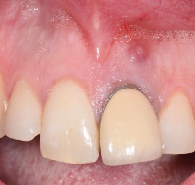
complexity of the treatment, the patient's ability to undergo care and the necessary IV or GA adjuncts required to facilitate such care. EXAMINATION Extra-oral examination will
identify the presence of swellings, sinuses or lymphadenopathy within the neck or face. These may all support the diagnosis of persistent endodontic disease (Figs 1 and 2). Intra-oral
examination should assess the patient's oral hygiene and occlusion. Details of any periodontal disease, caries or tooth surface loss should also be recorded. When assessing previously
endodontically treated teeth, particular attention should be paid to the following: * The presence of sinus tracts in buccal or palatal soft tissues * Tenderness within the buccal sulcus *
Mobility * Tenderness to percussion * Isolated deep pocketing suggestive of root fracture * Expansion of the alveolar bone * The quality of coronal restoration * The periodontal status of
the tooth. Table 3 discusses some of the factors at a tooth level which may influence patient management (Fig. 3). FURTHER INVESTIGATIONS ASSESSING PERIODONTAL HEALTH A basic periodontal
examination must be performed and it is essential to assess the tooth or teeth under suspicion in more detail. Horizontal pattern bone loss may contraindicate surgery, and when severe may
contraindicate any form of treatment especially when the diagnosis of a perio-endo lesion is suspected. Deep isolated pockets may represent draining sinuses for endodontic lesions. However,
they are also pathognomonic of root fractures, which often contraindicate any further endodontic treatment, be it surgical or non-surgical. Kim has suggested a classification for assessing
the perio-endo status of a tooth (Fig. 4). Classifications A-C represent favourable conditions for endodontic surgery. Classifications D-F represent progressive periodontal involvement. A
classification D reflects the coexistence of both periodontal and endodontic lesions. Classification E, the presence of a combined perio-endo lesion and F, a perio-endo lesion with loss of
buccal plate. Kim suggests that cases D-F carry uncertain outcomes and may be contraindications to treatment. This may be too simplistic. Firstly, the true nature of any defect may not be
fully understood until surgical exposure, at which time it is often reasonable to continue with planned therapy accepting a compromised outcome. Secondly, the biology of a primary endo
secondary periodontal lesion may have greater potential to heal than a primary perio secondary endodontic lesion; this distinction should be recognised rather than contraindicating all
perio-endo lesions. TESTING FOR FRACTURES Previously root treated teeth are usually heavily restored and fractures are not uncommon. Diagnosis of cracked teeth can be difficult but is
important, as the presence of a fracture may rule out any further complex treatment. The use of the Fract-finders or a Tooth Sleuth on an individual cusp can often recreate the vague,
painful symptoms caused by cracked teeth. Visualisation under magnification and good illumination is essential. Fibre-optic lights or disclosing solutions can also be used to highlight
fracture lines within teeth. If, radiographically, a lesion is not centred on the apex but rather the lateral aspect of the root, this may suggest a fracture. ASSESSING PULPAL HEALTH By
definition, a tooth that is suspected to need either retreatment or surgery will be non-vital. It is, however, sensible to test adjacent teeth to build the case that pathology stems from the
suspected tooth and ensure that the adjacent teeth are healthy. A variety of tools are available, but cold testing with a suitably cold medium consistently appears to be the most reliable.
Propane-butane mixes reach temperatures of 50 degrees Celsius and should be considered the gold standard, not ethyl chloride.13,14,15 RADIOLOGY The standard imaging required prior to
surgical endodontic treatment are plain film intra-oral radiographs. These can provide detailed information regarding the diagnosis of periapical lesions and should include at least 3 mm of
tissue beyond the apex (Fig. 5).16 The principles of parallax can be used assess multi-rooted teeth and gutta percha points may be placed down sinus tracts to identify the origin. Extra-oral
panoramic radiographs can also be of use for larger lesions, however superimposition of structures can sometimes make diagnosis difficult (Fig. 6). Increasingly, cone beam computed
tomography (CBCT) has been used to provide detailed radiographic examination and can be particularly useful in planning surgical endodontic treatment. Its use, however, should not be
routine, but reserved for cases in which conventional radiography has not provided sufficient information to make an accurate diagnosis or pre-surgical assessment for anatomical variances
and proximity to key structures. CBCT provides better assessment of the relationship of lesions to anatomical structures such as the maxillary antrum and mental nerve, when compared to
conventional radiography;17 such information can be invaluable when planning a surgical approach. CBCT can also provide greater information with regards to complications such as root
fracture18 and root resorption.19 Guidelines in the UK state that CBCT may be indicated in selected surgical cases, but that selection should be made on an assessment of potential
complicating factors (Fig. 7).20 Establishing the correct diagnosis is key in determining the most appropriate primary treatment strategy (Table 4). If there is doubt about the correct
diagnosis, referral to local ENT, maxillofacial surgery or facial pain services for a second opinion and possible management strategy is wise before undertaking any dental treatment.
INDICATIONS FOR RE-ROOT CANAL TREATMENT When undertaken to a high standard, endodontic treatment is associated with success rates in excess of 80% and survival rates over 90%.21,22 Primary
treatment can, however, fail for a number of reasons. Ultimately, this may be due to persistent intraradicular infection, extraradicular infection, foreign body response to extruded
materials, or the development of a cyst.23 It is essential to reflect upon the possible reason for persistent disease, as this will dictate the most appropriate treatment strategy. The most
common cause remains the persistence of bacteria within the root canal system.24 This may be due to a complex myriad of factors but the clinician should be aware of the most likely causes.
INADEQUATE PRIMARY ROOT CANAL TREATMENT RESULTING IN BACTERIAL PERSISTENCE SIMILAR TO A PRIMARY DISEASE Principally, this could be due to poor technique with inadequate shaping and cleaning
or reflect some of the challenges all endodontic treatments pose: complex micro- and macro-anatomy and the complex biofilm. If suspected, in these circumstances, retreatment should be
considered the most sensible option. If there is considerable iatrogenic damage to a tooth, the consent process should make the patient aware of the challenges in overcoming such problems.25
HIGH-QUALITY PRIMARY ROOT CANAL TREATMENT WITH PERSISTENT PRIMARY DISEASE Unless there is missed anatomy, the bacterial species in these cases may be more limited and more virulent.25
Retreatment may be more challenging and consideration must be given to the limitations of instrumentation and cleaning within complex anatomy. Nonetheless, retreatment should be considered
the first option unless there is confidence that the primary treatment cannot be improved upon. OTHER POSSIBLE AETIOLOGICAL FACTORS Irrespective of the quality of the root canal treatment,
there are several factors that may result in failure if not diagnosed. These include fractures, perforations, and failure of the coronal restoration and seal. In the case of fractures, it is
unlikely that retreatment or surgery will result in success. Where the coronal seal is compromised one should be optimistic about the likelihood of success. Perforations present more
challenges but ultimately success may be related to the size, duration and site of the perforation.26 Non-surgical retreatment is associated with success rates of over 80% when undertaken to
a high standard, with survival rates of up to 95%22,27 there is some evidence that it may offer a better prognosis than apical surgery alone.28 Furthermore the presence of an existing root
filling and adequate coronal seal are prognostic indicators in surgical endodontics.29,30 As such, the possibility of orthograde retreatment should always be considered before surgery or
extraction. INDICATIONS FOR ENDODONTIC SURGERY IATROGENIC OR DEVELOPMENTAL ANOMALIES WHICH PREVENT ORTHOGRADE TREATMENT Factors that may prevent the clinician accessing apical anatomy and
thus thoroughly decontaminating include: ledging, fractured instruments, restoration with posts, and heavily sclerosed canals. It is important to note that the above are not absolute
indications for surgery, as there are many techniques for retrieving fractured instruments31 and removing posts from canals.32 A clinician with specialist skill and access to a microscope
may be able to resolve such problems without resorting to a surgical approach (Figs 8 and 9). FAILURE OF ORTHOGRADE TREATMENT WHEN RETREATMENT IS UNLIKELY TO BE SUCCESSFUL If the original
treatment has been undertaken to a high standard and there is confidence that it cannot be improved, then surgery becomes the treatment of choice. Often the judgement about the quality of a
root canal treatment is based upon a two-dimensional radiograph, with only proxy information to the techniques and skill of the original treating clinician. Unless one actually performed the
original treatment and can objectively vouch for the technical merits of the protocol, it is impossible to know how well the canals are shaped, cleaned and obturated, and under what
conditions the care was provided. As such, even if the original treatment appears high-quality there may be a possibility to improve the care provided (Fig. 10). FAILURE OF ORTHOGRADE
TREATMENT WHEN CORONAL DISASSEMBLY WOULD JEOPARDISE THE PROGNOSIS OF THE TOOTH OR PROVE TOO COSTLY OR TIME CONSUMING. If the collateral damage incurred in disassembly would render the tooth
unrestorable or begin to incur costs that start to parallel other prosthetic modalities, an honest conversation should be held with the patient to explore the surgical endodontics or
alternatives to endodontic therapy (Fig. 11). BIOPSY There are occasions when the diagnosis of an apical lesion is not clear from the clinical history and radiographic investigations. In
such cases, a surgical approach may be indicated so that a specimen can be obtained for histopathological analysis. SURGICAL EXPLORATION In cases where there is a suspected fracture or
perforation, the diagnosis may remain unclear despite CBCT imaging. Surgical exploration in these cases can be invaluable (Fig. 12). RETRIEVAL OF DISPLACED MATERIALS OR INSTRUMENTS Foreign
body reactions or biofilm development around extruded materials/instruments contributes to non-healing lesions.33,34 If these cannot be retrieved via the canal system surgery may be the only
option available. PATIENT FACTORS PRECLUDING LENGTHY RETREATMENT These could include patients with trismus, patients who cannot tolerate rubber dam, or simply patients who, through
conditions of chronic pain, cannot remain seated comfortably for the two hours plus required to undertake retreatment. In these cases, though not the first line choice, surgical treatment
may be the most pragmatic option. CONTRAINDICATIONS TO ENDODONTIC SURGERY INTERNAL FACTORS Inadequate surgical skill, training or support. MEDICAL OR PSYCHOLOGICAL FACTORS PRECLUDING SURGERY
Factors such as dental anxiety, the patient's ability to maintain complex restorations and even an individual preference towards particular treatment options can all rule out the
provision of surgery. As discussed earlier, the patient's capacity to consent and any relevant medical history can contraindicate surgical treatment. DENTAL FACTORS UNSTABLE ORAL
DISEASE Endodontic surgery is complex and should be avoided in patients with unstable dentitions. Ownership of oral health and control of primary disease is an essential prerequisite for all
advanced care (Fig. 13). UNUSUAL ROOT CONFIGURATION Roots should be assessed for access and surgery avoided where the root is unlikely to be prepared adequately. Lower incisors, lingual
canals on lower posterior teeth, and palatal roots of upper teeth can be particularly difficult to access. Long roots or limited mouth opening can further complicate procedures. PROXIMITY TO
VITAL STRUCTURES The inferior dental, mental and lingual nerves, the maxillary sinuses and the roots of adjacent teeth may be in close proximity to apical lesions. Potential damage
involving these structures should be included in the consent and planning process, and surgery avoided if the risk is deemed too high. COMPROMISED PERIODONTAL STATUS If the apical amputation
renders the tooth without sufficient periodontal support, surgery must be avoided. COMPROMISED RESTORATIVE STATUS If the coronal restoration is so mutilated that restoration is impossible,
then surgery is futile. SUCCESS IN SURGICAL ENDODONTICS Success is usually measured on the basis of radiographic examination and the presence of symptoms (Table 5): * Success/favourable
outcomes: the outcome can be classed as successful or favourable if there is complete radiographic healing and an absence of symptoms * Failure/unfavourable outcomes: an unsuccessful outcome
is seen where there is no evidence of bony healing and clinical signs or symptoms suggest that endodontic disease is progressing * Uncertain outcomes: if a periapical lesion remains but is
asymptomatic, this could be regarded as an uncertain outcome. It may be that there is scar tissue apically rather than regeneration of periodontal ligament, therefore showing incomplete
radiographic healing whilst the patient remains asymptomatic. It is essential to recognise that these are proxy indicators of a cellular process apically rather than a definitive indication
of healing. Whilst good bony healing is generally a sign of success, it is impossible to say without histopathalogical examination whether a lesion which appears to have healed with scar
tissue remains inflamed.35 Nonetheless, it has been shown that certain radiographic features do correlate with histology indicative of success or failure of surgical endodontic treatment
(Table 5); in reality, a more pragmatic approach is often needed in defining success. To determine long-term success, it is necessary to follow-up the patient clinically and radiographically
at regular intervals.16 It has been suggested that, in the short-term, surgical treatment caries a greater success rate than non-surgical treatment, which then levels out between the two
treatments in the medium to long-term, most likely due to the persistence of an intraradicular infection.36 Molven _et al._ demonstrated that the majority of lesions managed with apical
surgery classified as having incomplete radiographic healing, remained asymptomatic during 8-12 years of observation.37 It has also been shown that, of cases considered to have healed
successfully after one year, 91% remain healed and symptom free at seven years of follow-up (Fig. 14).38 For these reasons, patients who have undergone endodontic surgery tend to be followed
up clinically and radiographically for one year. With the advancement of techniques, instruments and materials, success rates have been increasing. Endodontic microsurgery has been defined
as the use of an operating microscope or an endoscope with high-power magnification together with microsurgical instruments, ultrasonic root-end preparation, and more biocompatible filling
materials such as IRM, SuperEBA or MTA.1 Recent meta-analyses of these trends suggest a cumulative success rate of 89-93.5% for endodontic microsurgery.1,39,40,41 From these meta-analyses,
several factors have been associated with success. TREATMENT-RELATED FACTORS The outcomes of up-to-date endodontic microsurgery, using ultrasonic instruments, magnification and modern root
end filling materials, have been compared to those of traditional endodontic surgery which utilises a bur to prepare the root end before filling with amalgam. The use of modern techniques
appears to increase the success rate from 59% to 93.5%.1 A further study compared endodontic microsurgery to the use of modern techniques without a microscope. Without the use of higher
magnification, the success rate fell from 94% to 88% (Table 6).40 TOOTH-RELATED FACTORS Numerous studies have looked for correlations between tooth-related factors and the success of
surgical treatment.42,43,44,45,46 The following factors were identified as having a greater chance of success: periapical lesions <5 mm in size, an absence of pre-operative pain, good
radiographic density of the root end filling, and pre-operative probing depths of <3 mm. Maxillary teeth generally had a greater success rate than mandibular teeth, and teeth treated
surgically for the second time had a significantly worse prognosis.47 CONCLUSION Non-surgical retreatment should always be considered in the first instance when a patient presents with
persistent periapical disease. There are certain cases where surgical endodontics may be a more appropriate, if not the only available modality, but careful assessment of both the patient
and the tooth is required to ensure that such treatment is appropriate. When indicated, the use of a microsurgical technique will give the greatest opportunity for a successful outcome, with
success rates over 90%. Paper two in this series will review techniques, instruments and materials that may contribute to these higher success rates. REFERENCES * Setzer F C, Shah S B,
Kohli M R, Karabucak B, Kim S. Outcome of endodontic surgery: A meta-analysis of the literature - part 1: Comparison of traditional root-end surgery and endodontic microsurgery. _J Endod_
2010; 36: 1757-1765. * Ghurye S, McMillan R. Orofacial pain - an update on diagnosis and management. _Br Dent J_ 2017; 223: 639-647. * Hauman C H J, Chandler N P, Tong D C. Endodontic
implications of the maxillary sinus: a review. _Int Endod J_ 2002; 35: 127-141. * American Association of Anaesthesiologists. ASA Physical Status Classification System. 2014. Available at
https://www.asahq.org/standards-and-guidelines/asa-physical-status-classification-system (accessed May 2019). * Scottish Dental Clinical Effectiveness Programme. Management of Dental
Patients Taking Anticoagulants or Antiplatelet Drugs. 2015. Available at http://www.sdcep.org.uk/wp-content/uploads/2015/09/SDCEP-Anticoagulants-Guidance.pdf (accessed May 2019). * National
Institute for Health and Care Excellence. Anticoagulation - oral. 2015. Available at https://cks.nice.org.uk/anticoagulation-oral (accessed May 2019). * Borromeo G L, Tsao C E, Darby I B,
Ebeling P R. A review of the clinical implications of bisphosphonates in dentistry. _Aust Dent J_ 2011; 56: 2-9. * Gibson N, Ferguson J W. Steroid cover for dental patients on long-term
steroid medication: proposed clinical guidelines based upon a critical review of the literature. _Br Dent J_ 2004; 197: 681-685. * UK Medicines Information. What steroid supplementation is
required for a patient with primary adrenal insufficiency undergoing a dental procedure? 2016. Available at
https://www.sps.nhs.uk/wp-content/uploads/2014/04/NW-QA-323.3-Steroid-supplementation-for-patients-with-adrenal-insufficiency-.pdf (accessed May 2019). * National Institute for Health and
Care Excellence. Prophylaxis against infective endocarditis: antimicrobial prophylaxis against infective endocarditis in adults and children undergoing interventional procedures. 2008.
Available at https://www.nice.org.uk/guidance/cg64 (accessed May 2019). * Cahill T J, Dayer M, Prendergast B, Thornhill M. Do patients at risk of infective endocarditis need antibiotics
before dental procedures? _BMJ_ 2017; 358: j3942. * Scottish Dental Clinical Effectiveness Programme. Antibiotic Prophylaxis Against Invective Endocarditis Implementation Advice. 2018.
Available at http://www.sdcep.org.uk/wp-content/uploads/2018/08/SDCEP-Antibiotic-Prophylaxis-Implementation-Advice.pdf (accessed May 2019) * Lin J, Chandler N P. Electric pulp testing: a
review. _Int Endod J_ 2008; 41: 365-374. * Jafarzadeh H, Abbott P V. Review of pulp sensibility tests. Part I: general information and thermal tests. _Int Endod J_ 2010; 43: 738-762. *
Alghaithy R A, Qualtrough A J. Pulp sensibility and vitality tests for diagnosing pulpal health in permanent teeth: a critical review. _Int Endod J_ 2017; 50: 135-142. * Evans G, Bishop K,
Renton T. _Guidelines for Surgical Endodontics_. London: The Royal College of Surgeons of England, 2012. Available at
https://www.rcseng.ac.uk/-/media/files/rcs/fds/publications/surgical_endodontics_2012.pdf (accessed May 2019). * Venskutonis T, Plotino G, Juodzbalys G, Mickevičiene L. The importance of
cone-beam computed tomography in the management of endodontic problems: a review of the literature. _J Endod_ 2014; 40: 1895-1901. * Varshosaz M, Tavakoli M A, Mostafavi M, Baghban A A.
Comparison of conventional radiography with cone beam computed tomography for detection of vertical root fractures: an in vitro study. _J Oral Sci_ 2010; 52: 593-597. * Bernardes R A, de
Paulo R S, Pereira L O, Duarte M A, Ordinola-Zapata R, de Azevedo J R. Comparative study of cone beam computed tomography and intraoral periapical radiographs in diagnosis of
lingual-simulated external root resorptions. _Dent Traumatol_ 2012; 28: 268-272. * Horner K. Radiographic selection criteria: new guidelines, old challenges. _Br Dent J_ 2013; 214: 201-213.
* Ng Y L, Mann V, Rahbaran S, Lewsey J, Gulabivala K. Outcome of primary root canal treatment: systematic review of the literature - Part 2. Influence of clinical factors. _Int Endod J_
2008; 41: 6-31. * Ng Y L, Mann V, Gulabivala K. A prospective study of the factors affecting outcomes of nonsurgical root canal treatment: part 1: periapical health. _Int Endod J_ 2011; 44:
583-609. * Nair P N. On the causes of persistent apical periodontitis: a review. _Int Endod J_ 2006; 39: 249-281. * Siqueira J F Jr. Aetiology of root canal treatment failure: why
well-treated teeth can fail. _Int Endod J_ 2001; 34: 1-10. * Hancock H H 3rd, Sigurdsson A, Trope M, Moiseiwitsch J. Bacteria isolated after unsuccessful endodontic treatment in a North
American population. _Oral Surg Oral Med Oral Pathol Oral Radiol Endod_ 2001; 91: 579-586. * Saed S M, Ashley M P, Darcey J. Root perforations: aetiology, management strategies and outcomes.
The hole truth. _Br Dent J_ 2016; 220: 171-180. * Ng Y L, Mann V, Gulabivala K. A prospective study of the factors affecting outcomes of non-surgical root canal treatment: part 2: tooth
survival. _Int Endod J_ 2011; 44: 610-625. * Ørstavik D, Pitt Ford T R. _Essential Endodontology: Prevention and Treatment of Apical Periodontitis_. 2nd ed. Oxford: Wiley-Blackwell, 2007. *
Rahbaran S, Gilthorpe M S, Harrison S D, Gulabivala K. Comparison of clinical outcome of periapical surgery in endodontic and oral surgery units of a teaching dental hospital: A
retrospective study. _Oral Surg Oral Med Oral Pathol Oral Radiol Endod_ 2001; 91: 700-709. * Serrano-Giménez M, Sánchez-Torres A, Gay-Escoda C. Prognostic factors on periapical surgery: A
systematic review. _Med Oral Patol Oral Cir Bucal_ 2015; 20: e715-e722. * McGuigan M B, Louca C, Duncan H F. Clinical decision-making after endodontic instrument fracture. _Br Dent J_ 2013;
214: 395-400. * Dickie J, McCrosson J. Post removal techniques part 1. _Dent Update_ 2014; 41: 490-492, 495-498. * Love R M, Firth N. Histopathological profile of surgically removed
persistent periapical radiolucent lesions of endodontic origin. _Int Endod J_ 2009; 42: 198-202. * Ricucci D, Langeland K. Apical limit of root canal instrumentation and obturation, part 2.
A histological study. _Int Endod J_ 1998; 31: 394-409. * Andreasen J O, Rud J. Correlation between histology and radiography in the assessment of healing after endodontic surgery. _Int J
Oral Surg_ 1972; 1: 161-173. * Del Fabbro M, Taschieri S, Testori T, Francetti L, Weinstein R L. Surgical versus non-surgical endodontic re-treatment for periradicular lesions. _Cochrane
Database Syst Rev_ 2007; 18: CD005511. * Molven O, Halse a, Grung B. Incomplete healing (scar tissue) after periapical surgeryradiographic findings 8 to 12 years after treatment. _J Endod_
1996; 22: 264-268. * Rubinstein R A, Kim S. Long-term follow-up of cases considered healed one year after apical microsurgery. _J Endod_ 2002; 28: 378-383. * Tsesis I, Faivishevsky V, Kfir
A, Rosen E. Outcome of Surgical Endodontic Treatment Performed by a Modern Technique: A Meta-analysis of Literature. _J Endod_ 2009; 35: 1505-1511. * Setzer F C, Kohli M R, Shah S B,
Karabucak B, Kim S. Outcome of endodontic surgery: a meta-analysis of the literature - Part 2: Comparison of endodontic microsurgical techniques with and without the use of higher
magnification. _J Endod_ 2012; 38: 1-10. * Tsesis I, Rosen E, Taschieri S, Telishevsky Strauss Y, Ceresoli V, Del Fabbro M. Outcomes of surgical endodontic treatment performed by a modern
technique: An updated meta-analysis of the literature. _J Endod_ 2013; 39: 332-339. * Lui J N, Khin M M, Krishnaswamy G, Chen N N. Prognostic factors relating to the outcome of endodontic
microsurgery. _J Endod_ 2014; 40: 1071-1076. * García-Guerrero C, Guauque S Q, Molano N, Pineda G A, Nino-Barrera J L, Marín-Zuluaga D J. Predictors of clinical outcomes in endodontic
microsurgery: a systematic review and meta-analysis. _G Ital Endod_ 2017; 31: 2-13. * Testori T, Capelli M, Milani S, Weinstein R L. Success and failure in periradicular surgery: a
longitudinal retrospective analysis. _Oral Surg Oral Med Oral Pathol Oral Radiol Endod_ 1999; 87: 493-498. * von Arx T, Peñarrocha M, Jensen S. Prognostic factors in apical surgery with
root-end filling: a meta-analysis. _J Endod_ 2010; 36: 957-973. * Friedman S. The prognosis and expected outcome of apical surgery. _Endod Topics_ 2005; 11: 219-262. * Peterson J, Gutmann J
L. The outcome of endodontic resurgery: a systematic review. _Int Endod J_ 2001; 34: 169-175. Download references AUTHOR INFORMATION AUTHORS AND AFFILIATIONS * University Dental Hospital of
Manchester, Orthodontic Department, Higher Cambridge Street, Manchester, UK Liam Monaghan * University Dental Hospital of Manchester, Oral Surgery, Higher Cambridge Street, Manchester, UK
Sarah Jadun * University Dental Hospital of Manchester, Restorative Department, Higher Cambridge Street, Manchester, UK James Darcey Authors * Liam Monaghan View author publications You can
also search for this author inPubMed Google Scholar * Sarah Jadun View author publications You can also search for this author inPubMed Google Scholar * James Darcey View author publications
You can also search for this author inPubMed Google Scholar CORRESPONDING AUTHOR Correspondence to James Darcey. RIGHTS AND PERMISSIONS Reprints and permissions ABOUT THIS ARTICLE CITE THIS
ARTICLE Monaghan, L., Jadun, S. & Darcey, J. Endodontic microsurgery. Part one: diagnosis, patient selection and prognoses. _Br Dent J_ 226, 940–948 (2019).
https://doi.org/10.1038/s41415-019-0415-3 Download citation * Published: 28 June 2019 * Issue Date: June 2019 * DOI: https://doi.org/10.1038/s41415-019-0415-3 SHARE THIS ARTICLE Anyone you
share the following link with will be able to read this content: Get shareable link Sorry, a shareable link is not currently available for this article. Copy to clipboard Provided by the
Springer Nature SharedIt content-sharing initiative