- Select a language for the TTS:
- UK English Female
- UK English Male
- US English Female
- US English Male
- Australian Female
- Australian Male
- Language selected: (auto detect) - EN
Play all audios:
ABSTRACT The mechanisms underlying the role of oxytocin (OT) as a regulator of social behavior in mammals are only partly understood. Recently, it has been proposed that OT increases the
salience of social stimuli. We carried out a randomized, double-blind, cross-over study of the effects of OT on binocular rivalry, a visual phenomenon underpinned by the interplay of
excitation and inhibition in the cortex. A final sample of 45 participants viewed images of social stimuli (faces with different emotional expressions) and non-social stimuli (houses and
Gabor patches). We demonstrate a robust effect that intranasal OT increases the salience of human faces in binocular rivalry, such that dominance durations of faces are longer—this effect is
not modulated by the facial expression. We tentatively show that OT treatment increases dominance durations for non-social stimuli. Our results lend support to the social salience
hypothesis of OT, and in addition offer provisional support for the role of OT in influencing excitation-inhibition balance in the brain. SIMILAR CONTENT BEING VIEWED BY OTHERS IN THE NOSE
OR ON THE TONGUE? CONTRASTING MOTIVATIONAL EFFECTS OF ORAL AND INTRANASAL OXYTOCIN ON AROUSAL AND REWARD DURING SOCIAL PROCESSING Article Open access 04 February 2021 GAZE DIRECTION AND FACE
ORIENTATION MODULATE PERCEPTUAL SENSITIVITY TO FACES UNDER INTEROCULAR SUPPRESSION Article Open access 10 May 2022 ERPS RESPONSES TO DOMINANCE FEATURES FROM HUMAN FACES Article Open access
02 December 2022 INTRODUCTION The neuropeptide oxytocin (OT) has long been implicated in the regulation of social behavior in mammals, including humans1. The exact role of OT in this
regulation, and the mechanisms behind it, are only partly understood. The picture is complicated by the wealth of studies reporting varying results of the administration of intranasal OT,
including both prosocial and antisocial effects2,3. Several hypotheses have been proposed to explain the influence of OT on social behavior. The social salience hypothesis suggests that OT
increases the salience of social cues, which in turn can increase or modulate the influence of context and inter-individual factors on social behavior4,5. While the effects of exogenously
administered OT are not confined solely to social behavior or social cognition, many studies have indicated that exogenously administered OT affects for example, memory for faces6, detection
of social words7, and learning from social feedback8. Such effects have been observed for both positive and negative stimuli9,10,11,12,13. While individual studies have reported selective
effects on stimuli with either positive or negative valence, taken together evidence is in line with a general increase of the salience of social stimuli, regardless of the valence of those
stimuli. Thus, while OT presumably has several different effects on social cognition, the social salience hypothesis would explain much of the results of studies on exogenously administered
OT. Binocular rivalry is a visual phenomenon that occurs when dissimilar images are presented to different eyes (e.g., a face to one eye and a house to the other). The images then compete
for visual awareness, i.e. the image that the viewer consciously perceives (dominant percept) is one of the presented images, while the other one is suppressed and unseen. After a few
seconds, the suppressed image become dominant and the previously dominant image becomes suppressed. This results in transitions between which image is consciously perceived, and these
transitions may be instant or contain a mix of the two images (piecemeal percept). This phenomenon has been extensively used to study conscious visual awareness, where a potentially salient
visual stimulus can be dominant or suppressed for relatively long periods of time (several seconds)14. Thus, studies using binocular rivalry may yield insights regarding information
processing and awareness—what enters our conscious minds, and what influences this salience? Previous studies have investigated binocular rivalry in relation to altered social cognition in
psychiatric disorders, and shown reduced salience (shorter dominance durations) for positive social cues in the form of smiling faces in social anxiety disorder15, increased initial salience
(initial dominant percept) for fearful faces in anxiety disorders16, and altered binocular rivalry alternation rates for emotional faces with increased depressive symptoms17,18. Simple
characteristics of the stimuli, such as contrast, spatial frequency, and color, can also influence their strength19,20. In addition, the strength of a stimulus can be influenced by what that
stimulus is associated with, such as neutral faces paired with negative gossip being more dominant than those paired with positive or neutral gossip21—demonstrating clear top-down
influences on visual consciousness. Classical accounts of the neural underpinnings of binocular rivalry proposed a reciprocal and fluctuating lateral inhibition of the visual cortices22.
Later models incorporated inhibitory and excitatory components, as well as top-down influences14,23,24, such as attentional aspects, and meaningfulness of the stimulus. While the specific
mechanics of this top-down influence remain to be determined, the reciprocal action in the primary visual cortices is believed to depend on the balance of excitation23,25 and
inhibition26,27. A link between binocular rivalry and excitation-inhibition balance is also supported by findings in individuals with autism spectrum disorder (ASD)28,29,30, where that
balance is thought to be disturbed31,32. Accordingly, binocular rivalry offers a so far untapped opportunity to investigate how OT may influence the salience of social cues in humans. Using
for the first time binocular rivalry in combination with intranasal OT treatment, and in line with the social salience hypothesis, we hypothesized that the dominance of a face (i.e., a
visual stimulus rich in social cues) would increase when exogenous OT is administered, and that this effect should be specific to social stimuli, as well as independent of the affective
valence of the face. METHODS PARTICIPANTS A total of 50 healthy male volunteers (mean age = 27.9 ± 6.2 years) were recruited through advertising online and at the local campus, and were
monetarily compensated with 300 NOK (approximately 34 USD). All participants had normal visual acuity, either unaided or with correction, and reported no eye conditions (such as amblyopia,
strabismus, or diplopia) or any pertinent medical conditions. Color blindness was screened for using Ishihara color plates. Following a description of the aim of the study and the procedures
involved, written consent was obtained. The study was approved by the Regional Ethics Committee (2009/208/REK sør-øst C). DRUG PROTOCOL OT (Syntocinon®, Novartis) and placebo (normal
saline, 0.9%, Miwana) were administered in the form of nasal spray in identical conventional pump-actuated bottles (five puffs in each nostril). OT was administered in a dose of 40 IUs, as
in our previous study33. The study used a randomized, double-blind, placebo-controlled, cross-over design, such that all subjects received both OT and placebo treatment over the course of
two visits on separate days. Using Latin squares, participants were randomly assigned to receive either OT or placebo on their first visit. The participants as well as the experimenter were
blinded as to whether they received OT or placebo on their separate visits. The average length of time between visits was 5.73 days (range 3–12 days). Participants were verbally instructed
to self-administer the intranasal spray in each nostril, and were explicitly instructed to sniff well and to not tilt their head back during the administration. At the end of each visit, the
participants were asked whether or not they thought they received OT, and did not perform better than chance (57% correct; _χ_2(1) = 1.089; _p_ > 0.05). Since the test described below
was part of a battery of four brief tests, participants were randomized for the order of these tests, and this order was then kept constant between sessions. The test described below would
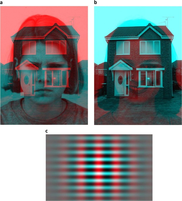
thus take place ~30–60 min after administration of OT or placebo. STIMULI During each of the two visits, participants viewed a series of 30 stationary anaglyphs—please see Fig. 1 for
examples34. Of these, 28 consisted of an image of a female face (sourced from the Karolinska Directed Emotional Faces) and a superimposed house image (public domain image available online),
where the face would be either red or cyan, and the house would have the opposite color. Half of the anaglyphs had a red face and half had a cyan face. The face displayed one of six
emotional expressions—joy, anger, sadness, surprise, fear, or disgust—or had a neutral expression. Thus, four anaglyphs out of the 28 would display each expression. Two final anaglyphs
consisted of Gabor patches, i.e., intersecting vertical and horizontal low spatial frequency lines. Gabor patches are thus visual stimuli with low complexity and no social information. One
of the pictures had red vertical lines and cyan horizontal lines, and one had the opposite arrangement. See Fig. 1 for examples of face/house and Gabor anaglyphs. The anaglyphs were
presented using SMI Experiment Center (SensoMotoric Instruments, RED500, SMI© Berlin, Germany) on a stationary computer. The anaglyphs were presented at an approximate viewing angle of 38
degrees, with participants positioned at a distance of 70 cm from the computer screen. EXPERIMENTAL PROCEDURE Each participant was provided with colored anaglyph glasses (red and cyan), and
received instructions prior to a brief practice trial. Each experimental run consisted of the 28 face/house anaglyphs being presented in sequence to the participant. Each anaglyph would be
presented for 30 s, with one anaglyph directly following the next. This sequence of 28 anaglyphs were then followed by a brief one minute break, after which the participant then viewed the
two Gabor anaglyphs, which would be presented for 60 s each. The participant self-reported dominant or piecemeal percepts by pressing one of three buttons to indicate what they perceived.
Button presses were registered with a single time stamp, regardless of how long the button was pressed. While viewing the face/house anaglyphs, the left button would indicate face dominance,
the right one house dominance, and the middle one piecemeal percept. During viewing of the Gabor anaglyphs, the left and right buttons would indicate dominant red or cyan percept,
respectively, and the middle one piecemeal percept. DATA ANALYSIS AND STATISTICS Data retrieved from BeGaze software (SMI© Berlin, Germany) contained timed key presses for each of the
separate anaglyphs. Data were extracted using an in-house script written in Python 3.4 (see Supplementary Material). We extracted durations of dominance (ms) of uninterrupted percepts for
all anaglyphs, categorized as face, house, and Gabor dominance durations, respectively. The time before the initial button press for each anaglyph was excluded, since there was no way to
accurately determine whether the indicated percept before initial key press was in fact what the participant was seeing, or if it was caused by bleed-over from the previous anaglyph, or
simply reaction time latency. Similarly, the time following the ultimate button press for each anaglyph was also excluded, to avoid artificially shortened durations. All dominance duration
means were calculated following an initial exclusion of dominance durations shorter than 250 milliseconds or longer than the mean plus three standard deviations across all participants for
the two types of anaglyph (face/house and Gabor, respectively). We extracted average durations of face dominance duration for all separate emotions (seven variables per session), and also
averaged durations across all emotions for an overall mean duration of face dominance duration (one variable per session). House dominance durations were averaged across all emotions (one
variable per session). Gabor dominance was averaged across both dominance percepts (one variable per session). We also calculated alternation rates for face/house anaglyphs and Gabor
anaglyphs, respectively, since this has been previously used to investigate excitation-inhibition balance30. An alternation was defined as a complete transition from one dominant percept to
another, and alternation rate was quantified as the number of alternations per 30 s. Five participants were excluded from statistical analyses for consistently displaying no meaningful
rivalry (i.e., less than two key presses for more than 50% of stimuli in either or both of the sessions) or misunderstanding the instructions. This yielded a final sample of 45 participants.
In order to ensure good quality data, results from individual stimuli were excluded if they contained less than two key presses, leading to a data loss of 3.9% from the _n_ = 45 sample. The
data were analyzed using SPSS 23. We utilized repeated measures analysis of variance (RM-ANOVA) to analyze average dominance durations between different dominant percepts (face, house, and
Gabor), where percept type and drug were within-subject factors. RM-ANOVA was also used to investigate the modulation of dominance durations for face percepts and face predominance by
emotional content, where drug and emotion were within-subject factors. Post-hoc tests were paired t-tests. Alternation rates were tested using paired _t_-tests for the average rates across
face/house and Gabor anaglyphs, respectively. All tests were two-tailed. We did not conduct a specific power analysis prior to data collection for the binocular rivalry task. With 45 people
per group in a between-subjects study, for a two-tailed contrast (_t_-test) G*power estimates 80% power to detect a medium effect, _d_ = 0.6. However, in within-subjects studies of drug
effects on behavioral task outcomes, correlations between drug and placebo are typically 0.6 or higher35,36,37,38,39. In our data, the correlation coefficient of average dominance durations
of faces, houses, and gabors between drug conditions ranged from _r_ = 0.687 to _r_ = 0.829. Assuming _r_ = 0.6 between oxytocin and placebo condition measures of binocular rivalry based on
previous literature only, our within-subjects study is at least as sensitive as a between-subjects study with 75 participants per group, i.e. has 80% power to detect _d_ = 0.46. This means
that even considering the boost in statistical power yielded by our design, the study lacked the sensitivity to detect small effects. RESULTS Please see Table 1 for all pairwise comparisons
mentioned below. DOMINANCE DURATIONS Average dominance durations (ms) of uninterrupted percepts were calculated for face (across all emotions), house, and Gabor percepts. Using RM-ANOVA with
a 2 (oxytocin/placebo) × 3 (face, house, or Gabor percept) design, we found a significant main effect of drug (_F_(1,42) = 10.64; _p_ = 0.002; _ηp_2 = 0.202) and of percept (_F_(2,41) =
45.74; _p_ < 0.001; _ηp_2 = 0.691), as well as an interaction between drug and percept (_F_(2,41) = 3.50; _p_ = 0.039; _ηp_2 = 0.146). Subsequent post-hoc analyses revealed that compared
to the placebo treatment, OT significantly increased face dominance durations for all emotions combined (_t_(44) = 3.86; _p_ < 0.001; _ηp_2 = 0.253; Fig. 2a). The similar combined measure
for house dominance durations was on the border of statistical significance for OT treatment increasing dominance durations for houses as well (_t_(44) = 1.69; _p_ = 0.098; _ηp_2 = 0.061;
Fig. 2b), compared to placebo. Similarly, Gabor dominance durations showed a trend approaching significance for being increased in the OT condition compared to the placebo condition (_t_(42)
= 1.86; _p_ = 0.069; _ηp_2 = 0.076; Fig. 2c). Dominance durations for faces were also averaged separately for the different emotions. RM-ANOVA analysis with a 2 (oxytocin/placebo) × 7
(emotions) was performed, with Mauchly’s test of sphericity indicating that sphericity was violated for drug × emotion (_W_ = 0.221; _p_ < 0.001). The degrees of freedom were thus
corrected using Huyhn-Feldt estimates of sphericity. There was no significant interaction between drug and emotion of the face perceived on dominance durations (_F_(5,200) = 0.975; _p_ =
0.431). The face dominance durations were higher with oxytocin for all emotional face categories. Post-hoc paired _t_-tests comparing oxytocin and placebo reached significance for sadness
and disgust. The mean difference in dominance durations was least apparent for the neutral condition (Fig. 2d). There were no statistically significant differences in piecemeal durations,
for face/house stimuli or for Gabor stimuli (all _p_ > 0.05). ALTERNATION RATES We included alternation rates as an outcome since this has been previously utilized in order to investigate
excitation-inhibition balance with binocular rivalry30. There was, however, no significant difference between intranasal oxytocin and placebo with regards to alternation rate between the
face and house percept, defined as alternations/30 s (_t_(44) = 1.18; _p_ = 0.25). There was also no significant difference between the oxytocin and placebo condition with regards to
alternation rate between dominant Gabor percepts, defined as switches/30 s (_t_(42) = 1.45; _p_ = 0.16). DISCUSSION To our knowledge, this is the first study to utilize binocular rivalry to
investigate the effects of intranasal OT on visual processing. Longer dominance durations for faces during binocular rivalry confirmed our hypothesis that intranasal OT increases the
salience of faces. The higher effect size for faces rather than non-social stimuli is congruent with the social salience hypothesis4; we however, did not find that emotional content
significantly modulated the response to OT administration. Our data thus provides support for the idea that OT increases the salience of social signals of positive and negative affective
valence. Nevertheless, we also found non-significant but intriguing trends that intranasal OT increases the dominance durations for both Gabor and house percepts—both non-social in nature.
We did not find any significant differences in alternation rates between placebo and oxytocin for the face/house anaglyphs, nor for the Gabor anaglyphs. The increase in salience across
valences is in line with studies where the outcome measure can be assumed to depend on the salience of stimuli, for instance showing that intranasal OT increases memory retention of
faces6,12 and emotion recognition accuracy11,40. The role of OT in face and emotion recognition has been stressed by the above mentioned pharmacological as well as genetic
studies41,42,43,44; however, other studies report incongruent results39. We would argue that our results are novel in their methodological approach, and lend support to the social salience
hypothesis, through a psychophysiological measurement. There are several potential mechanisms by which OT may influence rivalry dynamics and the processing of visual social information.
Firstly, OT may have increased face dominance during binocular rivalry via attentional mechanisms45,46,47,48. Recent neuroimaging studies using intranasal OT are in line with enhanced visual
attention, e.g., enhanced early visual activity for faces49, and increased functional connectivity in the nucleus basalis of Meynert, which regulates selective attention50. This
interpretation of our results is in line with the social salience hypothesis, where attention, as guided by oxytocinergic influences on dopaminergic circuitry, is central4. Secondly,
previous pharmacological studies on the effects of the serotonin receptor agonists psilocybin and tandospirone demonstrated increased dominance durations for Gabor patches51,52, suggesting
that serotonin acts to modulate binocular rivalry. OT has been shown to directly influence the release of serotonin53, in for instance the nucleus accumbens54. Serotonergic signaling,
therefore, constitutes a potential mediator of OT’s effects on rivalry dynamics. Thirdly, binocular rivalry is a phenomenon based on interplay between inhibitory and excitatory neurons, and
there is ample evidence that OT is able to influence excitation-inhibition balance in various parts of the brain. For example, it has been implicated in the initial transient switch in
γ-aminobutyric acid (GABA) receptors from depolarizing to hyperpolarizing55,56,57. OT receptor knockout mice display a decreased ratio of GABA-ergic to total presynapses in the hippocampus,
and increased seizure susceptibility58, and OT has been shown to influence glutamate neurotransmission in brain slices of mice59. Intriguingly, in the sensory cortices, OT rapidly alters the
excitation-inhibition balance in the auditory cortex to enable maternal behavior in female mice60, as well as enhance pre- and postsynaptic glutamatergic signaling in the rat olfactory
bulb61. While we cannot, based on these three lines of reasoning, pinpoint from our data where OT acts to achieve its effects on rivalry dynamics, our results provide the support that OT
does influence the excitation-inhibition balance phenomenon of binocular rivalry, by top-down effects and/or directly in the visual cortex. In addition, these proposed mechanisms may in part
explain the fact that we see strong trends towards increased dominance durations for non-social stimuli as well. Our tentative findings on non-social stimuli are congruent with previous
literature indicating that the effects of OT are not restricted to only social contexts, as it may mediate for instance approach and avoidance behavior in non-social contexts as well62. The
approach-avoidance hypothesis of OT has not been specifically addressed here, and our non-social stimuli were arguably simplistic. This makes it difficult to draw any firm conclusions on how
our findings may relate to this hypothesis, but provides an intriguing avenue of further research. OT administration in mammals has been shown to rescue social behavior deficits63. Hence,
it is not surprising that OT has been suggested as a possible treatment for social impairments in several psychiatric and neurological diagnoses, such as developmental prosopagnosia64,
borderline personality disorder65, and ASD66. This therapeutic option is appealing, given the relatively mild side effect profile of OT, and the ease of intranasal administration; however,
the effect duration is relatively short. Substantial controversy remains about how and how much intranasally administered oxytocin reaches the brain in humans67, and the few human studies
applying variable doses have yet to produce conclusive results68,69,70. The dose applied here (40IU) is the largest dose typically applied in human research. Studies attempting treatment
with intranasal OT for individuals with ASD have had encouraging, although somewhat mixed, results63. Our findings encourage future studies on exogenously administered OT as a treatment for
social impairments, for example, in combination with behavioral therapy to fully utilize the effect duration. Limitations to the study include that it was not powered to detect small
effects, although many intranasal oxytocin studies report small effects. Another potential weakness of our study is that it included only men. Given the well-known sex-specific
idiosyncrasies of the OT system71, we would urge future studies to include women as well. Furthermore, this study included only female faces, and future studies would do well to investigate
if early visual processing of same-sex and opposite-sex faces is different. In addition, this study did not include any measure of whether each emotional expression was recognized, which
would have allowed a deeper analysis of the possible correlation between a change in early visual processing and aspects of social cognition. Lastly, the participants were not asked whether
they had any conditions that may have obstructed the nasal cavity (e.g., past nasal surgery, current cold/flu), which could possibly influence the response to intranasal OT. In conclusion,
we have demonstrated that intranasal OT administration leads to altered visual processing of social stimuli, by increasing dominance durations for faces in a binocular rivalry paradigm. This
is an easily administered and non-invasive test, which can be further modified to probe aspects of visual awareness, with additional pharmacological manipulations, which will allow more
detailed investigations of the effects of OT on perception and processing of social stimuli. Our results lend support to the hypothesis that OT increases the salience of social stimuli, and
suggests that this increased salience happens across stimuli of different valences. CHANGE HISTORY * _ 23 OCTOBER 2020 The original HTML version of this Article was updated shortly after
publication to add the University of Gothenburg Open Access funding acknowledgement. _ REFERENCES * Lee, H. J., Macbeth, A. H., Pagani, J. H. & Young, W. S. III. Oxytocin: the great
facilitator of life. _Prog. Neurobiol._ 88, 127–151 (2009). CAS PubMed PubMed Central Google Scholar * Bartz, J. A., Zaki, J., Bolger, N. & Ochsner, K. N. Social effects of oxytocin
in humans: context and person matter. _Trends Cogn. Sci._ 15, 301–309 (2011). CAS PubMed Google Scholar * Olff, M. et al. The role of oxytocin in social bonding, stress regulation and
mental health: an update on the moderating effects of context and interindividual differences. _Psychoneuroendocrinology_ 38, 1883–1894 (2013). CAS PubMed Google Scholar * Shamay-Tsoory,
S. G. & Abu-Akel, A. The social salience hypothesis of oxytocin. _Biol. Psychiatry_ 79, 194–202 (2016). CAS PubMed Google Scholar * Shamay-Tsoory, S. G. et al. Intranasal
administration of oxytocin increases envy and schadenfreude (gloating). _Biol. Psychiatry_ 66, 864–870 (2009). CAS PubMed Google Scholar * Rimmele, U., Hediger, K., Heinrichs, M. &
Klaver, P. Oxytocin makes a face in memory familiar. _J. Neurosci._ 29, 38–42 (2009). CAS PubMed PubMed Central Google Scholar * Unkelbach, C., Guastella, A. J. & Forgas, J. P.
Oxytocin selectively facilitates recognition of positive sex and relationship words. _Psychol. Sci._ 19, 1092–1094 (2008). PubMed Google Scholar * Hu, J. et al. Oxytocin selectively
facilitates learning with social feedback and increases activity and functional connectivity in emotional memory and reward processing regions. _Hum. Brain Mapp._ 36, 2132–2146 (2015).
PubMed PubMed Central Google Scholar * Gamer, M., Zurowski, B. & Buchel, C. Different amygdala subregions mediate valence-related and attentional effects of oxytocin in humans. _Proc.
Natl Acad. Sci. USA_ 107, 9400–9405 (2010). CAS PubMed PubMed Central Google Scholar * Leppanen, J., Ng, K. W., Tchanturia, K. & Treasure, J. Meta-analysis of the effects of
intranasal oxytocin on interpretation and expression of emotions. _Neurosci. Biobehav Rev._ 78, 125–144 (2017). CAS PubMed Google Scholar * Shahrestani, S., Kemp, A. H. & Guastella,
A. J. The impact of a single administration of intranasal oxytocin on the recognition of basic emotions in humans: a meta-analysis. _Neuropsychopharmacology_ 38, 1929–1936 (2013). CAS
PubMed PubMed Central Google Scholar * Savaskan, E., Ehrhardt, R., Schulz, A., Walter, M. & Schachinger, H. Post-learning intranasal oxytocin modulates human memory for facial
identity. _Psychoneuroendocrinology_ 33, 368–374 (2008). CAS PubMed Google Scholar * Xu, L. et al. Oxytocin enhances attentional bias for neutral and positive expression faces in
individuals with higher autistic traits. _Psychoneuroendocrinology_ 62, 352–358 (2015). CAS PubMed Google Scholar * Blake, R. & Logothetis, N. Visual competition. _Nat. Rev.
Neurosci._ 3, 13–21 (2002). CAS PubMed Google Scholar * Anderson, E. C. et al. Smiles may go unseen in generalized social anxiety disorder: evidence from binocular rivalry for reduced
visual consciousness of positive facial expressions. _J. Anxiety Disord._ 27, 619–626 (2013). PubMed Google Scholar * Singer, N., Eapen, M., Grillon, C., Ungerleider, L. G. & Hendler,
T. Through the eyes of anxiety: dissecting threat bias via emotional-binocular rivalry. _Emotion_ 12, 960–969 (2012). PubMed PubMed Central Google Scholar * Anderson, E., Siegel, E. H.
& Barrett, L. F. What you feel influences what you see: the role of affective feelings in resolving binocular rivalry. _J. Exp. Soc. Psychol._ 47, 856–860 (2011). PubMed PubMed Central
Google Scholar * Yoon, K. L., Hong, S. W., Joormann, J. & Kang, P. Perception of facial expressions of emotion during binocular rivalry. _Emotion_ 9, 172–182 (2009). PubMed Google
Scholar * Brascamp, J. W., Klink, P. C. & Levelt, W. J. The ‘laws’ of binocular rivalry: 50 years of Levelt’s propositions. _Vis. Res._ 109, 20–37 (2015). CAS PubMed Google Scholar *
Walle, K. M., Kyler, H. L., Nordvik, J. E., Becker, F. & Laeng, B. Binocular rivalry after right-hemisphere stroke: effects of attention impairment on perceptual dominance patterns.
_Brain Cogn._ 117, 84–96 (2017). PubMed Google Scholar * Anderson, E., Siegel, E. H., Bliss-Moreau, E. & Barrett, L. F. The visual impact of gossip. _Science_ 332, 1446–1448 (2011).
CAS PubMed PubMed Central Google Scholar * Blake, R. A neural theory of binocular rivalry. _Psychol. Rev._ 96, 145–167 (1989). CAS PubMed Google Scholar * Dayan, P. A hierarchical
model of binocular rivalry. _Neural Comput._ 10, 1119–1135 (1998). CAS PubMed Google Scholar * Tong, F., Meng, M. & Blake, R. Neural bases of binocular rivalry. _Trends Cogn. Sci._
10, 502–511 (2006). PubMed Google Scholar * Said, C. P. & Heeger, D. J. A model of binocular rivalry and cross-orientation suppression. _PLoS Comput Biol._ 9, e1002991 (2013). CAS
PubMed PubMed Central Google Scholar * van Loon, A. M. et al. GABA shapes the dynamics of bistable perception. _Curr. Biol._ 23, 823–827 (2013). PubMed Google Scholar * Seely, J. &
Chow, C. C. Role of mutual inhibition in binocular rivalry. _J. Neurophysiol._ 106, 2136–2150 (2011). PubMed PubMed Central Google Scholar * Freyberg, J., Robertson, C. E. &
Baron-Cohen, S. Reduced perceptual exclusivity during object and grating rivalry in autism. _J. Vis._ 15, 11 (2015). PubMed Google Scholar * Robertson, C. E., Kravitz, D. J., Freyberg, J.,
Baron-Cohen, S. & Baker, C. I. Slower rate of binocular rivalry in autism. _J. Neurosci._ 33, 16983–16991 (2013). CAS PubMed PubMed Central Google Scholar * Robertson, C. E., Ratai,
E. M. & Kanwisher, N. Reduced GABAergic action in the autistic brain. _Curr. Biol._ 26, 80–85 (2016). CAS PubMed Google Scholar * Rubenstein, J. L. & Merzenich, M. M. Model of
autism: increased ratio of excitation/inhibition in key neural systems. _Genes Brain Behav._ 2, 255–267 (2003). CAS PubMed PubMed Central Google Scholar * Nelson, S. B. & Valakh, V.
Excitatory/inhibitory balance and circuit homeostasis in autism spectrum disorders. _Neuron_ 87, 684–698 (2015). CAS PubMed PubMed Central Google Scholar * Leknes, S. et al. Oxytocin
enhances pupil dilation and sensitivity to ‘hidden’ emotional expressions. _Soc. Cogn. Affect Neurosci._ 8, 741–749 (2013). PubMed Google Scholar * Duane, K. R. A brief history of
stereoscopy. _Wiley Interdiscip. Rev.: Computational Stat._ 5, 334–340 (2013). Google Scholar * Auyeung, B. et al. Oxytocin increases eye contact during a real-time, naturalistic social
interaction in males with and without autism. _Transl. Psychiat._ 5, e507 (2015). CAS Google Scholar * Eckstein, M. et al. Oxytocin increases eye-gaze towards novel social and non-social
stimuli. _Soc. Neurosci._ 14, 594–607 (2019). PubMed Google Scholar * Eikemo, M. et al. Sweet taste pleasantness is modulated by morphine and naltrexone. _Psychopharmacol. (Berl.)._ 233,
3711–3723 (2016). CAS Google Scholar * Loseth, G. E., Eikemo, M. & Leknes, S. Effects of opioid receptor stimulation and blockade on touch pleasantness: a double-blind randomised
trial. _Soc. Cogn. Affect Neurosci._ 14, 411–422 (2019). PubMed PubMed Central Google Scholar * Hubble, K. et al. Oxytocin reduces face processing time but leaves recognition accuracy and
eye-gaze unaffected. _J. Int Neuropsychol. Soc._ 23, 23–33 (2017). PubMed Google Scholar * Domes, G., Kumbier, E., Heinrichs, M. & Herpertz, S. C. Oxytocin promotes facial emotion
recognition and amygdala reactivity in adults with Asperger syndrome. _Neuropsychopharmacology_ 39, 698–706 (2014). CAS PubMed Google Scholar * Hovey, D. et al. Emotion recognition
associated with polymorphism in oxytocinergic pathway gene ARNT2. _Soc. Cogn. Affect Neurosci._ 13, 173–181 (2018). PubMed Google Scholar * Westberg, L. et al. Variation in the oxytocin
receptor gene is associated with face recognition and its neural correlates. _Front Behav. Neurosci._ 10, 178 (2016). PubMed PubMed Central Google Scholar * Cattaneo, Z. et al. Congenital
prosopagnosia is associated with a genetic variation in the oxytocin receptor (OXTR) gene: an exploratory study. _Neuroscience_ 339, 162–173 (2016). CAS PubMed Google Scholar * Skuse, D.
H. et al. Common polymorphism in the oxytocin receptor gene (OXTR) is associated with human social recognition skills. _Proc. Natl Acad. Sci. USA_ 111, 1987–1992 (2014). CAS PubMed Google
Scholar * Dieter, K. C., Brascamp, J., Tadin, D. & Blake, R. Does visual attention drive the dynamics of bistable perception? _Atten. Percept. Psychophys._ 78, 1861–1873 (2016). PubMed
PubMed Central Google Scholar * Domes, G. et al. Intranasal oxytocin increases covert attention to positive social cues. _Psychol. Med._ 43, 1747–1753 (2013). CAS PubMed Google Scholar
* Guastella, A. J., Mitchell, P. B. & Dadds, M. R. Oxytocin increases gaze to the eye region of human faces. _Biol. Psychiatry_ 63, 3–5 (2008). CAS PubMed Google Scholar *
Tollenaar, M. S., Chatzimanoli, M., van der Wee, N. J. & Putman, P. Enhanced orienting of attention in response to emotional gaze cues after oxytocin administration in healthy young men.
_Psychoneuroendocrinology_ 38, 1797–1802 (2013). CAS PubMed Google Scholar * Andari, E., Richard, N., Leboyer, M. & Sirigu, A. Adaptive coding of the value of social cues with
oxytocin, an fMRI study in autism spectrum disorder. _Cortex_ 76, 79–88 (2016). PubMed Google Scholar * Rilling, J. K., Chen, X., Chen, X. & Haroon, E. Intranasal oxytocin modulates
neural functional connectivity during human social interaction. _Am. J. Primatol._ 80, e22740 (2018). PubMed PubMed Central Google Scholar * Nagamine, M., Yoshino, A., Miyazaki, M.,
Takahashi, Y. & Nomura, S. Effects of selective 5-HT1A agonist tandospirone on the rate and rhythmicity of binocular rivalry. _Psychopharmacol. (Berl.)._ 198, 279–286 (2008). CAS Google
Scholar * Carter, O. L. et al. Modulating the rate and rhythmicity of perceptual rivalry alternations with the mixed 5-HT2A and 5-HT1A agonist psilocybin. _Neuropsychopharmacology_ 30,
1154–1162 (2005). CAS PubMed Google Scholar * Yoshida, M. et al. Evidence that oxytocin exerts anxiolytic effects via oxytocin receptor expressed in serotonergic neurons in mice. _J.
Neurosci._ 29, 2259–2271 (2009). CAS PubMed PubMed Central Google Scholar * Dolen, G., Darvishzadeh, A., Huang, K. W. & Malenka, R. C. Social reward requires coordinated activity of
nucleus accumbens oxytocin and serotonin. _Nature_ 501, 179–184 (2013). PubMed PubMed Central Google Scholar * Tyzio, R. et al. Maternal oxytocin triggers a transient inhibitory switch in
GABA signaling in the fetal brain during delivery. _Science_ 314, 1788–1792 (2006). CAS PubMed Google Scholar * Tyzio, R. et al. Oxytocin-mediated GABA inhibition during delivery
attenuates autism pathogenesis in rodent offspring. _Science_ 343, 675–679 (2014). CAS PubMed Google Scholar * Leonzino, M. et al. The timing of the excitatory-to-inhibitory GABA switch
is regulated by the oxytocin receptor via KCC2. _Cell Rep._ 15, 96–103 (2016). CAS PubMed PubMed Central Google Scholar * Sala, M. et al. Pharmacologic rescue of impaired cognitive
flexibility, social deficits, increased aggression, and seizure susceptibility in oxytocin receptor null mice: a neurobehavioral model of autism. _Biol. Psychiatry_ 69, 875–882 (2011). CAS
PubMed Google Scholar * Ninan, I. Oxytocin suppresses basal glutamatergic transmission but facilitates activity-dependent synaptic potentiation in the medial prefrontal cortex. _J.
Neurochem._ 119, 324–331 (2011). CAS PubMed Google Scholar * Marlin, B. J., Mitre, M., D’Amour, J. A., Chao, M. V. & Froemke, R. C. Oxytocin enables maternal behaviour by balancing
cortical inhibition. _Nature_ 520, 499–504 (2015). CAS PubMed PubMed Central Google Scholar * Osako, Y., Otsuka, T., Taniguchi, M., Oka, T. & Kaba, H. Oxytocin enhances presynaptic
and postsynaptic glutamatergic transmission between rat olfactory bulb neurones in culture. _Neurosci. Lett._ 299, 65–68 (2001). CAS PubMed Google Scholar * Harari-Dahan, O. &
Bernstein, A. A general approach-avoidance hypothesis of oxytocin: accounting for social and non-social effects of oxytocin. _Neurosci. Biobehav Rev._ 47, 506–519 (2014). CAS PubMed Google
Scholar * DeMayo, M. M., Song, Y. J. C., Hickie, I. B. & Guastella, A. J. A review of the safety, efficacy and mechanisms of delivery of nasal oxytocin in children: therapeutic
potential for autism and prader-willi syndrome, and recommendations for future research. _Paediatr. Drugs_ 19, 391–410 (2017). PubMed Google Scholar * Bate, S. et al. Intranasal inhalation
of oxytocin improves face processing in developmental prosopagnosia. _Cortex_ 50, 55–63 (2014). PubMed Google Scholar * Servan, A., Brunelin, J. & Poulet, E. The effects of oxytocin
on social cognition in borderline personality disorder. _Encephale_ 44, 46–51 (2018). CAS PubMed Google Scholar * Benner, S. & Yamasue, H. Clinical potential of oxytocin in autism
spectrum disorder: current issues and future perspectives. _Behav. Pharmacol._ 29, 1–12 (2018). CAS PubMed Google Scholar * Leng, G. & Ludwig, M. Intranasal oxytocin: myths and
delusions. _Biol. Psychiatry_ 79, 243–250 (2016). CAS PubMed Google Scholar * Quintana, D. S. et al. Low-dose intranasal oxytocin delivered with breath powered device modulates pupil
diameter and amygdala activity: a randomized controlled pupillometry and fMRI study. _Neuropsychopharmacology_ 44, 306–313 (2019). CAS PubMed Google Scholar * Spengler, F. B. et al.
Kinetics and dose dependency of intranasal oxytocin effects on amygdala reactivity. _Biol. Psychiatry_ 82, 885–894 (2017). CAS PubMed Google Scholar * Quintana, D. S., Outhred, T.,
Westlye, L. T., Malhi, G. S. & Andreassen, O. A. The impact of oxytocin administration on brain activity: a systematic review and meta-analysis protocol. _Syst. Rev._ 5, 205 (2016).
PubMed PubMed Central Google Scholar * Carter, C. S. Oxytocin pathways and the evolution of human behavior. _Annu Rev. Psychol._ 65, 17–39 (2014). PubMed Google Scholar Download
references ACKNOWLEDGEMENTS The authors would like to thank Dr. Nouchine Hadjikhani and Dr. Soaleha Shams for help with critiquing and editing the paper. This research was supported by
funding from the Swedish Research Council, an ALF-grant from Region Västra Götaland, and the Wilhelm and Martina Lundgren Foundation. AUTHOR INFORMATION AUTHORS AND AFFILIATIONS * Department
of Pharmacology, Institute of Neuroscience and Physiology, The Sahlgrenska Academy, University of Gothenburg, Gothenburg, Sweden Daniel Hovey & Lars Westberg * Department of Psychiatry
and Psychotherapy, University of Tübingen, Tübingen, Germany Louise Martens * Max Planck Institute for Biological Cybernetics, Tübingen, Germany Louise Martens * Department of Psychology,
University of Oslo, Oslo, Norway Bruno Laeng & Siri Leknes * RITMO Centre for Interdisciplinary Studies in Rhythm, Time and Motion, Oslo, Norway Bruno Laeng Authors * Daniel Hovey View
author publications You can also search for this author inPubMed Google Scholar * Louise Martens View author publications You can also search for this author inPubMed Google Scholar * Bruno
Laeng View author publications You can also search for this author inPubMed Google Scholar * Siri Leknes View author publications You can also search for this author inPubMed Google Scholar
* Lars Westberg View author publications You can also search for this author inPubMed Google Scholar CORRESPONDING AUTHOR Correspondence to Daniel Hovey. ETHICS DECLARATIONS CONFLICT OF
INTEREST The authors declare that they have no conflict of interest. ADDITIONAL INFORMATION PUBLISHER’S NOTE Springer Nature remains neutral with regard to jurisdictional claims in published
maps and institutional affiliations. Open access funding provided by University of Gothenburg. SUPPLEMENTARY INFORMATION SUPPLEMENTARY DATA 1 RIGHTS AND PERMISSIONS OPEN ACCESS This article
is licensed under a Creative Commons Attribution 4.0 International License, which permits use, sharing, adaptation, distribution and reproduction in any medium or format, as long as you
give appropriate credit to the original author(s) and the source, provide a link to the Creative Commons license, and indicate if changes were made. The images or other third party material
in this article are included in the article’s Creative Commons license, unless indicated otherwise in a credit line to the material. If material is not included in the article’s Creative
Commons license and your intended use is not permitted by statutory regulation or exceeds the permitted use, you will need to obtain permission directly from the copyright holder. To view a
copy of this license, visit http://creativecommons.org/licenses/by/4.0/. Reprints and permissions ABOUT THIS ARTICLE CITE THIS ARTICLE Hovey, D., Martens, L., Laeng, B. _et al._ The effect
of intranasal oxytocin on visual processing and salience of human faces. _Transl Psychiatry_ 10, 318 (2020). https://doi.org/10.1038/s41398-020-00991-3 Download citation * Received: 10
August 2018 * Revised: 22 June 2020 * Accepted: 24 June 2020 * Published: 19 September 2020 * DOI: https://doi.org/10.1038/s41398-020-00991-3 SHARE THIS ARTICLE Anyone you share the
following link with will be able to read this content: Get shareable link Sorry, a shareable link is not currently available for this article. Copy to clipboard Provided by the Springer
Nature SharedIt content-sharing initiative