- Select a language for the TTS:
- UK English Female
- UK English Male
- US English Female
- US English Male
- Australian Female
- Australian Male
- Language selected: (auto detect) - EN
Play all audios:
ABSTRACT BACKGROUND The scientific imaging of works of art is crucial for the assessment of the presence and distribution of pigments and other materials on surfaces. It is known that some
ancient pigments are luminescent: these include pink red-lakes and the blue and purple pigments Egyptian Blue (CaCuSi4O10), Han blue (BaCuSi4O10) and Han purple (BaCuSi2O6). Indeed, the
unique near-infrared luminescence emission of Egyptian blue allows the imaging of its distribution on surfaces. RESULTS We focus on the imaging of the time-resolved photoluminescence of
ancient Egyptian objects in the Burri Collection from the Civic Museum of Crema and of the Cremasco (Italy). Time-resolved photoluminescence images have been acquired using excitation at 355
nm for detecting the ns-emission of red lakes and binding media; by employing 532 nm excitation Egyptian blue is probed, and the spatial distribution of its long-lived microsecond emission
is imaged. For the first time we provide data on the photoluminescence lifetime of Egyptian blue directly from objects. Moreover, we demonstrate that the use of a pulsed laser emitting at
two different wavelengths increases the effectiveness of the lifetime imaging technique for mapping the presence of emissions from pigments on painted surfaces. Laser-induced luminescence
spectra from different areas of the objects and traditional digital imaging, using led-based lamps, long pass filters and a commercial photographic camera, complement the results from
photoluminescence lifetime imaging. We demonstrate the versatility of a new instrumental setup, capable of recording decay emission kinetics with lifetimes from nanosecond to microseconds.
CONCLUSIONS While the combined wavelength approach for the imaging of emissions from different materials has been demonstrated for the study of ancient Egyptian pigments (both organic and
inorganic), the method could be extended to the analysis of modern pigments and paintings. SIMILAR CONTENT BEING VIEWED BY OTHERS OBJECTIVE INTERPRETATION OF ULTRAVIOLET-INDUCED LUMINESCENCE
FOR CHARACTERIZING PICTORIAL MATERIALS Article Open access 19 November 2023 A TIME-CORRELATED SINGLE PHOTON COUNTING SPAD ARRAY CAMERA WITH A BESPOKE DATA-PROCESSING ALGORITHM FOR
LIGHTSHEET FLUORESCENCE LIFETIME IMAGING (FLIM) AND FLIM VIDEOS Article Open access 27 March 2024 BLUE-SHIFT PHOTOCONVERSION OF NEAR-INFRARED FLUORESCENT PROTEINS FOR LABELING AND TRACKING
IN LIVING CELLS AND ORGANISMS Article Open access 19 December 2023 BACKGROUND The imaging of paintings and painted objects relies on the interaction between radiation and matter and the
detection of reflected or emitted photons. Technical examination and analysis of works of artworks typically relies on the integration of non-invasive imaging and suitable spectroscopic
analysis, often followed by taking and analysing samples. In this work we report an imaging study of Egyptian objects with a novel portable setup for time-resolved photoluminescence (TRPL)
imaging and spectroscopy, with a focus on the analysis of Egyptian Blue and lake pigments on artefacts. Photoluminescence (PL) of pigments and paint has received significant attention due to
its practical use in the assessment of the condition of works of art: for example, aged organic varnishes may appear more luminescent than freshly applied paint and some pigments, including
semiconductor materials, emit characteristic PL signals [1]. Among ancient Egyptian pigments, Egyptian blue and madder-based red lakes are luminescent and are routinely identified with the
aid of microscopic and spectroscopic analysis [2]. While the fluorescence observed in madder lakes is due to the anthraquinone molecules purpurin and alizarin, the optical emission of
Egyptian blue has been ascribed to Cu2+ in a solid calcium-silica matrix [3–5]. Recent research on the mineral pigment has reported that synthetic cuprorivaite has a radiative emission with
a maximum peaked at 910 nm and a lifetime of 107 μs and a quantum yield of 10 % [6]. The imaging of PL from painted surfaces is often carried out to detect the colour of the emission, for
which various approaches have been suggested. Whereas ultraviolet (UV) light sources based on Mercury lamps require filtering of spurious radiation emitted in the visible to render them
useful for imaging and photography, innovative uses of Xenon-flashes and digital photography have demonstrated the peculiar and noteworthy infrared emissions from Egyptian blue on paintings
and painted objects from the British Museum and, more recently, on Fayoum Portraits [7–9]. Many other examples of digital imaging of Egyptian blue from objects have been documented. In
addition to digital photographic imaging, PL spectroscopic imaging techniques have found multiple applications for the analysis of paintings, including those carried out with a scanning
Lidar approach [10, 11] and with light emitting diodes (LED) [12]. With these methods, the detection of PL can be carried out with imaging spectrometers or suitably filtered imaging
detectors [13, 14]. Another approach to the imaging of the PL from works of art relies on the detection of the emission lifetime, which can be achieved by combining pulsed laser excitation
with a time-gated imaging detector, known as fluorescence lifetime imaging or, more generally, TRPL Imaging [15]. Applications of TRPL imaging for the detection of organic materials on stone
sculptures [16], wall paintings [17], and semiconductor pigments on paper [18] have been published, and demonstrate how TRPL imaging can map the chemical composition of surfaces based on
significant differences in emission lifetime. In previous applications a ns Q-switched Nd:YAG laser emitting at 355 nm was employed as this wavelength can excite emissions from binding media
and organic polymers and aforementioned fluorescent pigments. In this work we have modified our TRPL imaging set-up by adding excitation at 532 nm from the same Nd:YAG laser to allow the
excitation of Egyptian blue. The combined wavelength approach has been applied to the imaging of painted objects from Carla Maria Burri Collection in the Civic Museum of Crema and the
Cremasco (see sample description). Complementary laser-induced PL spectroscopy from selected points on the objects has been carried out and digital photography using LED excitation
complement images acquired with TRPL imaging. RESULTS AND DISCUSSION Preliminary PL spectroscopy analysis of a model sample painted with the commercially available Egyptian blue pigment
reveals a characteristic emission spectrum with a maximum emission at 920 ± 6 nm and a mono-exponential emission lifetime of 138 ± 4 µs (95 % confidence) (Fig. 1), in good agreement with the
spectral data reported by others from pigments from the same supplier [7, 19]. The emission lifetime measured in our commercial pigment is in agreement with Borozov et al. [19] but differs
with respect to that reported for the synthetic mineral calculated using single photon counting and 637 nm excitation (τ = 107 μs [6]). It is possible that the discrepancy in the PL lifetime
is due to grinding of the pigment rather than to differences related to the methods used for the estimation of the PL decay [19]. PL imaging of the ancient Egyptian cartonnage (Fig. 2a)
reveals the presence of different luminescent painted areas, effectively probed with the two lamp-based excitations: following UV-excitation (Fig. 2b) an orange emission on red-pink areas
and an intense bluish emission in white areas are observed. Conversely, excitation in the green and following inspection of the emission in the near-infrared reveals the use of an infrared
luminescent pigment in most, but not all, of the areas painted with dark colours (Fig. 2c). Some of the areas painted dark are black and do not contain luminescent pigments. Results of PL
spectroscopy of a dark painted area following excitation at 532 nm (point 3 in Fig. 2a), is clear evidence of the use of the Egyptian blue pigment, as the detected emission spectrum closely
resembles that of the Egyptian blue model sample (Fig. 3). TRPL imaging of details of the cartonnage painted with some of the dark colours reveals a monoexponential microsecond decay kinetic
with a mean lifetime in the analysed area of 119 µs (7.7 µs interquartile range) and a negative skewness of −3.04, a reflection of the distribution of lifetime values that tend to be
shorter than the mean (Fig. 4b). UV-induced optical emission of the cartonnage is associated with a nanosecond decay kinetic, typically ascribed to emission from organic molecules: following
analysis of the first ten nanoseconds of the emission decay profile, red-pink painted areas, white painted areas (as the face and the decorative heat) and the yellow painted background have
an effective lifetime close to 3.4, 3.8 and 3.1 ns, respectively (Fig. 4). Although the reconstructed lifetime values differ by hundreds of picoseconds, the lifetime map allows the rapid
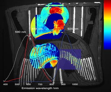
discrimination between the different painted areas, suggesting the presence of different organic pigments and binders. Specifically, the emission spectrum recorded on the red-pink painted
areas (point 1 in Fig. 2a), peaked at 615 nm, resembles the spectral features of a red lake pigment, likely madder-based [4, 20] (Fig. 3). The broad spectrum recorded in the white painted
area, with an emission peak at 590 nm, (Fig. 3) suggests the presence of a complex organic material, and is ascribed to emissions from binding media [1]. TRPL imaging of the stone mask,
following visible excitation, suggests the presence of traces of the Egyptian blue pigment for decorating the areas of hair, beard and irises of the eyes in Fig. 5. Blue used to paint the
irises in polychromy has also been reported by Verri [7]. The clear identification of Egyptian Blue has been achieved through PL spectroscopy on a point on the hair of the mask: an emission
spectrum comparable to that recorded on the commercial sample of Egyptian blue was recorded (data not shown). In terms of lifetime analysis, these painted areas show a microsecond decay
kinetic close to that found from details on the cartonnage painted with Egyptian blue: here, we have detected a mean lifetime of 121.3 µs (6.6 µs of interquartile range) and a negative skew
of −2.79 (Fig. 5). CONCLUSIONS The dual wavelength-excitation PL lifetime imaging approach reported here for the first time has been demonstrated to be valuable for probing the optical
emission of different materials, of organic and inorganic nature, for the study of Ancient Egyptian artefacts. The same approach could be extended to the analysis of modern pigments and
paintings, allowing an in-depth investigation of the emission properties of organic and inorganic artist materials, including Cd-based pigments, in a combined way [13, 21]. Whereas the PL
properties of the naturally occurring cuprorivaite mineral have been widely investigated in terms of both spectrum and decay kinetics [3, 6] and its brilliant emission has been exploited for
the rapid detection of the presence of the pigment in artworks from the Ancient Egypt [7], the near-infrared emission in Egyptian blue-painted ancient objects is not completely understood
[19]. The intense and long emission recorded with PL imaging which is on the order of 120 μs is nonetheless diagnostic for the presence of Egyptian blue. Differences in lifetime in objects
with respect to that reported and detected from that of the pure mineral and the synthetic commercial pigment are noted. The reasons behind lifetime differences are beyond the scope of this
work and require more refined analysis. Research in the future should address different degrees of crystallinity of the pigment in ancient objects following ancient synthesis processes,
grinding or sintering of the pigments and determine if chemical interactions between the pigment and the surrounding matrix in objects affects the emission lifetimes. Further analyses with
complementary methods sensitive to impurities which could account for differences in lifetime would be required to better refine these hypotheses. METHODS DESCRIPTION OF THE COLLECTION AND
OBJECTS The antiquities collection of the late Carla Maria Burri (1935–2009), Director of the Italian Cultural Institute in Egypt from 1993 and 1999, was donated to the Museo Civico di Crema
e del Cremasco in Winter 2010/2011 [22]. In 2013, Christian Orsenigo was charged with the study of the artefacts, under the supervision of Dr. Francesco Muscolino of the Soprintendenza
Archeologia della Lombardia. The collection consists of about eighty objects and covers a vast time span; ranging from the most ancient item—a faience tile once belonging to the decoration
of the walls of the galleries underneath Djoser’s Step Pyramid at Saqqara (ca. 2630–2611 B.C.E.)—excluding some flints actually under study—to Islamic glass dating to the 11th C. (Orsenigo
2016, forthcoming). The collection includes objects belonging to different typologies, such as funerary statuettes (shabtis) from the late New Kingdom, masks and parts of sarcophagi, bronzes
and amulets, other than Hellenistic and Roman terracotta figurines and lamps. In this work we examine a Cartonnage and a Mask. THE CARTONNAGE The polychrome cartonnage fragment probably
comes from the back terminal of a mask, depicting a human-headed ba-bird, crowned with solar disc and holding two maat-feathers at each wing. Unfortunately nothing is known about the
provenance of this object, as is the case for a very similar fragment kept at the Petrie Museum, UCL (accession number UC45900), that is a particularly good comparandum [23] The material is
linen covered with plaster and then painted. Cartonnages such as this can be dated from Ptolemaic to Roman Periods [24] THE MASK Even if preserved only in its upper front part, the object is
likely a miniature painted terracotta comic mask of an actor playing a slave character in the New Comedy. It shows a broad nose, brows flying up to the sides and wrinkled forehead. It can
be dated to the Ptolemaic Period. See references for comparisons with similar objects [25, 26]. REFERENCE SAMPLE A painted model sample of Egyptian blue pigment (Kremer pigmente, GmbH) was
prepared and analysed. The pigment, dispersed in Plextol, was applied as a painted layer on a glass substrate. TIME-RESOLVED PHOTOLUMINESCENCE IMAGING The TRPL imaging device is, described
in detail elsewhere, and is summarized below. A schematic diagrame of the setup can be found elsewhere [16]. The device is comprised of a ns laser excitation source combined with a
time-gated intensified camera (C9546-03, Hamamatsu Photonics, Hamamatsu City, Japan), capable of high speed gating to capture images of transient phenomena. A custom-built trigger unit and a
precision delay generator (DG535 Stanford Research System, Sunnyvale, CA, USA) complete the system, which has a net temporal jitter close to 0.5 ns. The Q-switching laser source (FTSS
355-50 Crylas GmbH, Berlin, Germany), based on the third harmonic of a diode-pumped Nd:YAG crystal (λ = 355 nm, Pulse energy = 70 μJ, Pulse duration = 1.0 ns, repetition frequency = 100 Hz),
has been modified in order to obtain the second harmonic of the same source emission (λ = 532 nm, Pulse energy = 60 μJ, Pulse duration = 1.0 ns). By using both wavelengths we probe the PL
emission of materials with different absorption spectra. The two emission lines of the laser source are collinear; hence it is easy to switch between them during measurements. In order to
spectrally clean laser emissions, proper bandpass filters (FL355-10 or FL532-10, Thorlabs GmbH Germany) are employed at the exit of the laser source. The laser beam, coupled to a silica
optical fibre, is magnified with suitable silica optics in order to illuminate a circular area of about 25 cm diameter on the surface of the object, with a typical fluence per pulse below
140 nJcm−2. This very low power density does not lead to detectable changes in the intensity of emission due to photooxidation in samples following typical measurements. The kinetics of the
emission is detected by the gated intensified camera, which is based on a GaAs photocathode with spectral sensitivity from 380 to 900 nm. The gate width of the camera is adjustable from 3 ns
to continuous mode, depending on the kinetic properties of the surface under investigation. In this work a gate width of 5 ns was employed to detect the nanosecond kinetics of the emission
from organic materials. Long-lived decay kinetics ascribed to emissions from areas painted in Egyptian blue have been effectively sampled by increasing the width to 100 µs. A proper optical
highpass filter w placed in front of the camera lens in order to remove excitation light: the B + W UV/IR Cut 486 M MRC filter (Schneider Optics), with high transmission from 380 to 720 nm,
was employed for lifetime analysis following 355 nm excitation, whereas the Kodak Wratten 23A filter transmitting light beyond 550 nm was employed for measurements at 532 nm excitation. As
has been shown in previous research, the short temporal jitter of the system is key to the estimation of ns lifetimes which may be used to differentiate organic binding media and pigments
[16]. TRPL imaging is achieved through the reconstruction of the effective lifetime map based on a simple mono-exponential decay model [14]. LASER-INDUCED PHOTOLUMINESCENCE SPECTROSCOPY A
compact spectrometer and the same dual-wavelength laser source employed in the TRPL imaging device were used for detecting emission spectra from selected points on objects. The compact
spectrometer (TM-CCD C10083CA-2100, Hamamatsu Photonics) mounts a back thinned CCD image sensor and a transmission-type grating, recording spectra between 320–1100 nm with a spectral
resolution of 6 nm. Through fibre optics both the laser and the spectrometer are remotely connected to an optical probe, working in the 45–0° configuration mode. Proper transmission
high-pass filters (FEL420 or FEL550, Thorlabs GmbH Germany) are mounted on the probe which, that allow excitation and collection of photons from a point on surface of approximately 1 mm
diameter at a distance of 35 mm. The probe is equipped with a proper transmission high-pass filter (FGL420 or FELH550, Thorlabs GmbH Germany) depending on the employed laser wavelength.
Spectra are reported following background subtraction (mainly related to the sensor read and dark noise) and correction for the spectral efficiency of the device. DIGITAL IMAGING As proposed
in past research [7, 8], a commercial Nikon D7100 digital camera (D60) was employed for recording UV-induced digital images of the PL emission of objects. Excitation was provided from a
Xenon-based flash equipped with an UV bandpass filter (DUG11, Schott AG), whereas a transmission filter blocking light in the UV and in the NIR spectral range (W UV/IR Cut 486 M MRC filter,
Schneider Optics) was mounted in front of a 50-mm focal camera lens [7, 8]. Similarly, infrared digital photography of the emission of Egyptian Blue painted objects was performed using the
commercial digital camera without the infrared blocking filter (supplied by Advanced Camera Services, UK) and mounting an). An infrared transmission filter (R72, HOYA) was placed in front of
the camera lens [8] for detection only of the infrared emission. Excitation of PL emission was achieved using a 15 W Watt green LED-based lamp with emission in the green (4000 lx @ 1 m)
(FLAT PAR CAN RGB 10 IR WH, Cameo (Germany)). REFERENCES * Nevin A, Spoto G, Anglos A. Laser spectroscopies for elemental and molecular analysis in art and archaeology. Appl Phys A.
2012;106:339–61. Article Google Scholar * Scott DA, Warlander S, Mazurek J, Quirke S. Examination of some pigments, grounds and media from Egyptian cartonnage fragments in the Petrie
Museum, University College London. J Archaeol Sci. 2009;36(3):923–32. Article Google Scholar * Pozza G, Ajò D, Chiari G, De Zuane F, Favaro M. Photoluminescence of the inorganic pigments
Egyptian blue, Han blue and Han purple. J Cult Herit. 2000;1(4):393–8. Article Google Scholar * Clementi C, Doherty B, Gentili PL, Miliani C, Romani A, Brunetti BG, Sgamellotti A.
Vibrational and electronic properties of painting lakes. Appl Phys A. 2008;92(1):25–33. Article Google Scholar * Claro A, Melo MJ, Schäfer S, de Melo JSS, Pina F, van den Berg KJ,
Burnstock A. The use of microspectrofluorimetry for the characterization of lake pigments. Talanta. 2008;74(4):922–9. Article Google Scholar * Accorsi G, Verri G, Bolognesi M, Armaroli N,
Clementi C, Miliani C, Romani A. The exceptional near-infrared luminescence properties of cuprorivaite (Egyptian blue). Chem Commun. 2009;23:3392–4. Article Google Scholar * Verri G. The
spatially resolved characterisation of Egyptian blue, Han blue and Han purple by photo-induced luminescence digital imaging. Anal Bioanal Chem. 2008;394(4):1011–21. Article Google Scholar
* Verri G, Saunders D. Xenon flash for Reflectance and luminescence (multispectral) imaing in cultural heritage applications. Br Mus Tech Bull. 2014;8:87–92. Google Scholar * Ganio M,
Salvant J, Williams J, Lee L, Cossairt O, Walton M. Investigating the use of Egyptian blue in Roman Egyptian portraits and panels from Tebtunis, Egypt. Appl Phys A. 2015;121(3):813–21.
Article Google Scholar * Spizzichino V, Angelini F, Caneve L, Colao F, Corrias R, Ruggiero L. In situ study of modern synthetic materials and pigments in contemporary paintings by
laser-induced fluorescence scanning. Stud Conserv. 2015;60(S1):78–84. Google Scholar * Raimondi V, Lognoli D, Palombi L. A fluorescence LIDAR combining spectral, lifetime and imaging
capabilities for the remote sensing of cultural heritage assets. Proc SPIE Int Soc Opt Eng. 2014;. doi:10.1117/12.2067388. Google Scholar * Daveri A, Vagnin M, Nucera F, Azzarelli M, Romani
A, Clementi C. Visible-induced luminescence imaging: a user-friendly method based on a system of interchangeable and tunable LED light sources. Microchem J. 2014;.
doi:10.1016/j.microc.2015.11.019 (IN PRESS). Google Scholar * Thoury M, Delaney JK, De La Rie ER, Palmer M, Morales K, Krueger J. Near-infrared luminescence of cadmium pigments: in situ
identification and mapping in paintings. Appl Spectrosc. 2011;65(8):939–51. Article Google Scholar * Comelli D, Valentini G, Nevin A, Farina A, Toniolo L, Cubeddu R. A portable
UV-fluorescence multispectral imaging system for the analysis of painted surfaces. Rev Sci Instrum. 2008;79(8):086112. Article Google Scholar * Nevin A, Cesaratto A, Bellei S, D’Andrea C,
Toniolo L, Valentini G, Comelli D. Time-resolved photoluminescence spectroscopy and imaging: new approaches to the analysis of cultural heritage and its degradation. Sensors.
2014;14(4):6338–55. Article Google Scholar * Comelli D, D’Andrea C, Valentini G, Cubeddu R, Colombo C, Toniolo L. Fluorescence lifetimeimaging and spectroscopy as tools for nondestructive
analysis of works of art. Appl Opt. 2004;43:2175–83. Article Google Scholar * Comelli D, Nevin A, Valentini G, Osticioli I, Castellucci EM, Toniolo L, Gulotta D, Cubeddu R. Insights into
Masolino’s wall paintings in Castiglione Olona: advanced reflectance and fluorescence imaging analysis. J Cult Herit. 2011;12(1):11–8. Article Google Scholar * Comelli D, Nevin A,
Brambilla A, Osticioli I, Valentini G, Toniolo L, Fratelli M, Cubeddu R. On the discovery of an unusual luminescent pigment in Van Gogh’s painting “Les Bretonnes et le pardon de Pont Aven”.
Appl Phys A. 2012;106(1):25–34. Article Google Scholar * Borozov SM, Würth C, Resch-Genger U, Klimant I. New life of ancient pigments: application in high-performance optical sensing
materials. Anal Chem. 2013;85:9371–7. Article Google Scholar * Grazia C, Clementi C, Miliani C, Romani A. Photophysical properties of alizarin and purpurin Al(III) complexes in solution
and in solid state. Photochem Photobiol Sci. 2011;10:1249–55. Article Google Scholar * Cesaratto A, D’Andrea C, Nevin A, Valentini G, Tassone F, Alberti R, Frizzi T, Comelli D. Analysis of
cadmium-based pigments with time-resolved photoluminescence. Anal Methods. 2014;6:130–8. Article Google Scholar * Gallo Carrabba D. Carla Maria Burri: l’Egitto mi ha aperto le sue
braccia. Crema: Gruppo Antropologico Cremasco; 2012. Google Scholar * Petrie museum. http://petriecat.museums.ucl.ac.uk/. Accessed 1 Dec 2015. * D’Auria S, Lacovara P, Roehrig CH. Mummies
and magic: the funerary arts of ancient Egypt. Boston: Museum of Fine Arts; 1988. Google Scholar * Webster TBL, Green JR, Seeberg A. Monuments illustrating new comedy. London: Institute of
Classical Studies, University of London School of Advanced Study; 1995. Google Scholar * British museum collection database. http://www.britishmuseum.org/research/collection_online.
Accessed 1 Dec 2015. Download references AUTHORS’ CONTRIBUTIONS Analysis was carried out by DC, AN, VC and SM. Historical context and information has been provided by CO. All authors read
and approved the final manuscript. ACKNOWLEDGEMENTS Research was partially funded through the Bilateral Project between Italy and Egypt coordinated by Daniela Comelli (Politecnico di Milano,
Italy) and Abdelrazek Elnaggar (Univeristy of Fayoum, Egypt) (Progetti di Grande Rilevanza, Protocollo Esecutivo–EGITTO, PGR 00101.) Authors thank Daniela Gallo Carrabba and Fiorenzo Gnesi
(Associazione Carla Maria Burri), Francesca Moruzzi and Simone Riboldi (Museo civico di Crema e del Cremasco), Francesco Muscolino (Soprintendenza Archeologia della Lombardia) and Patrizia
Piacentini (Università degli Studi di Milano, Chair of Egyptology). COMPETING INTERESTS The authors declare that they have no competing interests. AUTHOR INFORMATION Author notes AUTHORS AND
AFFILIATIONS * Diparitmento di Fisica, Politecnico di Milano, Piazza Leonardo da Vinci 32, 20133, Milan, Italy Daniela Comelli & Valentina Capogrosso * Istituto di Fotonica e
Nanotecnologie-Consiglio Nazionale delle Ricerche (IFN-CNR), Piazza Leonardo da Vinci 32, 20133, Milan, Italy Valentina Capogrosso & Austin Nevin * Dipartimento di Studi letterari,
filologici e linguistici, Biblioteca e Archivi di Egittologia, Università degli Studi di Milano, via Festa del Perdono 3, 20122, Milan, Italy Christian Orsenigo Authors * Daniela Comelli
View author publications You can also search for this author inPubMed Google Scholar * Valentina Capogrosso View author publications You can also search for this author inPubMed Google
Scholar * Christian Orsenigo View author publications You can also search for this author inPubMed Google Scholar * Austin Nevin View author publications You can also search for this author
inPubMed Google Scholar CORRESPONDING AUTHOR Correspondence to Austin Nevin. ADDITIONAL INFORMATION Daniela Comelli and Austin Nevin contributed equally to this work RIGHTS AND PERMISSIONS
OPEN ACCESS This article is distributed under the terms of the Creative Commons Attribution 4.0 International License (http://creativecommons.org/licenses/by/4.0/), which permits
unrestricted use, distribution, and reproduction in any medium, provided you give appropriate credit to the original author(s) and the source, provide a link to the Creative Commons license,
and indicate if changes were made. The Creative Commons Public Domain Dedication waiver (http://creativecommons.org/publicdomain/zero/1.0/) applies to the data made available in this
article, unless otherwise stated. Reprints and permissions ABOUT THIS ARTICLE CITE THIS ARTICLE Comelli, D., Capogrosso, V., Orsenigo, C. _et al._ Dual wavelength excitation for the
time-resolved photoluminescence imaging of painted ancient Egyptian objects. _Herit Sci_ 4, 21 (2016). https://doi.org/10.1186/s40494-016-0090-5 Download citation * Received: 14 December
2015 * Accepted: 30 May 2016 * Published: 15 August 2016 * DOI: https://doi.org/10.1186/s40494-016-0090-5 SHARE THIS ARTICLE Anyone you share the following link with will be able to read
this content: Get shareable link Sorry, a shareable link is not currently available for this article. Copy to clipboard Provided by the Springer Nature SharedIt content-sharing initiative
KEYWORDS * Photoluminescence imaging * Egyptian blue * Lifetime * Laser induced fluorescence * Pigments * Ancient Egyptian materials * Paintings