- Select a language for the TTS:
- UK English Female
- UK English Male
- US English Female
- US English Male
- Australian Female
- Australian Male
- Language selected: (auto detect) - EN
Play all audios:
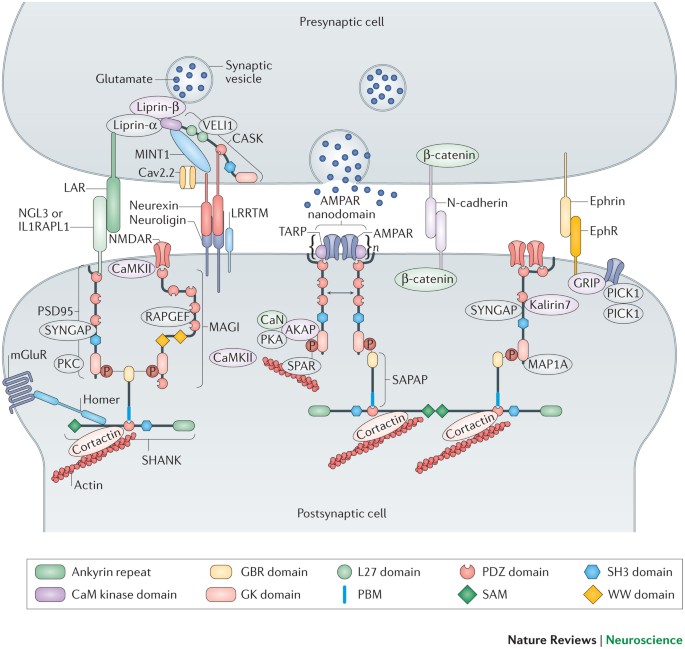
KEY POINTS * The postsynaptic density (PSD) is a large, dynamic protein assembly that orchestrates the densities and activities of both AMPA-type and NMDA-type glutamate receptors in
excitatory synapses. * The membrane-associated guanylate kinase (MAGUK) family of scaffold proteins, including Discs large homologues (DLGs) and membrane-associated guanylate kinase inverted
(MAGI) proteins, are key organizers of PSDs. They act as an interface between the upstream membrane-spanning glutamate receptors and cell adhesion proteins and the downstream
synapse-associated protein 90/postsynaptic density protein 95 (PSD95)-associated protein (SAPAP)–SRC homology 3 (SH3) and multiple ankyrin repeat domains protein (SHANK) complexes and the
cytoskeleton. * The guanylate kinase-like (GK) domain of MAGUKs binds to target proteins in a phosphorylation-dependent manner. This phosphorylation-dependent target recognition by MAGUKs
suggests that the assembly of PSD is activity-dependent. * Each MAGUK contains a highly conserved domain organization with PSD95–DLG1–Zonula occludens 1 (PDZ)–SH3–GK domains arranged in
tandem. The PDZ–SH3–GK tandem forms a supramodule allowing the MAGUK scaffolds to bind to target proteins with high specificity as well as to cluster transmembrane ion channels and
receptors. * Biochemical and structural studies of MAGUKs have offered insights into why mutations affecting genes encoding MAGUKs and their target proteins may alter synaptic protein
organization and lead to defects of synaptic development and signalling. ABSTRACT Membrane-associated guanylate kinases (MAGUKs) are a family of scaffold proteins that are highly enriched in
synapses and are responsible for organizing the numerous protein complexes required for synaptic development and plasticity. Mutations in genes encoding MAGUKs and their interacting
proteins can cause a broad spectrum of human psychiatric disorders. Here, we review MAGUK-mediated synaptic protein complex formation and regulation by focusing on findings from recent
biochemical and structural investigations. These mechanistic-based studies show that the formation of MAGUK-organized complexes is often directly regulated by protein phosphorylation,
suggesting a close connection between neuronal activity and the assembly of dynamic protein complexes in synapses. Access through your institution Buy or subscribe This is a preview of
subscription content, access via your institution ACCESS OPTIONS Access through your institution Subscribe to this journal Receive 12 print issues and online access $189.00 per year only
$15.75 per issue Learn more Buy this article * Purchase on SpringerLink * Instant access to full article PDF Buy now Prices may be subject to local taxes which are calculated during checkout
ADDITIONAL ACCESS OPTIONS: * Log in * Learn about institutional subscriptions * Read our FAQs * Contact customer support SIMILAR CONTENT BEING VIEWED BY OTHERS NEUREXINS: MOLECULAR CODES
FOR SHAPING NEURONAL SYNAPSES Article 08 January 2021 TAU FORMS SYNAPTIC NANO-BIOMOLECULAR CONDENSATES CONTROLLING THE DYNAMIC CLUSTERING OF RECYCLING SYNAPTIC VESICLES Article Open access
10 November 2023 ACTIVITY DEPENDENT DISSOCIATION OF THE HOMER1 INTERACTOME Article Open access 25 February 2022 ACCESSION CODES ACCESSIONS PROTEIN DATA BANK * 1NF3 * 2KA9 * 2LC7 * 2QKT *
3R0H * 3UAT * 4DC2 * 4WSI REFERENCES * Funke, L., Dakoji, S. & Bredt, D. S. Membrane-associated guanylate kinases regulate adhesion and plasticity at cell junctions. _Annu. Rev.
Biochem._ 74, 219–245 (2005). CAS PubMed Google Scholar * Oliva, C., Escobedo, P., Astorga, C., Molina, C. & Sierralta, J. Role of the MAGUK protein family in synapse formation and
function. _Dev. Neurobiol._ 72, 57–72 (2012). CAS PubMed Google Scholar * Zhu, J., Shang, Y., Chen, J. & Zhang, M. Structure and function of the guanylate kinase-like domain of the
MAGUK family scaffold proteins. _Front. Biol._ 7, 379–396 (2012). CAS Google Scholar * Feng, W. & Zhang, M. Organization and dynamics of PDZ-domain-related supramodules in the
postsynaptic density. _Nat. Rev. Neurosci._ 10, 87–99 (2009). CAS PubMed Google Scholar * Chen, K. & Featherstone, D. E. Pre and postsynaptic roles for _Drosophila_ CASK. _Mol. Cell.
Neurosci._ 48, 171–182 (2011). CAS PubMed Google Scholar * Danielson, E. et al. S-SCAM/MAGI-2 is an essential synaptic scaffolding molecule for the GluA2-containing maintenance pool of
AMPA receptors. _J. Neurosci._ 32, 6967–6980 (2012). CAS PubMed PubMed Central Google Scholar * Zheng, C. Y., Seabold, G. K., Horak, M. & Petralia, R. S. MAGUKs, synaptic
development, and synaptic plasticity. _Neuroscientist_ 17, 493–512 (2011). CAS PubMed PubMed Central Google Scholar * Grant, S. G. Synaptopathies: diseases of the synaptome. _Curr. Opin.
Neurobiol._ 22, 522–529 (2012). CAS PubMed Google Scholar * Palay, S. L. Synapses in the central nervous system. _J. Biophys. Biochem. Cytol._ 2, 193–202 (1956). CAS PubMed PubMed
Central Google Scholar * Gray, E. G. Axo-somatic and axo-dendritic synapses of the cerebral cortex: an electron microscope study. _J. Anat._ 93, 420–433 (1959). CAS PubMed PubMed Central
Google Scholar * Blomberg, F., Cohen, R. S. & Siekevitz, P. The structure of postsynaptic densities isolated from dog cerebral cortex. II. Characterization and arrangement of some of
the major proteins within the structure. _J. Cell Biol._ 74, 204–225 (1977). CAS PubMed PubMed Central Google Scholar * Carlin, R. K., Grab, D. J., Cohen, R. S. & Siekevitz, P.
Isolation and characterization of postsynaptic densities from various brain regions: enrichment of different types of postsynaptic densities. _J. Cell Biol._ 86, 831–845 (1980). CAS PubMed
Google Scholar * Cohen, R. S., Blomberg, F., Berzins, K. & Siekevitz, P. The structure of postsynaptic densities isolated from dog cerebral cortex. I. Overall morphology and protein
composition. _J. Cell Biol._ 74, 181–203 (1977). CAS PubMed PubMed Central Google Scholar * Harris, K. M. & Weinberg, R. J. Ultrastructure of synapses in the mammalian brain. _Cold
Spring Harb. Perspect. Biol._ 4, pii: a005587 (2012). Google Scholar * Chen, X. et al. Organization of the core structure of the postsynaptic density. _Proc. Natl Acad. Sci. USA_ 105,
4453–4458 (2008). CAS PubMed Google Scholar * Sheng, M. & Hoogenraad, C. C. The postsynaptic architecture of excitatory synapses: a more quantitative view. _Annu. Rev. Biochem._ 76,
823–847 (2007). CAS PubMed Google Scholar * Triller, A. & Choquet, D. New concepts in synaptic biology derived from single-molecule imaging. _Neuron_ 59, 359–374 (2008). CAS PubMed
Google Scholar * Dani, A., Huang, B., Bergan, J., Dulac, C. & Zhuang, X. Superresolution imaging of chemical synapses in the brain. _Neuron_ 68, 843–856 (2010).THIS PAPER REPORTS A
SYSTEMATIC THREE-COLOUR, 3D STOCHASTIC OPTICAL RECONSTRUCTION MICROSCOPY IMAGING TOOL THAT CAN DETERMINE THE SPATIAL ORGANIZATION OF SUB-SYNAPTIC COMPONENTS IN BOTH PRE- AND POST-SYNAPSES
WITH NANOMETRE PRECISION. CAS PubMed PubMed Central Google Scholar * O'Rourke, N. A., Weiler, N. C., Micheva, K. D. & Smith, S. J. Deep molecular diversity of mammalian
synapses: why it matters and how to measure it. _Nat. Rev. Neurosci._ 13, 365–379 (2012). CAS PubMed PubMed Central Google Scholar * MacGillavry, H. D. & Hoogenraad, C. C. The
internal architecture of dendritic spines revealed by super-resolution imaging: What did we learn so far? _Exp. Cell Res._ 335, 180–186 (2015). CAS PubMed Google Scholar * Sigrist, S. J.
& Sabatini, B. L. Optical super-resolution microscopy in neurobiology. _Curr. Opin. Neurobiol._ 22, 86–93 (2012). CAS PubMed Google Scholar * Maglione, M. & Sigrist, S. J. Seeing
the forest tree by tree: super-resolution light microscopy meets the neurosciences. _Nat. Neurosci._ 16, 790–797 (2013).REFERENCES 19–22 SUMMARIZE FINDINGS IN MOLECULAR ARCHITECTURE AND THE
DYNAMIC BEHAVIOUR OF SYNAPTIC COMPONENTS FROM RECENT SUPER-RESOLUTION IMAGING STUDIES. CAS PubMed Google Scholar * Nair, D. et al. Super-resolution imaging reveals that AMPA receptors
inside synapses are dynamically organized in nanodomains regulated by PSD95. _J. Neurosci._ 33, 13204–13224 (2013).THIS PAPER SHOWS THAT, IN RAT HIPPOCAMPAL NEURONS, AMPARS ARE OFTEN
CONCENTRATED INTO SEVERAL CLUSTERS (THE SO-CALLED AMPAR NANODOMAINS), EACH WITH A WIDTH OF ∼70 NM AND CONTAINING ∼20 RECEPTORS. MOREOVER, AMPAR NANODOMAINS ARE OFTEN CO-CLUSTERED WITH PSD95.
THE LEVEL OF PSD95 EXPRESSION CORRELATES WITH THE AMPAR NANODOMAIN SIZE AND NEURONAL ACTIVITY. CAS PubMed PubMed Central Google Scholar * Cheng, D. et al. Relative and absolute
quantification of postsynaptic density proteome isolated from rat forebrain and cerebellum. _Mol. Cell. Proteom._ 5, 1158–1170 (2006). CAS Google Scholar * Caroni, P., Donato, F. &
Muller, D. Structural plasticity upon learning: regulation and functions. _Nat. Rev. Neurosci._ 13, 478–490 (2012). CAS PubMed Google Scholar * Choquet, D. & Triller, A. The dynamic
synapse. _Neuron_ 80, 691–703 (2013). CAS PubMed Google Scholar * Bosch, M. et al. Structural and molecular remodeling of dendritic spine substructures during long-term potentiation.
_Neuron_ 82, 444–459 (2014). CAS PubMed PubMed Central Google Scholar * Nishiyama, J. & Yasuda, R. Biochemical computation for spine structural plasticity. _Neuron_ 87, 63–75 (2015).
CAS PubMed PubMed Central Google Scholar * Huganir, R. L. & Nicoll, R. A. AMPARs and synaptic plasticity: the last 25 years. _Neuron_ 80, 704–717 (2013). CAS PubMed PubMed Central
Google Scholar * Fukata, Y. et al. Local palmitoylation cycles define activity-regulated postsynaptic subdomains. _J. Cell Biol._ 202, 145–161 (2013). CAS PubMed PubMed Central Google
Scholar * MacGillavry, H. D., Song, Y., Raghavachari, S. & Blanpied, T. A. Nanoscale scaffolding domains within the postsynaptic density concentrate synaptic AMPA receptors. _Neuron_
78, 615–622 (2013).TOGETHER WITH REFERENCE 23, THIS WORK DEMONSTRATES THE CRITICAL ROLE OF PSD95 IN THE POSITIONING AND CLUSTERING OF AMPARS. CAS PubMed PubMed Central Google Scholar *
de Mendoza, A., Suga, H. & Ruiz-Trillo, I. Evolution of the MAGUK protein gene family in premetazoan lineages. _BMC Evol. Biol._ 10, 93 (2010). PubMed PubMed Central Google Scholar *
Woods, D. F., Hough, C., Peel, D., Callaini, G. & Bryant, P. J. Dlg protein is required for junction structure, cell polarity, and proliferation control in _Drosophila_ epithelia. _J.
Cell Biol._ 134, 1469–1482 (1996). CAS PubMed Google Scholar * Elias, G. M. & Nicoll, R. A. Synaptic trafficking of glutamate receptors by MAGUK scaffolding proteins. _Trends Cell
Biol._ 17, 343–352 (2007). CAS PubMed Google Scholar * Nithianantharajah, J. et al. Synaptic scaffold evolution generated components of vertebrate cognitive complexity. _Nat. Neurosci._
16, 16–24 (2013). CAS PubMed Google Scholar * Butz, S., Okamoto, M. & Sudhof, T. C. A tripartite protein complex with the potential to couple synaptic vesicle exocytosis to cell
adhesion in brain. _Cell_ 94, 773–782 (1998). CAS PubMed Google Scholar * Hsueh, Y. P. et al. Direct interaction of CASK/LIN-2 and syndecan heparan sulfate proteoglycan and their
overlapping distribution in neuronal synapses. _J. Cell Biol._ 142, 139–151 (1998). CAS PubMed PubMed Central Google Scholar * Nagashima, S., Kodaka, M., Iwasa, H. & Hata, Y.
MAGI2/S-SCAM outside brain. _J. Biochem._ 157, 177–184 (2015). CAS PubMed Google Scholar * Dudok, J. J., Sanz, A. S., Lundvig, D. M. & Wijnholds, J. MPP3 is required for maintenance
of the apical junctional complex, neuronal migration, and stratification in the developing cortex. _J. Neurosci._ 33, 8518–8527 (2013). CAS PubMed PubMed Central Google Scholar * Li, Y.
et al. Structure of Crumbs tail in complex with the PALS1 PDZ-SH3-GK tandem reveals a highly specific assembly mechanism for the apical Crumbs complex. _Proc. Natl Acad. Sci. USA_ 111,
17444–17449 (2014).THIS PAPER SHOWS THAT THE MPP5 PSG FORMS AN INTEGRAL STRUCTURAL UNIT IN BINDING TO CRB-CT. A STRUCTURAL ANALYSIS INDICATES THAT THE LIGAND-BINDING INDUCED DOMAIN-SWAPPED
HOMODIMER FORMATION OF MPP5 PSG MAY REPRESENT A GENERAL MODE FOR MAGUKS IN ORGANIZING LARGE PROTEIN COMPLEXES IN SYNAPSES. CAS PubMed Google Scholar * Najm, J. et al. Mutations of CASK
cause an X-linked brain malformation phenotype with microcephaly and hypoplasia of the brainstem and cerebellum. _Nat. Genet._ 40, 1065–1067 (2008). CAS PubMed Google Scholar * Zhang, M.
& Wang, W. Organization of signaling complexes by PDZ-domain scaffold proteins. _Acc. Chem. Res._ 36, 530–538 (2003). CAS PubMed Google Scholar * Hofmann, K., Bucher, P. &
Tschopp, J. The CARD domain: a new apoptotic signalling motif. _Trends Biochem. Sci._ 22, 155–156 (1997). CAS PubMed Google Scholar * Sudol, M., Chen, H. I., Bougeret, C., Einbond, A.
& Bork, P. Characterization of a novel protein-binding module—the WW domain. _FEBS Lett._ 369, 67–71 (1995). CAS PubMed Google Scholar * Wei, Z. et al. Liprin-mediated large signaling
complex organization revealed by the liprin-α/CASK and liprin-α/liprin-β complex structures. _Mol. Cell_ 43, 586–598 (2011). CAS PubMed Google Scholar * Mukherjee, K. et al. CASK
functions as a Mg2+-independent neurexin kinase. _Cell_ 133, 328–339 (2008). CAS PubMed PubMed Central Google Scholar * Xu, W. PSD-95-like membrane associated guanylate kinases
(PSD-MAGUKs) and synaptic plasticity. _Curr. Opin. Neurobiol._ 21, 306–312 (2011). CAS PubMed PubMed Central Google Scholar * Chen, X. et al. PSD-95 family MAGUKs are essential for
anchoring AMPA and NMDA receptor complexes at the postsynaptic density. _Proc. Natl Acad. Sci. USA_ 112, E6983–E6992 (2015). CAS PubMed Google Scholar * Kornau, H. C., Schenker, L. T.,
Kennedy, M. B. & Seeburg, P. H. Domain interaction between NMDA receptor subunits and the postsynaptic density protein PSD-95. _Science_ 269, 1737–1740 (1995). CAS PubMed Google
Scholar * Chetkovich, D. M., Chen, L., Stocker, T. J., Nicoll, R. A. & Bredt, D. S. Phosphorylation of the postsynaptic density-95 (PSD-95)/discs large/zona occludens-1 binding site of
stargazin regulates binding to PSD-95 and synaptic targeting of AMPA receptors. _J. Neurosci._ 22, 5791–5796 (2002). CAS PubMed PubMed Central Google Scholar * Schnell, E. et al. Direct
interactions between PSD-95 and stargazin control synaptic AMPA receptor number. _Proc. Natl Acad. Sci. USA_ 99, 13902–13907 (2002). CAS PubMed Google Scholar * Hafner, A. S. et al.
Lengthening of the Stargazin cytoplasmic tail increases synaptic transmission by promoting interaction to deeper domains of PSD-95. _Neuron_ 86, 475–489 (2015). CAS PubMed Google Scholar
* Bats, C., Groc, L. & Choquet, D. The interaction between Stargazin and PSD-95 regulates AMPA receptor surface trafficking. _Neuron_ 53, 719–734 (2007). CAS PubMed Google Scholar *
Levy, J. M., Chen, X., Reese, T. S. & Nicoll, R. A. Synaptic consolidation normalizes AMPAR quantal size following MAGUK loss. _Neuron_ 87, 534–548 (2015).THIS PAPER DEMONSTRATES THAT
PSD93, PSD95 AND SAP102 ARE ALL REQUIRED FOR SYNAPTIC LOCALIZATION OF AMPAR AND SYNAPTIC ACTIVITY. CAS PubMed PubMed Central Google Scholar * Benson, D. L. & Huntley, G. W. Synapse
adhesion: a dynamic equilibrium conferring stability and flexibility. _Curr. Opin. Neurobiol._ 22, 397–404 (2012). CAS PubMed Google Scholar * Thalhammer, A. & Cingolani, L. A. Cell
adhesion and homeostatic synaptic plasticity. _Neuropharmacology_ 78, 23–30 (2014). CAS PubMed Google Scholar * Gottmann, K. Transsynaptic modulation of the synaptic vesicle cycle by
cell-adhesion molecules. _J. Neurosci. Res._ 86, 223–232 (2008). CAS PubMed Google Scholar * Yamagata, M., Sanes, J. R. & Weiner, J. A. Synaptic adhesion molecules. _Curr. Opin. Cell
Biol._ 15, 621–632 (2003). CAS PubMed Google Scholar * Takahashi, H. & Craig, A. M. Protein tyrosine phosphatases PTPδ, PTPσ, and LAR: presynaptic hubs for synapse organization.
_Trends Neurosci._ 36, 522–534 (2013). CAS PubMed PubMed Central Google Scholar * Schreiner, D. et al. Targeted combinatorial alternative splicing generates brain region-specific
repertoires of neurexins. _Neuron_ 84, 386–398 (2014). CAS PubMed Google Scholar * Treutlein, B., Gokce, O., Quake, S. R. & Sudhof, T. C. Cartography of neurexin alternative splicing
mapped by single-molecule long-read mRNA sequencing. _Proc. Natl Acad. Sci. USA_ 111, E1291–1299 (2014). CAS PubMed Google Scholar * Arac, D. et al. Structures of neuroligin-1 and the
neuroligin-1/neurexin-1 β complex reveal specific protein–protein and protein–Ca2+ interactions. _Neuron_ 56, 992–1003 (2007). CAS PubMed Google Scholar * Iida, J., Hirabayashi, S., Sato,
Y. & Hata, Y. Synaptic scaffolding molecule is involved in the synaptic clustering of neuroligin. _Mol. Cell. Neurosci._ 27, 497–508 (2004). CAS PubMed Google Scholar * Irie, M. et
al. Binding of neuroligins to PSD-95. _Science_ 277, 1511–1515 (1997). CAS PubMed Google Scholar * Sudhof, T. C. The presynaptic active zone. _Neuron_ 75, 11–25 (2012). CAS PubMed
PubMed Central Google Scholar * Futai, K. et al. Retrograde modulation of presynaptic release probability through signaling mediated by PSD-95-neuroligin. _Nat. Neurosci._ 10, 186–195
(2007). CAS PubMed PubMed Central Google Scholar * Wittenmayer, N. et al. Postsynaptic Neuroligin1 regulates presynaptic maturation. _Proc. Natl Acad. Sci. USA_ 106, 13564–13569 (2009).
CAS PubMed Google Scholar * Hu, Z. et al. Neurexin and neuroligin mediate retrograde synaptic inhibition in _C. elegans_. _Science_ 337, 980–984 (2012). CAS PubMed PubMed Central
Google Scholar * Froyen, G. et al. Detection of genomic copy number changes in patients with idiopathic mental retardation by high-resolution X-array-CGH: important role for increased gene
dosage of XLMR genes. _Hum. Mutat._ 28, 1034–1042 (2007). CAS PubMed Google Scholar * Hayashi, S. et al. The CASK gene harbored in a deletion detected by array-CGH as a potential
candidate for a gene causative of X-linked dominant mental retardation. _Am. J. Med. Genet. A_ 146A, 2145–2151 (2008). CAS PubMed Google Scholar * Piluso, G. et al. A missense mutation in
CASK causes FG syndrome in an Italian family. _Am. J. Hum. Genet._ 84, 162–177 (2009). CAS PubMed PubMed Central Google Scholar * Tarpey, P. S. et al. A systematic, large-scale
resequencing screen of X-chromosome coding exons in mental retardation. _Nat. Genet._ 41, 535–543 (2009). CAS PubMed PubMed Central Google Scholar * Arking, D. E. et al. A common genetic
variant in the neurexin superfamily member CNTNAP2 increases familial risk of autism. _Am. J. Hum. Genet._ 82, 160–164 (2008). CAS PubMed PubMed Central Google Scholar * Sun, C. et al.
Identification and functional characterization of rare mutations of the neuroligin-2 gene (NLGN2) associated with schizophrenia. _Hum. Mol. Genet._ 20, 3042–3051 (2011). CAS PubMed PubMed
Central Google Scholar * Laumonnier, F. et al. X-linked mental retardation and autism are associated with a mutation in the _NLGN4_ gene, a member of the neuroligin family. _Am. J. Hum.
Genet._ 74, 552–557 (2004). CAS PubMed PubMed Central Google Scholar * Glessner, J. T. et al. Autism genome-wide copy number variation reveals ubiquitin and neuronal genes. _Nature_ 459,
569–573 (2009). CAS PubMed PubMed Central Google Scholar * Kim, J. H., Liao, D., Lau, L. F. & Huganir, R. L. SynGAP: a synaptic RasGAP that associates with the PSD-95/SAP90 protein
family. _Neuron_ 20, 683–691 (1998). CAS PubMed Google Scholar * Clement, J. P. et al. Pathogenic SYNGAP1 mutations impair cognitive development by disrupting maturation of dendritic
spine synapses. _Cell_ 151, 709–723 (2012). CAS PubMed PubMed Central Google Scholar * Berryer, M. H. et al. Mutations in SYNGAP1 cause intellectual disability, autism, and a specific
form of epilepsy by inducing haploinsufficiency. _Hum. Mutat._ 34, 385–394 (2013). CAS PubMed Google Scholar * Hamdan, F. F. et al. Mutations in SYNGAP1 in autosomal nonsyndromic mental
retardation. _N. Engl. J. Med._ 360, 599–605 (2009). CAS PubMed PubMed Central Google Scholar * Parker, M. J. et al. _De novo_, heterozygous, loss-of-function mutations in SYNGAP1 cause
a syndromic form of intellectual disability. _Am. J. Med. Genet. A_ 167A, 2231–2237 (2015). PubMed Google Scholar * Araki, Y., Zeng, M., Zhang, M. & Huganir, R. L. Rapid dispersion of
SynGAP from synaptic spines triggers AMPA receptor insertion and spine enlargement during LTP. _Neuron_ 85, 173–189 (2015). CAS PubMed PubMed Central Google Scholar * Penzes, P. et al.
The neuronal Rho-GEF Kalirin-7 interacts with PDZ domain-containing proteins and regulates dendritic morphogenesis. _Neuron_ 29, 229–242 (2001). CAS PubMed Google Scholar * Ma, X. M. et
al. Kalirin-7 is required for synaptic structure and function. _J. Neurosci._ 28, 12368–12382 (2008). CAS PubMed PubMed Central Google Scholar * Hotulainen, P. & Hoogenraad, C. C.
Actin in dendritic spines: connecting dynamics to function. _J. Cell Biol._ 189, 619–629 (2010). CAS PubMed PubMed Central Google Scholar * Frost, N. A., Kerr, J. M., Lu, H. E. &
Blanpied, T. A. A network of networks: cytoskeletal control of compartmentalized function within dendritic spines. _Curr. Opin. Neurobiol._ 20, 578–587 (2010). CAS PubMed PubMed Central
Google Scholar * Bosch, M. & Hayashi, Y. Structural plasticity of dendritic spines. _Curr. Opin. Neurobiol._ 22, 383–388 (2012). CAS PubMed Google Scholar * Urban, N. T., Willig, K.
I., Hell, S. W. & Nagerl, U. V. STED nanoscopy of actin dynamics in synapses deep inside living brain slices. _Biophys. J._ 101, 1277–1284 (2011). CAS PubMed PubMed Central Google
Scholar * Chazeau, A. et al. Nanoscale segregation of actin nucleation and elongation factors determines dendritic spine protrusion. _EMBO J._ 33, 2745–2764 (2014). CAS PubMed PubMed
Central Google Scholar * Kim, E. et al. GKAP, a novel synaptic protein that interacts with the guanylate kinase-like domain of the PSD-95/SAP90 family of channel clustering molecules. _J.
Cell Biol._ 136, 669–678 (1997). CAS PubMed PubMed Central Google Scholar * Zhu, J. et al. Guanylate kinase domains of the MAGUK family scaffold proteins as specific
phospho-protein-binding modules. _EMBO J._ 30, 4986–4997 (2011).BY DETERMINING THE STRUCTURE OF THE SAP97 SH3–GK IN COMPLEX WITH A PHOSPHO-LGN PEPTIDE, THIS STUDY, TOGETHER WITH THAT
REPORTED IN REFERENCE 127, REVEALED THAT THE MAGUK GK DOMAIN HAS EVOLVED FROM THE ANCIENT NUCLEOTIDE KINASE INTO A PHOSPHO-PROTEIN-BINDING DOMAIN. CAS PubMed PubMed Central Google Scholar
* Naisbitt, S. et al. Shank, a novel family of postsynaptic density proteins that binds to the NMDA receptor/PSD-95/GKAP complex and cortactin. _Neuron_ 23, 569–582 (1999). CAS PubMed
Google Scholar * Du, Y., Weed, S. A., Xiong, W. C., Marshall, T. D. & Parsons, J. T. Identification of a novel cortactin SH3 domain-binding protein and its localization to growth cones
of cultured neurons. _Mol. Cell. Biol._ 18, 5838–5851 (1998). CAS PubMed PubMed Central Google Scholar * Han, K. et al. SHANK3 overexpression causes manic-like behaviour with unique
pharmacogenetic properties. _Nature_ 503, 72–77 (2013). CAS PubMed PubMed Central Google Scholar * Niethammer, M. et al. CRIPT, a novel postsynaptic protein that binds to the third PDZ
domain of PSD-95/SAP90. _Neuron_ 20, 693–707 (1998). CAS PubMed Google Scholar * Brenman, J. E. et al. Localization of postsynaptic density-93 to dendritic microtubules and interaction
with microtubule-associated protein 1A. _J. Neurosci._ 18, 8805–8813 (1998). CAS PubMed Google Scholar * Peca, J. et al. Shank3 mutant mice display autistic-like behaviours and striatal
dysfunction. _Nature_ 472, 437–442 (2011). CAS PubMed PubMed Central Google Scholar * Schmeisser, M. J. et al. Autistic-like behaviours and hyperactivity in mice lacking ProSAP1/Shank2.
_Nature_ 486, 256–260 (2012). CAS PubMed Google Scholar * Won, H. et al. Autistic-like social behaviour in Shank2-mutant mice improved by restoring NMDA receptor function. _Nature_ 486,
261–265 (2012). CAS PubMed Google Scholar * Welch, J. M. et al. Cortico-striatal synaptic defects and OCD-like behaviours in Sapap3-mutant mice. _Nature_ 448, 894–900 (2007). CAS PubMed
PubMed Central Google Scholar * Ting, J. T., Peca, J. & Feng, G. Functional consequences of mutations in postsynaptic scaffolding proteins and relevance to psychiatric disorders.
_Annu. Rev. Neurosci._ 35, 49–71 (2012). CAS PubMed Google Scholar * Ye, F. & Zhang, M. Structures and target recognition modes of PDZ domains: recurring themes and emerging pictures.
_Biochem. J._ 455, 1–14 (2013).THIS REVIEW COMPREHENSIVELY SUMMARIZES THE MECHANISTIC BASES OF COMPLEX FORMATION BETWEEN THE EXTENDED PDZ DOMAIN AND THE EXTENDED PBM, AS WELL AS THE
REGULATED PDZ–TARGET INTERACTIONS. CAS PubMed Google Scholar * Hillier, B. J., Christopherson, K. S., Prehoda, K. E., Bredt, D. S. & Lim, W. A. Unexpected modes of PDZ domain
scaffolding revealed by structure of nNOS–syntrophin complex. _Science_ 284, 812–815 (1999). CAS PubMed Google Scholar * Wu, H. et al. PDZ domains of Par-3 as potential phosphoinositide
signaling integrators. _Mol. Cell_ 28, 886–898 (2007). CAS PubMed Google Scholar * Wang, C. K., Pan, L., Chen, J. & Zhang, M. Extensions of PDZ domains as important structural and
functional elements. _Protein Cell_ 1, 737–751 (2010). CAS PubMed PubMed Central Google Scholar * Wang, W., Weng, J., Zhang, X., Liu, M. & Zhang, M. Creating conformational entropy
by increasing interdomain mobility in ligand binding regulation: a revisit to N-terminal tandem PDZ domains of PSD-95. _J. Am. Chem. Soc._ 131, 787–796 (2009). CAS PubMed Google Scholar *
McCann, J. J., Zheng, L., Chiantia, S. & Bowen, M. E. Domain orientation in the N-terminal PDZ tandem from PSD-95 is maintained in the full-length protein. _Structure_ 19, 810–820
(2011). CAS PubMed PubMed Central Google Scholar * Eildal, J. N. et al. Rigidified clicked dimeric ligands for studying the dynamics of the PDZ1-2 supramodule of PSD-95. _ChemBioChem_
16, 64–69 (2015). CAS PubMed Google Scholar * Long, J. F. et al. Supramodular structure and synergistic target binding of the N-terminal tandem PDZ domains of PSD-95. _J. Mol. Biol._ 327,
203–214 (2003). CAS PubMed Google Scholar * Bach, A. et al. A high-affinity, dimeric inhibitor of PSD-95 bivalently interacts with PDZ1-2 and protects against ischemic brain damage.
_Proc. Natl Acad. Sci. USA_ 109, 3317–3322 (2012). CAS PubMed Google Scholar * Sainlos, M. et al. Biomimetic divalent ligands for the acute disruption of synaptic AMPAR stabilization.
_Nat. Chem. Biol._ 7, 81–91 (2011). CAS PubMed Google Scholar * Yau, K. W. & Hardie, R. C. Phototransduction motifs and variations. _Cell_ 139, 246–264 (2009). CAS PubMed PubMed
Central Google Scholar * Mishra, P. et al. Dynamic scaffolding in a G protein-coupled signaling system. _Cell_ 131, 80–92 (2007). CAS PubMed Google Scholar * Liu, W. et al. The INAD
scaffold is a dynamic, redox-regulated modulator of signaling in the _Drosophila_ eye. _Cell_ 145, 1088–1101 (2011). CAS PubMed Google Scholar * Petit, C. M., Zhang, J., Sapienza, P. J.,
Fuentes, E. J. & Lee, A. L. Hidden dynamic allostery in a PDZ domain. _Proc. Natl Acad. Sci. USA_ 106, 18249–18254 (2009). CAS PubMed Google Scholar * Bhattacharya, S. et al.
Ligand-induced dynamic changes in extended PDZ domains from NHERF1. _J. Mol. Biol._ 425, 2509–2528 (2013). CAS PubMed PubMed Central Google Scholar * Peterson, F. C., Penkert, R. R.,
Volkman, B. F. & Prehoda, K. E. Cdc42 regulates the Par-6 PDZ domain through an allosteric CRIB-PDZ transition. _Mol. Cell_ 13, 665–676 (2004). CAS PubMed Google Scholar * Garrard, S.
M. et al. Structure of Cdc42 in a complex with the GTPase-binding domain of the cell polarity protein, Par6. _EMBO J._ 22, 1125–1133 (2003). CAS PubMed PubMed Central Google Scholar *
McGee, A. W. et al. Structure of the SH3-guanylate kinase module from PSD-95 suggests a mechanism for regulated assembly of MAGUK scaffolding proteins. _Mol. Cell_ 8, 1291–1301 (2001). CAS
PubMed Google Scholar * Tavares, G. A., Panepucci, E. H. & Brunger, A. T. Structural characterization of the intramolecular interaction between the SH3 and guanylate kinase domains of
PSD-95. _Mol. Cell_ 8, 1313–1325 (2001). CAS PubMed Google Scholar * Deguchi, M. et al. BEGAIN (brain-enriched guanylate kinase-associated protein), a novel neuronal PSD-95/SAP90-binding
protein. _J. Biol. Chem._ 273, 26269–26272 (1998). CAS PubMed Google Scholar * Colledge, M. et al. Targeting of PKA to glutamate receptors through a MAGUK-AKAP complex. _Neuron_ 27,
107–119 (2000). CAS PubMed Google Scholar * Pak, D. T., Yang, S., Rudolph-Correia, S., Kim, E. & Sheng, M. Regulation of dendritic spine morphology by SPAR, a PSD-95-associated
RapGAP. _Neuron_ 31, 289–303 (2001). CAS PubMed Google Scholar * Siegrist, S. E. & Doe, C. Q. Microtubule-induced Pins/Galphai cortical polarity in _Drosophila_ neuroblasts. _Cell_
123, 1323–1335 (2005). CAS PubMed Google Scholar * Johnston, C. A., Hirono, K., Prehoda, K. E. & Doe, C. Q. Identification of an Aurora-A/PinsLINKER/Dlg spindle orientation pathway
using induced cell polarity in S2 cells. _Cell_ 138, 1150–1163 (2009). CAS PubMed PubMed Central Google Scholar * Hao, Y. et al. Par3 controls epithelial spindle orientation by
aPKC-mediated phosphorylation of apical Pins. _Curr. Biol._ 20, 1809–1818 (2010). CAS PubMed PubMed Central Google Scholar * Zhu, J. et al. Phosphorylation-dependent interaction between
tumor suppressors Dlg and Lgl. _Cell Res._ 24, 451–463 (2014). CAS PubMed PubMed Central Google Scholar * Betschinger, J., Mechtler, K. & Knoblich, J. A. The Par complex directs
asymmetric cell division by phosphorylating the cytoskeletal protein Lgl. _Nature_ 422, 326–330 (2003). CAS PubMed Google Scholar * Plant, P. J. et al. A polarity complex of mPar-6 and
atypical PKC binds, phosphorylates and regulates mammalian Lgl. _Nat. Cell Biol._ 5, 301–308 (2003). CAS PubMed Google Scholar * Bilder, D., Li, M. & Perrimon, N. Cooperative
regulation of cell polarity and growth by _Drosophila_ tumor suppressors. _Science_ 289, 113–116 (2000). CAS PubMed Google Scholar * Hirao, K. et al. A novel multiple PDZ
domain-containing molecule interacting with N-methyl-D-aspartate receptors and neuronal cell adhesion proteins. _J. Biol. Chem._ 273, 21105–21110 (1998). CAS PubMed Google Scholar *
Trinidad, J. C., Thalhammer, A., Specht, C. G., Schoepfer, R. & Burlingame, A. L. Phosphorylation state of postsynaptic density proteins. _J. Neurochem._ 92, 1306–1316 (2005). CAS
PubMed Google Scholar * Wang, C., Shang, Y., Yu, J. & Zhang, M. Substrate recognition mechanism of atypical protein kinase Cs revealed by the structure of PKCiota in complex with a
substrate peptide from Par-3. _Structure_ 20, 791–801 (2012). CAS PubMed Google Scholar * Daw, M. I. et al. PDZ proteins interacting with C-terminal GluR2/3 are involved in a
PKC-dependent regulation of AMPA receptors at hippocampal synapses. _Neuron_ 28, 873–886 (2000). CAS PubMed Google Scholar * Xia, J., Chung, H. J., Wihler, C., Huganir, R. L. &
Linden, D. J. Cerebellar long-term depression requires PKC-regulated interactions between GluR2/3 and PDZ domain-containing proteins. _Neuron_ 28, 499–510 (2000). CAS PubMed Google Scholar
* Fong, D. K., Rao, A., Crump, F. T. & Craig, A. M. Rapid synaptic remodeling by protein kinase C: reciprocal translocation of NMDA receptors and calcium/calmodulin-dependent kinase
II. _J. Neurosci._ 22, 2153–2164 (2002). CAS PubMed Google Scholar * Boehm, J. et al. Synaptic incorporation of AMPA receptors during LTP is controlled by a PKC phosphorylation site on
GluR1. _Neuron_ 51, 213–225 (2006). CAS PubMed Google Scholar * Lowenthal, M. S., Markey, S. P. & Dosemeci, A. Quantitative mass spectrometry measurements reveal stoichiometry of
principal postsynaptic density proteins. _J. Proteome Res._ 14, 2528–2538 (2015). CAS PubMed PubMed Central Google Scholar * te Velthuis, A. J., Admiraal, J. F. & Bagowski, C. P.
Molecular evolution of the MAGUK family in metazoan genomes. _BMC Evol. Biol._ 7, 129 (2007). PubMed PubMed Central Google Scholar * Pan, L., Chen, J., Yu, J., Yu, H. & Zhang, M. The
structure of the PDZ3-SH3-GuK tandem of ZO-1 protein suggests a supramodular organization of the membrane-associated guanylate kinase (MAGUK) family scaffold protein core. _J. Biol. Chem._
286, 40069–40074 (2011). CAS PubMed PubMed Central Google Scholar * Nomme, J. et al. The Src homology 3 domain is required for junctional adhesion molecule binding to the third PDZ
domain of the scaffolding protein ZO-1. _J. Biol. Chem._ 286, 43352–43360 (2011). CAS PubMed PubMed Central Google Scholar * Zhang, J., Lewis, S. M., Kuhlman, B. & Lee, A. L.
Supertertiary structure of the MAGUK core from PSD-95. _Structure_ 21, 402–413 (2013). CAS PubMed Google Scholar * McCann, J. J. et al. Supertertiary structure of the synaptic MAGuK
scaffold proteins is conserved. _Proc. Natl Acad. Sci. USA_ 109, 15775–15780 (2012). CAS PubMed Google Scholar * Hata, Y., Butz, S. & Sudhof, T. C. CASK: a novel dlg/PSD95 homolog
with an N-terminal calmodulin-dependent protein kinase domain identified by interaction with neurexins. _J. Neurosci._ 16, 2488–2494 (1996). CAS PubMed Google Scholar * Biederer, T. et
al. SynCAM, a synaptic adhesion molecule that drives synapse assembly. _Science_ 297, 1525–1531 (2002). CAS PubMed Google Scholar * Hara, H. et al. Clustering of CARMA1 through SH3-GUK
domain interactions is required for its activation of NF-κB signalling. _Nat. Commun._ 6, 5555 (2015). CAS PubMed Google Scholar * Moog, U. et al. Phenotypic and molecular insights into
CASK-related disorders in males. _Orphanet J. Rare Dis._ 10, 44 (2015). PubMed PubMed Central Google Scholar * Elias, G. M. et al. Synapse-specific and developmentally regulated targeting
of AMPA receptors by a family of MAGUK scaffolding proteins. _Neuron_ 52, 307–320 (2006). CAS PubMed Google Scholar * Schluter, O. M., Xu, W. & Malenka, R. C. Alternative N-terminal
domains of PSD-95 and SAP97 govern activity-dependent regulation of synaptic AMPA receptor function. _Neuron_ 51, 99–111 (2006). CAS PubMed Google Scholar Download references
ACKNOWLEDGEMENTS Research in the laboratory of M.Z. has been supported by grants from the Research Grants Council of Hong Kong (663811, 663812 and AoE-M09-12) and a 973 Program grant from
the Minister of Science and Technology of China (2014CB910204). J.Z. is supported by a grant from the National Natural Science Foundation of China (31470733). M.Z. is a Kerry Holdings
Professor in Science and a Senior Fellow of the Institute for Advanced Study (IAS) at the Hong Kong University of Science and Technology (HKUST). AUTHOR INFORMATION AUTHORS AND AFFILIATIONS
* Division of Life Science, State Key Laboratory of Molecular Neuroscience, Hong Kong University of Science and Technology, Clear Water Bay, Kowloon, Hong Kong, China Jinwei Zhu, Yuan Shang
& Mingjie Zhang * Center of Systems Biology and Human Health, School of Science and Institute for Advanced Study, Hong Kong University of Science and Technology, Clear Water Bay,
Kowloon, Hong Kong, China Mingjie Zhang * National Center for Protein Science Shanghai, Institute of Biochemistry and Cell Biology, Shanghai Institutes of Biological Sciences, Chinese
Academy of Sciences, 333 Haike Road, Pudong District, Shanghai, 201210, China Jinwei Zhu Authors * Jinwei Zhu View author publications You can also search for this author inPubMed Google
Scholar * Yuan Shang View author publications You can also search for this author inPubMed Google Scholar * Mingjie Zhang View author publications You can also search for this author
inPubMed Google Scholar CORRESPONDING AUTHOR Correspondence to Mingjie Zhang. ETHICS DECLARATIONS COMPETING INTERESTS The authors declare no competing financial interests. RELATED LINKS
DATABASES Protein Data Bank POWERPOINT SLIDES POWERPOINT SLIDE FOR FIG. 1 POWERPOINT SLIDE FOR FIG. 2 POWERPOINT SLIDE FOR FIG. 3 POWERPOINT SLIDE FOR FIG. 4 POWERPOINT SLIDE FOR FIG. 5
POWERPOINT SLIDE FOR FIG. 6 SUPPLEMENTARY INFORMATION SUPPLEMENTARY INFORMATION S1 (FIGURE) A | The structure of PKC-ι in complex with a substrate peptide derived from PAR3 (PDB code: 4DC2).
(PDF 532 kb) SUPPLEMENTARY INFORMATION S2 (FIGURE) A | Ribbon diagram of the SAP97 SH3–GK tandem (PDB code: 3UAT). (PDF 532 kb) GLOSSARY * Dendritic remodelling A biochemical process of
dendritic spines in which the actin cytoskeleton undergoes a rapid change in shape and structure in response to various stimuli. * Multivalency effect Binding avidity enhancement brought by
synergistic actions of individual interaction sites between multi-domain scaffold protein and multivalent target interactions. * Allosteric regulation Binding of an effector molecule to a
specific site of a protein causes conformational changes far away from the binding site and thus alters the function of the protein. * Phosphoprotein-binding modules Protein domains such as
the SH2 domain, the GK domain and the FHA domain that can specifically recognize phosphorylated proteins. * Supramodules Two or more protein modules arranged in tandem in a protein that
interact with each other to form a high-order structure with functions distinct from those of the individual or simple sum of the modules. * Domain-swapped dimer Two identical protein
molecules associate to form dimer by exchanging identical structural elements, such that native intramolecular interactions are replaced by their intermolecular counterparts. RIGHTS AND
PERMISSIONS Reprints and permissions ABOUT THIS ARTICLE CITE THIS ARTICLE Zhu, J., Shang, Y. & Zhang, M. Mechanistic basis of MAGUK-organized complexes in synaptic development and
signalling. _Nat Rev Neurosci_ 17, 209–223 (2016). https://doi.org/10.1038/nrn.2016.18 Download citation * Published: 18 March 2016 * Issue Date: April 2016 * DOI:
https://doi.org/10.1038/nrn.2016.18 SHARE THIS ARTICLE Anyone you share the following link with will be able to read this content: Get shareable link Sorry, a shareable link is not currently
available for this article. Copy to clipboard Provided by the Springer Nature SharedIt content-sharing initiative