- Select a language for the TTS:
- UK English Female
- UK English Male
- US English Female
- US English Male
- Australian Female
- Australian Male
- Language selected: (auto detect) - EN
Play all audios:
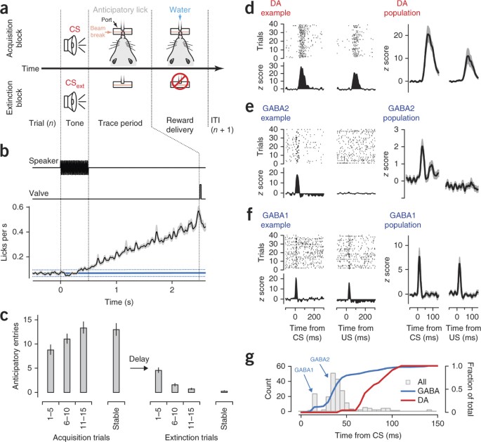
ABSTRACT Midbrain dopaminergic (DA) neurons are thought to guide learning via phasic elevations of firing in response to reward predicting stimuli. The mechanism for these signals remains
unclear. Using extracellular recording during associative learning, we found that inhibitory neurons in the ventral midbrain of mice responded to salient auditory stimuli with a burst of
activity that occurred before the onset of the phasic response of DA neurons. This population of inhibitory neurons exhibited enhanced responses during extinction and was anticorrelated with
the phasic response of simultaneously recorded DA neurons. Optogenetic stimulation revealed that this population was, in part, derived from inhibitory projection neurons of the substantia
nigra that provide a robust monosynaptic inhibition of DA neurons. Thus, our results elaborate on the dynamic upstream circuits that shape the phasic activity of DA neurons and suggest that
the inhibitory microcircuit of the midbrain is critical for new learning in extinction. Access through your institution Buy or subscribe This is a preview of subscription content, access via
your institution ACCESS OPTIONS Access through your institution Subscribe to this journal Receive 12 print issues and online access $209.00 per year only $17.42 per issue Learn more Buy
this article * Purchase on SpringerLink * Instant access to full article PDF Buy now Prices may be subject to local taxes which are calculated during checkout ADDITIONAL ACCESS OPTIONS: *
Log in * Learn about institutional subscriptions * Read our FAQs * Contact customer support SIMILAR CONTENT BEING VIEWED BY OTHERS VENTRAL TEGMENTAL AREA DOPAMINE PROJECTIONS TO THE
HIPPOCAMPUS TRIGGER LONG-TERM POTENTIATION AND CONTEXTUAL LEARNING Article Open access 21 May 2024 DOPAMINE FACILITATES ASSOCIATIVE MEMORY ENCODING IN THE ENTORHINAL CORTEX Article 22
September 2021 NIGROSTRIATAL DOPAMINE MODULATES THE STRIATAL-AMYGDALA PATHWAY IN AUDITORY FEAR CONDITIONING Article Open access 09 November 2023 REFERENCES * Schultz, W. Dopamine neurons and
their role in reward mechanisms. _Curr. Opin. Neurobiol._ 7, 191–197 (1997). Article CAS Google Scholar * Schultz, W., Dayan, P. & Montague, P.R. A neural substrate of prediction and
reward. _Science_ 275, 1593–1599 (1997). Article CAS Google Scholar * Schultz, W. Predictive reward signal of dopamine neurons. _J. Neurophysiol._ 80, 1–27 (1998). Article CAS Google
Scholar * Mirenowicz, J. & Schultz, W. Preferential activation of midbrain dopamine neurons by appetitive rather than aversive stimuli. _Nature_ 379, 449–451 (1996). Article CAS
Google Scholar * Waelti, P., Dickinson, A. & Schultz, W. Dopamine responses comply with basic assumptions of formal learning theory. _Nature_ 412, 43–48 (2001). Article CAS Google
Scholar * Roesch, M.R., Calu, D.J. & Schoenbaum, G. Dopamine neurons encode the better option in rats deciding between differently delayed or sized rewards. _Nat. Neurosci._ 10,
1615–1624 (2007). Article CAS Google Scholar * Matsumoto, M. & Hikosaka, O. Lateral habenula as a source of negative reward signals in dopamine neurons. _Nature_ 447, 1111–1115
(2007). Article CAS Google Scholar * Tobler, P.N., Dickinson, A. & Schultz, W. Coding of predicted reward omission by dopamine neurons in a conditioned inhibition paradigm. _J.
Neurosci._ 23, 10402–10410 (2003). Article CAS Google Scholar * Pan, W.-X., Schmidt, R., Wickens, J.R. & Hyland, B.I. Tripartite mechanism of extinction suggested by dopamine neuron
activity and temporal difference model. _J. Neurosci._ 28, 9619–9631 (2008). Article CAS Google Scholar * Pan, W.-X., Schmidt, R., Wickens, J.R. & Hyland, B.I. Dopamine cells respond
to predicted events during classical conditioning: evidence for eligibility traces in the reward-learning network. _J. Neurosci._ 25, 6235–6242 (2005). Article CAS Google Scholar *
Schultz, W. The phasic reward signal of primate dopamine neurons. _Adv. Pharmacol._ 42, 686–690 (1998). Article CAS Google Scholar * Stuber, G.D. et al. Reward-predictive cues enhance
excitatory synaptic strength onto midbrain dopamine neurons. _Science_ 321, 1690–1692 (2008). Article CAS Google Scholar * Matsumoto, M. & Hikosaka, O. Two types of dopamine neuron
distinctly convey positive and negative motivational signals. _Nature_ 459, 837–841 (2009). Article CAS Google Scholar * Hong, S., Jhou, T.C., Smith, M., Saleem, K.S. & Hikosaka, O.
Negative reward signals from the lateral habenula to dopamine neurons are mediated by rostromedial tegmental nucleus in primates. _J. Neurosci._ 31, 11457–11471 (2011). Article CAS Google
Scholar * Grace, A.A., Floresco, S.B., Goto, Y. & Lodge, D.J. Regulation of firing of dopaminergic neurons and control of goal-directed behaviors. _Trends Neurosci._ 30, 220–227 (2007).
Article CAS Google Scholar * Brazhnik, E., Shah, F. & Tepper, J.M. GABAergic afferents activate both GABAA and GABAB receptors in mouse substantia nigra dopaminergic neurons in vivo.
_J. Neurosci._ 28, 10386–10398 (2008). Article CAS Google Scholar * Tepper, J.M. & Lee, C.R. GABAergic control of substantia nigra dopaminergic neurons. _Prog. Brain Res._ 160,
189–208 (2007). Article CAS Google Scholar * Tepper, J.M., Martin, L.P. & Anderson, D.R. GABAA receptor–mediated inhibition of rat substantia nigra dopaminergic neurons by pars
reticulata projection neurons. _J. Neurosci._ 15, 3092–3103 (1995). Article CAS Google Scholar * Pan, W.-X. & Hyland, B.I. Pedunculopontine tegmental nucleus controls conditioned
responses of midbrain dopamine neurons in behaving rats. _J. Neurosci._ 25, 4725–4732 (2005). Article CAS Google Scholar * Redgrave, P. & Gurney, K. The short-latency dopamine signal:
a role in discovering novel actions? _Nat. Rev. Neurosci._ 7, 967–975 (2006). Article CAS Google Scholar * Bromberg-Martin, E.S., Matsumoto, M. & Hikosaka, O. Distinct tonic and
phasic anticipatory activity in lateral habenula and dopamine neurons. _Neuron_ 67, 144–155 (2010). Article CAS Google Scholar * Deniau, J.M., Mailly, P., Maurice, N. & Charpier, S.
The pars reticulata of the substantia nigra: a window to basal ganglia output. _Prog. Brain Res._ 160, 151–172 (2007). Article CAS Google Scholar * Fan, D., Rossi, M.A. & Yin, H.H.
Mechanisms of action selection and timing in substantia nigra neurons. _J. Neurosci._ 32, 5534–5548 (2012). Article CAS Google Scholar * Cohen, J.Y., Haesler, S., Vong, L., Lowell, B.B.
& Uchida, N. Neuron type–specific signals for reward and punishment in the ventral tegmental area. _Nature_ 482, 85–88 (2012). Article CAS Google Scholar * Gulley, J.M., Kosobud,
A.E.K. & Rebec, G.V. Behavior-related modulation of substantia nigra pars reticulata neurons in rats performing a conditioned reinforcement task. _Neuroscience_ 111, 337–349 (2002).
Article CAS Google Scholar * Bryden, D.W., Johnson, E.E., Diao, X. & Roesch, M.R. Impact of expected value on neural activity in rat substantia nigra pars reticulata. _Eur. J.
Neurosci._ 33, 2308–2317 (2011). Article Google Scholar * Parker, J.G. et al. Absence of NMDA receptors in dopamine neurons attenuates dopamine release, but not conditioned approach,
during Pavlovian conditioning. _Proc. Natl. Acad. Sci. USA_ 107, 13491–13496 (2010). Article CAS Google Scholar * Schultz, W., Apicella, P. & Ljungberg, T. Responses of monkey
dopamine neurons to reward and conditioned stimuli during successive steps of learning a delayed response task. _J. Neurosci._ 13, 900–913 (1993). Article CAS Google Scholar *
Nair-Roberts, R.G. et al. Stereological estimates of dopaminergic, GABAergic and glutamatergic neurons in the ventral tegmental area, substantia nigra and retrorubral field in the rat.
_Neuroscience_ 152, 1024–1031 (2008). Article CAS Google Scholar * Jin, X. & Costa, R.M. Start/stop signals emerge in nigrostriatal circuits during sequence learning. _Nature_ 466,
457–462 (2010). Article CAS Google Scholar * Bayer, H.M., Lau, B. & Glimcher, P.W. Statistics of midbrain dopamine neuron spike trains in the awake primate. _J. Neurophysiol._ 98,
1428–1439 (2007). Article Google Scholar * Paxinos, G. _The Rat Nervous System_, 3rd edn. (Elsevier, 2004). * Kolomiets, B.P. et al. Segregation and convergence of information flow through
the cortico-subthalamic pathways. _J. Neurosci._ 21, 5764–5772 (2001). Article CAS Google Scholar * Winer, J. & Schreiner, C. _The Inferior Colliculus_ (Springer, 2005). * Coizet, V.
et al. Short-latency visual input to the subthalamic nucleus is provided by the midbrain superior colliculus. _J. Neurosci._ 29, 5701–5709 (2009). Article CAS Google Scholar * Bevan,
M.D. & Bolam, J.P. Cholinergic, GABAergic and glutamate-enriched inputs from the mesopontine tegmentum to the subthalamic nucleus in the rat. _J. Neurosci._ 15, 7105–7120 (1995). Article
CAS Google Scholar * Condé, H., Dormont, J.F. & Farin, D. The role of the pedunculopontine tegmental nucleus in relation to conditioned motor performance in the cat. II. Effects of
reversible inactivation by intracerebral microinjections. _Exp. Brain Res._ 121, 411–418 (1998). Article Google Scholar * Yu, L., Stein, B.E. & Rowland, B.A. Adult plasticity in
multisensory neurons: short-term experience-dependent changes in the superior colliculus. _J. Neurosci._ 29, 15910–15922 (2009). Article CAS Google Scholar * Schultz, W. Behavioral
dopamine signals. _Trends Neurosci._ 30, 203–210 (2007). Article CAS Google Scholar * Bromberg-Martin, E.S., Matsumoto, M. & Hikosaka, O. Dopamine in motivational control: rewarding,
aversive and alerting. _Neuron_ 68, 815–834 (2010). Article CAS Google Scholar * Balcita-Pedicino, J.J., Omelchenko, N., Bell, R. & Sesack, S.R. The inhibitory influence of the
lateral habenula on midbrain dopamine cells: ultrastructural evidence for indirect mediation via the rostromedial mesopontine tegmental nucleus. _J. Comp. Neurol._ 519, 1143–1164 (2011).
Article CAS Google Scholar * Cebrián, C., Parent, A. & Prensa, L. Patterns of axonal branching of neurons of the substantia nigra pars reticulata and pars lateralis in the rat. _J.
Comp. Neurol._ 492, 349–369 (2005). Article Google Scholar * Dommett, E. et al. How visual stimuli activate dopaminergic neurons at short latency. _Science_ 307, 1476–1479 (2005). Article
CAS Google Scholar * Inglis, W.L. & Winn, P. The pedunculopontine tegmental nucleus: where the striatum meets the reticular formation. _Prog. Neurobiol._ 47, 1–29 (1995). Article
CAS Google Scholar * Shen, W., Flajolet, M., Greengard, P. & Surmeier, D.J. Dichotomous dopaminergic control of striatal synaptic plasticity. _Science_ 321, 848–851 (2008). Article
CAS Google Scholar * Pavlov, I. _Conditioned Reflexes: an Investigation of the Physiological Activity of the Cerebral Cortex_ (Oxford University Press, 1927). * Atasoy, D., Aponte, Y., Su,
H.H. & Sternson, S.M.A. FLEX switch targets Channelrhodopsin-2 to multiple cell types for imaging and long-range circuit mapping. _J. Neurosci._ 28, 7025–7030 (2008). Article CAS
Google Scholar * Gutkin, B.S. & Ermentrout, G.B. Dynamics of membrane excitability determine interspike interval variability: a link between spike generation mechanisms and cortical
spike train statistics. _Neural Comput._ 10, 1047–1065 (1998). Article CAS Google Scholar Download references ACKNOWLEDGEMENTS J. Paton, W. Denk, A. Karpova, A. Lee, J. Magee and G.
Murphy provided critical feedback at various stages of preparation of the manuscript and progression of the project. We are indebted to the extensive feedback from our colleagues following
presentation of this work at internal seminars on the Janelia Farm Research Campus. We thank members of our laboratory for critical reading and feedback on the manuscript. J.B. receives
funding from the Cambridge-Janelia Farm Graduate Program. This work was supported by funding from the Howard Hughes Medical Institute. AUTHOR INFORMATION AUTHORS AND AFFILIATIONS * Howard
Hughes Medical Institute, Janelia Farm Research Campus, Ashburn, Virginia, USA Wei-Xing Pan, Jennifer Brown & Joshua Tate Dudman * Department of Physiology, Development and Neuroscience,
University of Cambridge, Cambridge, UK Jennifer Brown Authors * Wei-Xing Pan View author publications You can also search for this author inPubMed Google Scholar * Jennifer Brown View
author publications You can also search for this author inPubMed Google Scholar * Joshua Tate Dudman View author publications You can also search for this author inPubMed Google Scholar
CONTRIBUTIONS W.-X.P. and J.T.D. designed the project. J.T.D., W.-X.P. and J.B. analyzed the data and wrote the manuscript. W.-X.P. performed the _in vivo_ recording and behavioral
experiments. J.B. performed the _in vitro_ experiments. J.T.D. implemented the computational model and performed a minority of the experiments. CORRESPONDING AUTHOR Correspondence to Joshua
Tate Dudman. ETHICS DECLARATIONS COMPETING INTERESTS The authors declare no competing financial interests. SUPPLEMENTARY INFORMATION SUPPLEMENTARY TEXT AND FIGURES Supplementary Figures 1–10
(PDF 5616 kb) RIGHTS AND PERMISSIONS Reprints and permissions ABOUT THIS ARTICLE CITE THIS ARTICLE Pan, WX., Brown, J. & Dudman, J. Neural signals of extinction in the inhibitory
microcircuit of the ventral midbrain. _Nat Neurosci_ 16, 71–78 (2013). https://doi.org/10.1038/nn.3283 Download citation * Received: 30 July 2012 * Accepted: 16 November 2012 * Published: 09
December 2012 * Issue Date: January 2013 * DOI: https://doi.org/10.1038/nn.3283 SHARE THIS ARTICLE Anyone you share the following link with will be able to read this content: Get shareable
link Sorry, a shareable link is not currently available for this article. Copy to clipboard Provided by the Springer Nature SharedIt content-sharing initiative


![[withdrawn] esfa update: 20 may 2020](https://www.gov.uk/assets/static/govuk-opengraph-image-03837e1cec82f217cf32514635a13c879b8c400ae3b1c207c5744411658c7635.png)