Ultramicroscopy: three-dimensional visualization of neuronal networks in the whole mouse brain

feature-image

Play all audios:

Loading...

ABSTRACT 
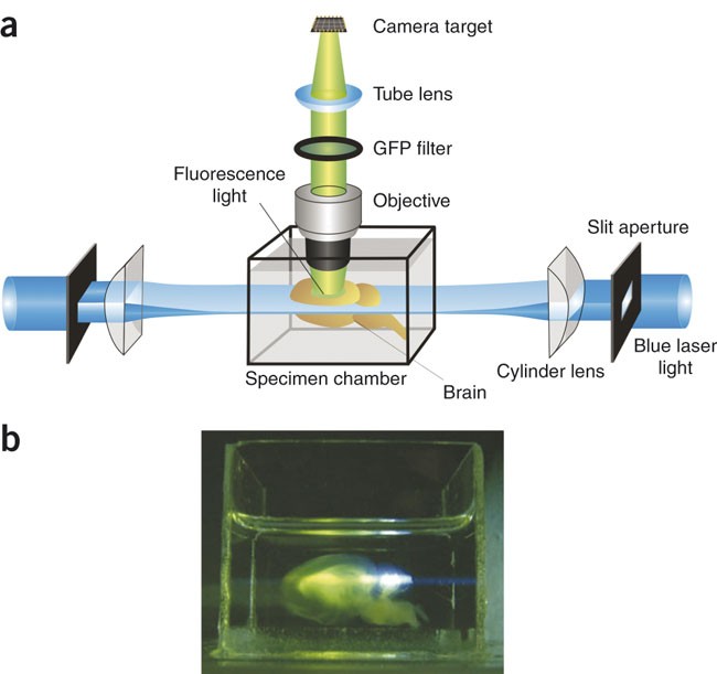
Visualizing entire neuronal networks for analysis in the intact brain has been impossible up to now. Techniques like computer tomography or magnetic resonance imaging (MRI) do not


yield cellular resolution, and mechanical slicing procedures are insufficient to achieve high-resolution reconstructions in three dimensions. Here we present an approach that allows imaging


of whole fixed mouse brains. We modified 'ultramicroscopy' by combining it with a special procedure to clear tissue. We show that this new technique allows optical sectioning of


fixed mouse brains with cellular resolution and can be used to detect single GFP-labeled neurons in excised mouse hippocampi. We obtained three-dimensional (3D) images of dendritic trees and


spines of populations of CA1 neurons in isolated hippocampi. Also in fruit flies and in mouse embryos, we were able to visualize details of the anatomy by imaging autofluorescence. Our


method is ideally suited for high-throughput phenotype screening of transgenic mice and thus will benefit the investigation of disease models. Access through your institution Buy or


subscribe This is a preview of subscription content, access via your institution ACCESS OPTIONS Access through your institution Subscribe to this journal Receive 12 print issues and online


access $259.00 per year only $21.58 per issue Learn more Buy this article * Purchase on SpringerLink * Instant access to full article PDF Buy now Prices may be subject to local taxes which


are calculated during checkout ADDITIONAL ACCESS OPTIONS: * Log in * Learn about institutional subscriptions * Read our FAQs * Contact customer support SIMILAR CONTENT BEING VIEWED BY OTHERS


MULTIPLEXED VOLUMETRIC CLEM ENABLED BY SCFVS PROVIDES INSIGHTS INTO THE CYTOLOGY OF CEREBELLAR CORTEX Article Open access 05 August 2024 FUNCTIONAL AND MULTISCALE 3D STRUCTURAL


INVESTIGATION OF BRAIN TISSUE THROUGH CORRELATIVE IN VIVO PHYSIOLOGY, SYNCHROTRON MICROTOMOGRAPHY AND VOLUME ELECTRON MICROSCOPY Article Open access 25 May 2022 HIGH-THROUGHPUT MAPPING OF A


WHOLE RHESUS MONKEY BRAIN AT MICROMETER RESOLUTION Article 26 July 2021 REFERENCES * Tyszka, J.M., Fraser, S.E. & Jacobs, R.E. Magnetic resonance microscopy: recent advantages and


applications. _Curr. Opin. Biotechnol._ 16, 93–99 (2005). Article  CAS  Google Scholar  * Kalender, W.A. CT: the unexpected evolution of an imaging modality. _Eur. Radiol._ 15 (Suppl. 4),


d21–d24 (2005). Article  Google Scholar  * Johnson, G.A., Cofer, G.P., Gewalt, S.L. & Hedlund, L.W. Morphological phenotyping with MR microscopy: The visible mouse. _Radiology_ 222,


789–793 (2002). Article  Google Scholar  * Helmchen, F. & Denk, W. Deep tissue two-photon microscopy. _Nat. Methods_ 2, 932–940 (2005). Article  CAS  Google Scholar  * Weninger, W.J.


& Mohun, T. Phenotyping transgenic embryos: a rapid 3-D screening method based on episcopic fluorescence image capturing. _Nat. Genet._ 30, 59–65 (2002). Article  CAS  Google Scholar  *


Streicher, J. et al. Computer-based three-dimensional visualization of developmental gene expression. _Nat. Genet._ 25, 147–152 (2000). Article  CAS  Google Scholar  * Sharpe, J. et al.


Optical projection tomography as a tool for 3D microscopy and gene expression studies. _Science_ 296, 541–545 (2002). Article  CAS  Google Scholar  * Huisken, J., Swoger, J., Del Bene, F.,


Wittbrodt, J. & Stelzer, E.H. Optical sectioning deep inside live embryos by selective plane illumination microscopy. _Science_ 305, 1007–1009 (2004). Article  CAS  Google Scholar  *


Siedentopf, H. & Zsigmondy, R. Über Sichtbarmachung und Größenbestimmung ultramikroskopischer Teilchen, mit besonderer Anwendung auf Goldrubingläser. _Annalen der Physik_ 10, 1–39


(1903). CAS  Google Scholar  * Spalteholz, W. Über das Durchsichtigmachen von menschlichen und tierischen Präparaten. (S. Hierzel, Leipzig, 1914). * Dent, J.A., Polson, A.G. &


Klymkowski, M.W. A whole-mount immunocytochemical analysis of the expression of the intermediate filament protein vimentin in _Xenopus_. _Development_ 105, 61–74 (1989). CAS  PubMed  Google


Scholar  * Feng, G. et al. Imaging neuronal subsets in transgenic mice expressing multiple spectral variants of GFP. _Neuron_ 28, 41–51 (2000). Article  CAS  Google Scholar  * Dickinson,


M.E., Bearman, G., Tille, S., Lansford, R. & Fraser, S.E. Multi-spectral imaging and linear unmixing add a whole new dimension to laser scanning fluorescence microscopy. _Biotechniques_


31, 1272–1278 (2001). Article  CAS  Google Scholar  * Jacobsen, H. & Hell, S.W. Effect of the specimen refractive index on the imaging of a confocal fluorescence microscope employing


high aperture oil immersion lenses. _Bioimaging_ 3, 39–47 (1995). Article  Google Scholar  * Ikegaya, Y. et al. Synfire chains and cortical songs: temporal modules of cortical activity.


_Science_ 304, 559–564 (2004). Article  CAS  Google Scholar  * Dalva, M.B. & Katz, L.C. Rearrangements of synaptic connections in visual cortex revealed by laser photostimulation.


_Science_ 265, 255–258 (1994). Article  CAS  Google Scholar  * Dodt, H.-U., Eder, M., Frick, A. & Zieglgänsberger, W. Precisely localized LTD in the neocortex revealed by infrared-guided


laser stimulation. _Science_ 286, 110–113 (1999). Article  CAS  Google Scholar  * Melles Griot Optics Guide 4, Gaussian beam optics, 17.15–17.20, (Melles Griot, Irvine, 1988). Download


references ACKNOWLEDGEMENTS We thank G. Ryseck for help with initial experiments and S. Espinoza, L. Luo, E. Kramer and C. Wotjak for specimens. This work was supported by grants of the


Hertie foundation and the SFB391. AUTHOR INFORMATION Author notes * Hans-Ulrich Dodt, Nina Jährling & Klaus Becker Present address: Present address: Department of Bioelectronics,


Institute of Solid State Electronics, Vienna University of Technology, Floragasse 7, 1040 Vienna, Austria., AUTHORS AND AFFILIATIONS * Max Planck Institute of Psychiatry, Kraepelinstr. 2,


Munich, 80804, Germany Hans-Ulrich Dodt, Ulrich Leischner, Anja Schierloh, Nina Jährling, Christoph Peter Mauch, Jan Michael Deussing, Matthias Eder, Walter Zieglgänsberger & Klaus


Becker * Department of Molecular Neurobiology, Max Planck Institute of Neurobiology, Am Klopferspitz 18, Martinsried, 82152, Germany Katrin Deininger Authors * Hans-Ulrich Dodt View author


publications You can also search for this author inPubMed Google Scholar * Ulrich Leischner View author publications You can also search for this author inPubMed Google Scholar * Anja


Schierloh View author publications You can also search for this author inPubMed Google Scholar * Nina Jährling View author publications You can also search for this author inPubMed Google


Scholar * Christoph Peter Mauch View author publications You can also search for this author inPubMed Google Scholar * Katrin Deininger View author publications You can also search for this


author inPubMed Google Scholar * Jan Michael Deussing View author publications You can also search for this author inPubMed Google Scholar * Matthias Eder View author publications You can


also search for this author inPubMed Google Scholar * Walter Zieglgänsberger View author publications You can also search for this author inPubMed Google Scholar * Klaus Becker View author


publications You can also search for this author inPubMed Google Scholar CORRESPONDING AUTHOR Correspondence to Hans-Ulrich Dodt. ETHICS DECLARATIONS COMPETING INTERESTS The authors declare


no competing financial interests. SUPPLEMENTARY INFORMATION SUPPLEMENTARY VIDEO 1 whole mouse brain reconstructed from 550 optical sections. (MOV 2686 kb) SUPPLEMENTARY VIDEO 2 Granule cells


with dendrites in the hippocampus of a thy-1 GFP mouse. (MOV 2732 kb) SUPPLEMENTARY VIDEO 3 Excised whole hippocampus. (MOV 1016 kb) SUPPLEMENTARY VIDEO 4 Optical sectioning of a whole


hippocampus. (MOV 1521 kb) SUPPLEMENTARY VIDEO 5 3D-reconstruction and animation of a part of a whole hippocampus. (MOV 2262 kb) SUPPLEMENTARY VIDEO 6 3D reconstruction and animation of


axonal bundles in the hippocampal alveus and dendritic spines of CA1 pyramidal neurons. (MOV 2134 kb) SUPPLEMENTARY VIDEO 7 Primary and secondary barrel field made visible by excitation of


autofluorescence in the whole brain of a 10 day old mouse. (MOV 1040 kb) SUPPLEMENTARY VIDEO 8 Optical sectioning of a mouse brain imaged by detection of scattered light. Note the appearance


of fibre tracts during the movement of the optical sectioning plane through the brain. (MOV 2115 kb) SUPPLEMENTARY METHODS (PDF 10 KB) RIGHTS AND PERMISSIONS Reprints and permissions ABOUT


THIS ARTICLE CITE THIS ARTICLE Dodt, HU., Leischner, U., Schierloh, A. _et al._ Ultramicroscopy: three-dimensional visualization of neuronal networks in the whole mouse brain. _Nat Methods_


4, 331–336 (2007). https://doi.org/10.1038/nmeth1036 Download citation * Received: 22 December 2006 * Accepted: 28 February 2007 * Published: 25 March 2007 * Issue Date: April 2007 * DOI:


https://doi.org/10.1038/nmeth1036 SHARE THIS ARTICLE Anyone you share the following link with will be able to read this content: Get shareable link Sorry, a shareable link is not currently


available for this article. Copy to clipboard Provided by the Springer Nature SharedIt content-sharing initiative