- Select a language for the TTS:
- UK English Female
- UK English Male
- US English Female
- US English Male
- Australian Female
- Australian Male
- Language selected: (auto detect) - EN
Play all audios:
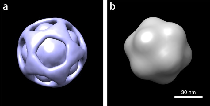
ABSTRACT Using a manifold-based analysis of experimental diffraction snapshots from an X-ray free electron laser, we determine the three-dimensional structure and conformational landscape of
the PR772 virus to a detector-limited resolution of 9 nm. Our results indicate that a single conformational coordinate controls reorganization of the genome, growth of a tubular structure
from a portal vertex and release of the genome. These results demonstrate that single-particle X-ray scattering has the potential to shed light on key biological processes. Access through
your institution Buy or subscribe This is a preview of subscription content, access via your institution ACCESS OPTIONS Access through your institution Access Nature and 54 other Nature
Portfolio journals Get Nature+, our best-value online-access subscription $29.99 / 30 days cancel any time Learn more Subscribe to this journal Receive 12 print issues and online access
$259.00 per year only $21.58 per issue Learn more Buy this article * Purchase on SpringerLink * Instant access to full article PDF Buy now Prices may be subject to local taxes which are
calculated during checkout ADDITIONAL ACCESS OPTIONS: * Log in * Learn about institutional subscriptions * Read our FAQs * Contact customer support SIMILAR CONTENT BEING VIEWED BY OTHERS
ROBUST TOTAL X-RAY SCATTERING WORKFLOW TO STUDY CORRELATED MOTION OF PROTEINS IN CRYSTALS Article Open access 03 March 2023 OBSERVATION OF A SINGLE PROTEIN BY ULTRAFAST X-RAY DIFFRACTION
Article Open access 12 January 2024 FORM FACTOR DETERMINATION OF BIOLOGICAL MOLECULES WITH X-RAY FREE ELECTRON LASER SMALL-ANGLE SCATTERING (XFEL-SAS) Article Open access 18 October 2023
REFERENCES * Neutze, R., Wouts, R., van der Spoel, D., Weckert, E. & Hajdu, J. Potential for biomolecular imaging with femtosecond X-ray pulses. _Nature_ 406, 752–757 (2000). Article
CAS PubMed Google Scholar * Breedlove, J.R. Jr. & Trammell, G.T. Molecular microscopy: fundamental limitations. _Science_ 170, 1310–1313 (1970). Article CAS PubMed Google Scholar
* Solem, J.C. & Baldwin, G.C. Microholography of living organisms. _Science_ 218, 229–235 (1982). Article CAS PubMed Google Scholar * Miao, J., Charalambous, P., Kirz, J. &
Sayre, D. Extending the methodology of X-ray crystallography to allow imaging of micrometre-sized non-crystalline specimens. _Nature_ 400, 342–344 (1999). Article CAS Google Scholar *
Fung, R., Shneerson, V., Saldin, D.K. & Ourmazd, A. Structure from fleeting illumination of faint spinning objects in flight. _Nat. Phys._ 5, 64–67 (2009). Article CAS Google Scholar
* Loh, N.T. & Elser, V. Reconstruction algorithm for single-particle diffraction imaging experiments. _Phys. Rev. E_ 80, 026705 (2009). Article CAS Google Scholar * Seibert, M.M. et
al. Single mimivirus particles intercepted and imaged with an X-ray laser. _Nature_ 470, 78–81 (2011). Article CAS PubMed PubMed Central Google Scholar * Hantke, M.F. et al.
High-throughput imaging of heterogeneous cell organelles with an X-ray laser. _Nat. Photonics_ 8, 943–949 (2014). Article CAS Google Scholar * Kuhn, R.J., Dowd, K.A., Beth Post, C. &
Pierson, T.C. Shake, rattle, and roll: impact of the dynamics of flavivirus particles on their interactions with the host. _Virology_ 479-480, 508–517 (2015). Article CAS PubMed Google
Scholar * Mateu, M.G. Assembly, stability and dynamics of virus capsids. _Arch. Biochem. Biophys._ 531, 65–79 (2013). Article CAS PubMed Google Scholar * Xu, R. et al. Single-shot
three-dimensional structure determination of nanocrystals with femtosecond X-ray free-electron laser pulses. _Nat. Commun._ 5, 4061 (2014). Article CAS PubMed Google Scholar * Ekeberg,
T. et al. Three-dimensional reconstruction of the giant mimivirus particle with an x-ray free-electron laser. _Phys. Rev. Lett._ 114, 098102 (2015). Article PubMed CAS Google Scholar *
Loh, N.D. et al. Cryptotomography: reconstructing 3D Fourier intensities from randomly oriented single-shot diffraction patterns. _Phys. Rev. Lett._ 104, 225501 (2010). Article CAS PubMed
Google Scholar * Moore, P.B. How should we think about the ribosome? _Annu. Rev. Biophys._ 41, 1–19 (2012). Article CAS PubMed Google Scholar * Changeux, J.P. & Edelstein, S.
Conformational selection or induced fit? 50 years of debate resolved. _F1000 Biol. Rep._ 3, 19 (2011). Article PubMed PubMed Central Google Scholar * Ye, L., Van Eps, N., Zimmer, M.,
Ernst, O.P. & Prosser, R.S. Activation of the A2A adenosine G-protein-coupled receptor by conformational selection. _Nature_ 533, 265–268 (2016). Article CAS PubMed Google Scholar *
Yu, I.M. et al. Structure of the immature dengue virus at low pH primes proteolytic maturation. _Science_ 319, 1834–1837 (2008). Article CAS PubMed Google Scholar * Henrich, B. et al.
The adaptive gain integrating pixel detector AGIP a detector for the European XFEL. _Nucl. Instrum. Methods Phys. Res. A_ 633, S11–S14 (2011). Article CAS Google Scholar * Jarzynski, C.
Nonequilibrium equality for free energy differences. _Phys. Rev. Lett._ 78, 2690–2693 (1997). Article CAS Google Scholar * Jarzynski, C. Equilibrium free energy differences from
nonequilibrium measurements. _Phys. Rev. E_ 56, 5018–5035 (1997). Article CAS Google Scholar * Crooks, G.E. Path-ensemble averages in systems driven far from equilibrium. _Phys. Rev. E_
61, 2361 (2000). Article CAS Google Scholar * Pressé, S. Principles of maximum entropy andn maximum caliber in statistical physics. _Rev. Mod. Phys._ 85, 1115–1141 (2013). Article CAS
Google Scholar * Schwander, P., Fung, R. & Ourmazd, A. Conformations of macromolecules and their complexes from heterogeneous datasets. _Phil. Trans. R. Soc. Lond. B_ 369, 20130567
(2014). Article CAS Google Scholar * Dashti, A. et al. Trajectories of the ribosome as a Brownian nanomachine. _Proc. Natl. Acad. Sci. USA_ 111, 17492–17497 (2014). Article CAS PubMed
PubMed Central Google Scholar * Frank, J. & Ourmazd, A. Continuous changes in structure mapped by manifold embedding of single-particle data in cryo-EM. _Methods_ 100, 61–67 (2016).
Article CAS PubMed PubMed Central Google Scholar * Reddy, H.K.N. et al. Coherent soft X-ray diffraction imaging of coliphage PR772 at the Linac coherent light source. _Sci. Data_ 4,
170079 (2017). Article CAS PubMed PubMed Central Google Scholar * Hosseinizadeh, A., Dashti, A., Schwander, P., Fung, R. & Ourmazd, A. Single-particle structure determination by
X-ray free-electron lasers: possibilities and challenges. _Struct. Dyn._ 2, 041601 (2015). Article CAS PubMed PubMed Central Google Scholar * Hosseinizadeh, A. et al. High-resolution
structure of viruses from random diffraction snapshots. _Phil. Trans. R. Soc. Lond. B_ 369, 20130326 (2014). Article CAS Google Scholar * Scheres, S.H. & Chen, S. Prevention of
overfitting in cryo-EM structure determination. _Nat. Methods_ 9, 853–854 (2012). Article CAS PubMed PubMed Central Google Scholar * Peralta, B. et al. Mechanism of membranous
tunnelling nanotube formation in viral genome delivery. _PLoS Biol._ 11, e1001667 (2013). Article CAS PubMed PubMed Central Google Scholar * Santos-Pérez, I. et al. Membrane-assisted
viral DNA ejection. _Biochim. Biophys. Acta_ 1861, 664–672 (2017). Article CAS Google Scholar * Incardona, N.L. & Kaesberg, P. A pH-induced structural change in bromegrass mosiac
virus. _Biophys. J._ 4, 11–21 (1964). Article CAS PubMed PubMed Central Google Scholar * Speir, J.A., Munshi, S., Wang, G., Baker, T.S. & Johnson, J.E. Structures of the native and
swollen forms of cowpea chlorotic mottle virus determined by X-ray crystallography and cryo-electron microscopy. _Structure_ 3, 63–78 (1995). Article CAS PubMed PubMed Central Google
Scholar * Ayyer, K., Lan, T.Y., Elser, V. & Loh, N.D. Dragonfly: an implementation of the expand-maximize-compress algorithm for single-particle imaging. _J. Appl. Crystallogr._ 49,
1320–1335 (2016). Article CAS PubMed PubMed Central Google Scholar * Frank, J. _Three-Dimensional Electron Microscopy of Macromolecular Assemblies_ 2nd edn. (Oxford University Press,
2006). * Coifman, R.R. et al. Geometric diffusions as a tool for harmonic analysis and structure definition of data: diffusion maps. _Proc. Natl. Acad. Sci. USA_ 102, 7426–7431 (2005).
Article CAS PubMed PubMed Central Google Scholar * Giannakis, D. & Majda, A.J. Nonlinear Laplacian spectral analysis for time series with intermittency and low-frequency
variability. _Proc. Natl. Acad. Sci. USA_ 109, 2222–2227 (2012). Article CAS PubMed PubMed Central Google Scholar * Giannakis, D., Schwander, P. & Ourmazd, A. The symmetries of
image formation by scattering. I. Theoretical framework. _Opt. Express_ 20, 12799–12826 (2012). Article PubMed Google Scholar * Schwander, P., Giannakis, D., Yoon, C.H. & Ourmazd, A.
The symmetries of image formation by scattering. II. Applications. _Opt. Express_ 20, 12827–12849 (2012). Article PubMed Google Scholar * White, T.A. CrystFEL: a software suite for
snapshot serial crystallography. _J. Appl. Crystallogr._ 45, 335–341 (2012). Article CAS Google Scholar * Fienup, J.R. Phase retrieval algorithms: a comparison. _Appl. Opt._ 21, 2758–2769
(1982). Article CAS PubMed Google Scholar * Coifman, R.R., Shkolnisky, Y., Sigworth, F.J. & Singer, A. Graph Laplacian tomography from unknown random projections. _IEEE Trans. Image
Process._ 17, 1891–1899 (2008). Article PubMed Google Scholar * Marchesini, S. et al. X-ray image reconstruction from a diffraction pattern alone. _Phys. Rev. B_ 68, 140101 (2003).
Article CAS Google Scholar * Dashti, A., Komarov, I. & D'Souza, R.M.D. Efficient computation of _k_-nearest neighbour graphs for large high-dimensional data sets on GPU clusters.
_PLoS One_ 8, e74113 (2013). Article CAS PubMed PubMed Central Google Scholar Download references ACKNOWLEDGEMENTS We acknowledge valuable discussions with H. Chapman, E. Lattman, J.
Spence and I. Vartaniants. The research conducted at University of Wisconsin–Milwaukee was supported by the US Department of Energy, Office of Science, Basic Energy Sciences, under contract
DE-SC0002164 (A.O., algorithm design and development) and by the US National Science Foundation (NSF) under contract STC 1231306 (A.O., numerical trial models and data analysis; M.S., data
analysis) and under contract number 1551489 (A.O., underlying analytical models). The research at Arizona State University was supported by the NSF under contract STC 1231306 (B.G.H.). Use
of the Linac Coherent Light Source (LCLS), SLAC National Accelerator Laboratory, was supported by the US Department of Energy, Office of Science, Office of Basic Energy Sciences under
contract DE-AC02-76SF00515. AUTHOR INFORMATION Author notes * Ahmad Hosseinizadeh, Ghoncheh Mashayekhi, Jeremy Copperman, Peter Schwander and Abbas Ourmazd: These authors contributed equally
to this work. AUTHORS AND AFFILIATIONS * Department of Physics, University of Wisconsin–Milwaukee, Milwaukee, Wisconsin, USA Ahmad Hosseinizadeh, Ghoncheh Mashayekhi, Jeremy Copperman,
Peter Schwander, Ali Dashti, Reyhaneh Sepehr, Russell Fung, Marius Schmidt & Abbas Ourmazd * Linac Coherent Light Source, SLAC National Accelerator Laboratory, Menlo Park, California,
USA Chun Hong Yoon & Andrew Aquila * Biodesign Center for Immunotherapy, Vaccines and Virotherapy, Arizona State University, Tempe, Arizona, USA Brenda G Hogue * Biodesign Center for
Applied Structural Discovery, Arizona State University, Tempe, Arizona, USA Brenda G Hogue * School of Life Sciences, Arizona State University, Tempe, Arizona, USA Brenda G Hogue *
Brookhaven National Laboratory, Upton, New York, USA Garth J Williams Authors * Ahmad Hosseinizadeh View author publications You can also search for this author inPubMed Google Scholar *
Ghoncheh Mashayekhi View author publications You can also search for this author inPubMed Google Scholar * Jeremy Copperman View author publications You can also search for this author
inPubMed Google Scholar * Peter Schwander View author publications You can also search for this author inPubMed Google Scholar * Ali Dashti View author publications You can also search for
this author inPubMed Google Scholar * Reyhaneh Sepehr View author publications You can also search for this author inPubMed Google Scholar * Russell Fung View author publications You can
also search for this author inPubMed Google Scholar * Marius Schmidt View author publications You can also search for this author inPubMed Google Scholar * Chun Hong Yoon View author
publications You can also search for this author inPubMed Google Scholar * Brenda G Hogue View author publications You can also search for this author inPubMed Google Scholar * Garth J
Williams View author publications You can also search for this author inPubMed Google Scholar * Andrew Aquila View author publications You can also search for this author inPubMed Google
Scholar * Abbas Ourmazd View author publications You can also search for this author inPubMed Google Scholar CONTRIBUTIONS A.H.: algorithmic design, data preprocessing, single-particle hit
finding, orientation and structural recovery, preparation of paper. G.M.: conformational analysis, preparation of paper. J.C.: conformational analysis and validation, preparation of paper.
P.S.: experimental and algorithmic design, code development, data preprocessing, data analysis, experiments at LCLS, preparation of paper. A.D., R.S. and R.F.: data-analytical contributions.
M.S.: analysis of results, preparation of paper. C.H.Y.: experimental design, data collection. B.G.H.: sample selection, preparation and characterization, experiments at LCLS, preparation
of paper. G.J.W.: planning and execution of experiment and discussions of data and analysis. A.A.: experimental design, data collection, preparation of paper. A.O.: experimental and
algorithmic design, data analysis and interpretation, preparation of paper. CORRESPONDING AUTHOR Correspondence to Abbas Ourmazd. ETHICS DECLARATIONS COMPETING INTERESTS The authors declare
no competing financial interests. INTEGRATED SUPPLEMENTARY INFORMATION SUPPLEMENTARY FIGURE 1 2D SNAPSHOTS OBTAINED BY DIRECT PHASING OF INDIVIDUAL DIFFRACTION SNAPSHOTS. Each row shows five
representative snapshots assigned to an extreme value of the conformational parameter τ. Note the reduction in the projected inner density between the two rows. (Rainbow color code, with
dark red corresponding to the highest density.) SUPPLEMENTARY FIGURE 2 RELIABILITY OF THE RECONSTRUCTED DIFFRACTION VOLUME. R-split as a function of the magnitude of the scattering vector
_q_, indicating reliable reconstruction up to the detector-limited resolution of 9 nm. The analysis was performed by random splitting, without substitution, of the 37,550 snapshots into two
disjoint datasets. Source data SUPPLEMENTARY FIGURE 3 SPATIAL RESOLUTION OF 3D DENSITY. Fourier Shell Correlation (FSC) vs. the magnitude of the scattering vector _q_ obtained from splitting
the 37,550 snapshots into two random disjoint subsets. The resolution is limited to 9 nm by the detector geometry. Source data SUPPLEMENTARY FIGURE 4 SPHERICALLY AVERAGED RADIAL DENSITIES
OF 3D STRUCTURES OBTAINED WITHOUT IMPOSING ICOSAHEDRAL SYMMETRY. The conformational coordinate ranges from 0 (black curve) to 1 (red curve). Averages (circles) and standard deviations (error
bars) pertain to twenty 3D iterative phasing calculations using all 37,550 single-particle snapshots. Spline fits are guides to the eye. Source data SUPPLEMENTARY FIGURE 5 A REPRESENTATIVE
EXPERIMENTAL XFEL DIFFRACTION SNAPSHOT OF THE PR772 VIRUS BEFORE AND AFTER PREPROCESSING. (A) A raw snapshot. (B) Same snapshot after background correction. The dark horizontal line in each
snapshot is due to the gap between the two detector panels. Intensity is shown in the rainbow color code, with red corresponding to the highest intensity. SUPPLEMENTARY FIGURE 6 DIFFUSION
MAP MANIFOLDS FORMED BY EXPERIMENTAL XFEL DIFFRACTION SNAPSHOTS OF THE PR772 VIRUS. (A) Manifold obtained by embedding 135,375 preprocessed XFEL snapshots (“hits”). (B) Distribution of
single- and multi-particle snapshots on the same manifold. The general parabolic shape stems from changes in the incident beam intensity intersecting the particles. (C) Manifold of
single-particle snapshots after outlier removal. A total of 37,550 single-particle snapshots were extracted by this analysis. Source data SUPPLEMENTARY FIGURE 7 COMPARISON OF ICOSAHEDRAL
WIGNER D FUNCTIONS WITH DIFFUSION MAP MANIFOLDS OBTAINED FROM SIMULATED AND EXPERIMENTAL DIFFRACTION SNAPSHOTS. (A) Icosahedral Wigner D-functions sampled in 30,000 randomly selected
orientations. (B) DM eigenfunctions obtained from 30,000 simulated, noise-free snapshots of an icosahedral capsid. (C) Manifold from 37,550 simulated snapshots of an icosahedral capsid with
experimental signal-to-noise ratio (SNR) and detector gap. (D) Manifold from 37,550 experimental single-particle PR772 snapshots. (E) Eigenvalue spectrum for an icosahedral capsid simulated
at the experimental SNR. (F) Eigenvalue spectrum for the PR772 virus. Source data SUPPLEMENTARY FIGURE 8 DIFFUSION MAP EIGENVALUE SPECTRA FOR EXPERIMENTAL AND SIMULATED DIFFRACTION SNAPSHOTS
OF PR772 VIRUS. (A) Spectrum for 37,550 single-particle experimental snapshots of PR772 virus. (B) Spectrum resulting from the same number of simulated snapshots of an icosahedral capsid
stretched by 10% along one 5-fold axis. Note the differences between the eigenvalue spectra. Source data SUPPLEMENTARY INFORMATION SUPPLEMENTARY TEXT AND FIGURES Supplementary Figures 1–8
and Supplementary Note (PDF 1339 kb) LIFE SCIENCES REPORTING SUMMARY Life Sciences Reporting Summary (PDF 160 kb) SUPPLEMENTARY DATA Single-particle indices (XLSX 1153 kb) 3D CONFORMATIONAL
MOVIE WITH IMPOSED ICOSAHEDRAL SYMMETRY. Evolution of the 3D structure of the PR772 virus, and the occupancy along the dominant conformational reaction coordinate. These results were
obtained assuming icosahedral symmetry. (MOV 800 kb) 3D CONFORMATIONAL MOVIE WITHOUT IMPOSING ICOSAHEDRAL SYMMETRY. Evolution of the 3D structure of the PR772 virus, and the occupancy along
the conformational reaction coordinate without imposing icosahedral symmetry. Note the protrusion of a tubular structure, and the concentration of the genome toward the tube. (MOV 418 kb)
SOURCE DATA SOURCE DATA TO FIG. 1 SOURCE DATA TO FIG. 2 SOURCE DATA TO SUPPLEMENTARY FIG. 3 SOURCE DATA TO SUPPLEMENTARY FIG. 4 SOURCE DATA TO SUPPLEMENTARY FIG. 5 SOURCE DATA TO
SUPPLEMENTARY FIG. 6 SOURCE DATA TO SUPPLEMENTARY FIG. 7 SOURCE DATA TO SUPPLEMENTARY FIG. 8 RIGHTS AND PERMISSIONS Reprints and permissions ABOUT THIS ARTICLE CITE THIS ARTICLE
Hosseinizadeh, A., Mashayekhi, G., Copperman, J. _et al._ Conformational landscape of a virus by single-particle X-ray scattering. _Nat Methods_ 14, 877–881 (2017).
https://doi.org/10.1038/nmeth.4395 Download citation * Received: 03 February 2017 * Accepted: 05 July 2017 * Published: 14 August 2017 * Issue Date: 01 September 2017 * DOI:
https://doi.org/10.1038/nmeth.4395 SHARE THIS ARTICLE Anyone you share the following link with will be able to read this content: Get shareable link Sorry, a shareable link is not currently
available for this article. Copy to clipboard Provided by the Springer Nature SharedIt content-sharing initiative