- Select a language for the TTS:
- UK English Female
- UK English Male
- US English Female
- US English Male
- Australian Female
- Australian Male
- Language selected: (auto detect) - EN
Play all audios:
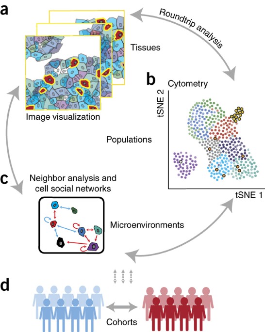
ABSTRACT Single-cell, spatially resolved omics analysis of tissues is poised to transform biomedical research and clinical practice. We have developed an open-source, computational histology
topography cytometry analysis toolbox (histoCAT) to enable interactive, quantitative, and comprehensive exploration of individual cell phenotypes, cell–cell interactions, microenvironments,
and morphological structures within intact tissues. We highlight the unique abilities of histoCAT through analysis of highly multiplexed mass cytometry images of human breast cancer
tissues. Access through your institution Buy or subscribe This is a preview of subscription content, access via your institution ACCESS OPTIONS Access through your institution Access Nature
and 54 other Nature Portfolio journals Get Nature+, our best-value online-access subscription $29.99 / 30 days cancel any time Learn more Subscribe to this journal Receive 12 print issues
and online access $259.00 per year only $21.58 per issue Learn more Buy this article * Purchase on SpringerLink * Instant access to full article PDF Buy now Prices may be subject to local
taxes which are calculated during checkout ADDITIONAL ACCESS OPTIONS: * Log in * Learn about institutional subscriptions * Read our FAQs * Contact customer support SIMILAR CONTENT BEING
VIEWED BY OTHERS THREE-DIMENSIONAL IMAGING MASS CYTOMETRY FOR HIGHLY MULTIPLEXED MOLECULAR AND CELLULAR MAPPING OF TISSUES AND THE TUMOR MICROENVIRONMENT Article Open access 24 December 2021
MCMICRO: A SCALABLE, MODULAR IMAGE-PROCESSING PIPELINE FOR MULTIPLEXED TISSUE IMAGING Article Open access 25 November 2021 SEMI-AUTOMATED APPROACHES FOR INTERROGATING SPATIAL HETEROGENEITY
OF TISSUE SAMPLES Article Open access 29 February 2024 REFERENCES * Tirosh, I. et al. _Science_ 352, 189–196 (2016). Article CAS Google Scholar * Bendall, S.C. et al. _Science_ 332,
687–696 (2011). Article CAS Google Scholar * Bodenmiller, B. et al. _Nat. Biotechnol._ 30, 858–867 (2012). Article CAS Google Scholar * Tabassum, D.P. & Polyak, K. _Nat. Rev.
Cancer_ 15, 473–483 (2015). Article CAS Google Scholar * Lee, J.H. et al. _Science_ 343, 1360–1363 (2014). Article CAS Google Scholar * Chen, K.H., Boettiger, A.N., Moffitt, J.R.,
Wang, S. & Zhuang, X. _Science_ 348, aaa6090 (2015). Article Google Scholar * Lin, J.-R., Fallahi-Sichani, M. & Sorger, P.K. _Nat. Commun._ 6, 8390 (2015). Article CAS Google
Scholar * Schubert, W. et al. _Nat. Biotechnol._ 24, 1270–1278 (2006). Article CAS Google Scholar * Gerdes, M.J. et al. _Proc. Natl. Acad. Sci. USA_ 110, 11982–11987 (2013). Article CAS
Google Scholar * Angelo, M. et al. _Nat. Med._ 20, 436–442 (2014). Article CAS Google Scholar * Giesen, C. et al. _Nat. Methods_ 11, 417–422 (2014). Article CAS Google Scholar *
Bodenmiller, B. _Cell Syst._ 2, 225–238 (2016). Article CAS Google Scholar * Ding, H., Wang, C., Huang, K. & Machiraju, R. _BMC Bioinformatics_ 16, S10 (2015). Article Google Scholar
* Beck, A.H. et al. _Sci. Transl. Med._ 3, 108ra113 (2011). Article Google Scholar * Jones, T.R. et al. _BMC Bioinformatics_ 9, 482 (2008). Article Google Scholar * Amir, A.D. et al.
_Nat. Biotechnol._ 31, 545–552 (2013). Article CAS Google Scholar * Shekhar, K., Brodin, P., Davis, M.M. & Chakraborty, A.K. _Proc. Natl. Acad. Sci. USA_ 111, 202–207 (2014). Article
CAS Google Scholar * Rieckmann, J.C. et al. _Nat. Immunol._ 18, 583–593 (2017). Article CAS Google Scholar * Levine, J.H. et al. _Cell_ 162, 184–197 (2015). Article CAS Google
Scholar * Ostuni, R., Kratochvill, F., Murray, P.J. & Natoli, G. _Trends Immunol._ 36, 229–239 (2015). Article CAS Google Scholar * Kononen, J. et al. _Nat. Med._ 4, 844–847 (1998).
Article CAS Google Scholar * Catena, R., Özcan, A., Jacobs, A., Chevrier, S. & Bodenmiller, B. _Genome Biol._ 17, 142 (2016). Article Google Scholar * Wang, H.A.O. et al. _Anal.
Chem._ 85, 10107–10116 (2013). Article CAS Google Scholar * Sommer, C., Straehle, C., Köthe, U. & Hamprecht, F.A. in Proc. 2011 8th IEEE International Symposium on. _Biomedical
Imaging: From Nano to Macro_ 230–233 (IEEE, 2011). * Schüffler, P.J. et al. _Cytometry A_ 87, 936–942 (2015). Article Google Scholar * Weber, L.M. & Robinson, M.D. _Cytometry A_ 89,
1084–1096 (2016). Article CAS Google Scholar Download references ACKNOWLEDGEMENTS We would like to thank the Bodenmiller lab for support and fruitful discussions. Thank you to open-source
softwares like “cyt”, “CellProfiler,” and many others. This work was supported by the Swiss National Science Foundation (SNSF) R'Equip grant 316030-139220, an SNSF Assistant
Professorship grant PP00P3-144874, a Swiss Cancer League grant, the PhosphonetPPM and MetastasiX SystemsX grants, and funding from the European Research Council (ERC) under the European
Union′s Seventh Framework Programme (FP/2007-2013)/ERC Grant Agreement no. 336921. D. Schapiro was supported by the Forschungskredit of the University of Zurich, grant FK-74419-01-01, and
the BioEntrepreneur-Fellowship of the University of Zurich, reference no. BIOEF-17-001. H.W.J. and D. Schulz are supported by European Molecular Biology Organization (EMBO) Long Term
Fellowships cofunded by the European Commission (LTFCOFUND2013 and 2014), grants ALTF-711 2015 and ALTF-970 2014, respectively. H.W.J. was also supported by a Transition Postdoc Fellowship
from the Swiss SystemsX.ch initiative ref. 2015/344, evaluated by the Swiss National Science Foundation. AUTHOR INFORMATION Author notes * Swetha Raghuraman Present address: Institute of
Cell Biology, ZMBE, University of Münster, Münster, Germany * Charlotte Giesen Present address: F. Hoffmann-La Roche Ltd, Kaiseraugst, Switzerland * Denis Schapiro and Hartland W Jackson:
These authors contributed equally to this work. AUTHORS AND AFFILIATIONS * Institute of Molecular Life Sciences, University of Zurich, Zurich, Switzerland Denis Schapiro, Hartland W Jackson,
Swetha Raghuraman, Jana R Fischer, Vito R T Zanotelli, Daniel Schulz, Charlotte Giesen, Raúl Catena & Bernd Bodenmiller * Life Science Zurich Graduate School, ETH Zurich and University
of Zurich, Zurich, Switzerland Denis Schapiro & Vito R T Zanotelli * Institute of Surgical Pathology, University Hospital Zurich, Zurich, Switzerland Zsuzsanna Varga Authors * Denis
Schapiro View author publications You can also search for this author inPubMed Google Scholar * Hartland W Jackson View author publications You can also search for this author inPubMed
Google Scholar * Swetha Raghuraman View author publications You can also search for this author inPubMed Google Scholar * Jana R Fischer View author publications You can also search for this
author inPubMed Google Scholar * Vito R T Zanotelli View author publications You can also search for this author inPubMed Google Scholar * Daniel Schulz View author publications You can
also search for this author inPubMed Google Scholar * Charlotte Giesen View author publications You can also search for this author inPubMed Google Scholar * Raúl Catena View author
publications You can also search for this author inPubMed Google Scholar * Zsuzsanna Varga View author publications You can also search for this author inPubMed Google Scholar * Bernd
Bodenmiller View author publications You can also search for this author inPubMed Google Scholar CONTRIBUTIONS D. Schapiro, H.W.J., and B.B. conceived of the project and software. H.W.J.,
C.G., and R.C. collected samples and validated antibodies. Z.V. assembled, classified, and provided tumor samples. H.W.J. completed the staining and image acquisition. D. Schapiro, S.R., and
J.R.F. wrote the code. D. Schapiro, H.W.J., and D. Schulz tested software on multiple data sources. D. Schapiro, H.W.J., and V.R.T.Z. analyzed the images and single-cell data. D. Schapiro,
H.W.J., and B.B. prepared the figures and wrote the manuscript. B.B. directed the project. CORRESPONDING AUTHOR Correspondence to Bernd Bodenmiller. ETHICS DECLARATIONS COMPETING INTERESTS
The authors declare no competing financial interests. SUPPLEMENTARY INFORMATION SUPPLEMENTARY TEXT AND FIGURES Supplementary Figures 1–9, Supplementary Table 1 and Supplementary Notes 1–4.
(PDF 69995 kb) LIFE SCIENCES REPORTING SUMMARY Life Sciences Reporting Summary (PDF 129 kb) SUPPLEMENTARY TABLE 2 Patient Metadata (XLSX 47 kb) SUPPLEMENTARY DATASET 1 Source Data for Figure
2 (TXT 12656 kb) SUPPLEMENTARY DATASET 2 Source Data for Supplementary Figure 3 (XLS 80 kb) SUPPLEMENTARY SOFTWARE 1 histoCAT_MacOS12 (ZIP 17932 kb) SUPPLEMENTARY SOFTWARE 2
histoCAT_Windows7 (EXE 20372 kb) SUPPLEMENTARY SOFTWARE 3 histoCAT_Windows10 (EXE 19986 kb) RIGHTS AND PERMISSIONS Reprints and permissions ABOUT THIS ARTICLE CITE THIS ARTICLE Schapiro, D.,
Jackson, H., Raghuraman, S. _et al._ histoCAT: analysis of cell phenotypes and interactions in multiplex image cytometry data. _Nat Methods_ 14, 873–876 (2017).
https://doi.org/10.1038/nmeth.4391 Download citation * Received: 07 February 2017 * Accepted: 07 July 2017 * Published: 07 August 2017 * Issue Date: 01 September 2017 * DOI:
https://doi.org/10.1038/nmeth.4391 SHARE THIS ARTICLE Anyone you share the following link with will be able to read this content: Get shareable link Sorry, a shareable link is not currently
available for this article. Copy to clipboard Provided by the Springer Nature SharedIt content-sharing initiative