- Select a language for the TTS:
- UK English Female
- UK English Male
- US English Female
- US English Male
- Australian Female
- Australian Male
- Language selected: (auto detect) - EN
Play all audios:
ABSTRACT Epithelial folding is a critical process underlying many morphogenetic events including vertebrate neural tube closure, however, its spatial regulation is largely unknown. Here we
show that during neural tube formation Rab11-positive recycling endosomes acquire bilaterally symmetric distribution in the _Xenopus_ neural plate, being enriched at medial apical cell
junctions. This mediolateral polarization was under the control of planar cell polarity (PCP) signalling, was necessary for neural plate folding and was accompanied by the polarization of
the exocyst component Sec15. Our further experiments demonstrate that similar PCP-dependent polarization of Rab11 is essential for ectopic apical constriction driven by the actin-binding
protein Shroom and during embryonic wound repair. We propose that anisotropic membrane trafficking has key roles in diverse morphogenetic behaviours of individual cells and propagates in a
tissue by a common mechanism that involves PCP. You have full access to this article via your institution. Download PDF SIMILAR CONTENT BEING VIEWED BY OTHERS WNT4 AND EPHRINB2 INSTRUCT
APICAL CONSTRICTION VIA DISHEVELLED AND NON-CANONICAL SIGNALING Article Open access 20 January 2023 APICAL–BASAL POLARITY AND THE CONTROL OF EPITHELIAL FORM AND FUNCTION Article 19 April
2022 CD13 ORIENTS THE APICAL-BASAL POLARITY AXIS NECESSARY FOR LUMEN FORMATION Article Open access 04 August 2021 INTRODUCTION Epithelial tissue folding is a universal mechanism for germ
layer rearrangements at gastrulation, neural tube closure and organogenesis. During vertebrate neurulation, dorsal ectoderm forms the neural plate that bends to generate the neural
groove1,2,3. At a later stage, the bilateral neural folds fuse at the midline into the neural tube. Actomyosin contractility and cell junction remodelling are believed to be major driving
forces for epithelial folding in _Drosophila_ embryos2,3,4,5, apical constriction in _C. elegans_ endoderm progenitors6 and during vertebrate neural tube closure7,8,9,10. In addition,
mutations in genes encoding core planar cell polarity (PCP) proteins Frizzled, Dishevelled, Vangl2/Strabismus, Flamingo/Celsr and Prickle reveal neural tube defects (reviewed by Bayly and
Axelrod11, Zallen11 and Wallingford _et al._13). Although PCP signalling has been linked to Rho signalling and actomyosin contractility in gastrulation and neurulation9,14,15,16, molecular
mechanisms underlying neural tube defects in PCP mutants are still unknown. In _Xenopus_ neural plate explants, deep layer cells display monopolar protrusive activity towards the midline17,
yet molecular markers of this polarization have not been identified and whether this polarity relates to apical constriction that occurs at the hinge regions of the bending neural plate is
unclear. As membrane trafficking has essential roles in diverse morphogenetic events, including cell intercalation15, epithelial polarization18,19 and neural tube formation20, we examined
potential functions for endocytic proteins that might be associated with morphological changes during _Xenopus_ neural plate closure. We were especially interested in Rab11, a recycling
endosome marker with roles in cell polarity and cell migration21,22,23,24,25. Our results reveal unique planar polarization of Rab11 in the neural plate, which is regulated by PCP signalling
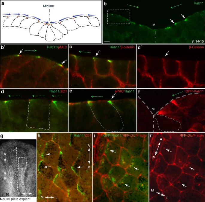
and which is essential for neural tube formation. RESULTS PLANAR POLARIZATION OF RAB11 IN THE NEURAL PLATE Immunostaining of sectioned early neurula embryos at stages 14–15 revealed
striking polarization of Rab11 in the plane of the neuroepithelium (Fig. 1a–c, Supplementary Fig. 1a–d). Even before neural plate folding became morphologically apparent, Rab11 became
localized to one apical corner of neuroepithelial cells that is closer to the dorsal midline, thereby establishing mirror-image planar polarity in the early neural plate. The bilateral
staining progressively changed towards apical midline staining at the neural fold stages (Fig. 1a,b, Supplementary Fig. 1a,b). Other apical or basolateral markers, such as atypical PKC, ZO1
and β-catenin, did not reveal similar polarization (Fig. 1c–e). The observed pattern was often detected as comet-shape cytoplasmic staining directed towards a cell junction (marked by ZO1,
Fig. 1d). To independently confirm that Rab11 is indeed unilaterally polarized in individual cells, we examined early embryos injected with RNA-encoding Rab11-green fluorescent protein (GFP;
Fig. 1f). Mosaically expressed exogenous Rab11-GFP was similarly distributed in a gradient with the highest concentration at the medial apical corner of the cell (Fig. 1f). Lack of staining
in the neighboring cells, which did not receive Rab11-GFP RNA, allowed us to unequivocally define the position of Rab11-GFP relative to the cell border. _En face_ views of the neural plate
immunostained for both endogenous and exogenous Rab11 confirmed the polarization of Rab11 in the plane of the tissue, in a manner reminiscent of the distribution of PCP proteins in
_Drosophila_ epithelia11 (Fig. 1g–i). Interestingly, Diversin, a vertebrate homologue of the fly PCP protein Diego, revealed a similar polarization (Fig. 1i,i′), providing insight into the
role of PCP proteins in neural tube closure. These findings suggest that anisotropic Rab11-dependent recycling is an early event in neural tube closure. RAB11 IS REQUIRED FOR MYOSIN
ACTIVATION AND NEURAL FOLDING To test whether Rab11-mediated membrane trafficking is required for neural plate folding, we studied the phenotype of embryos microinjected with RNA-encoding
Rab11S25N, a dominant negative construct23, which is distributed throughout the cytoplasm (Supplementary Fig. 1e,f). In the majority of injected embryos (85%, _n_=53), the neural plate
remained open at the injected side, yet this defect was rescued by wild-type Rab11 RNA (Fig. 2a,b). Similar results have been obtained using a specific morpholino antisense oligonucleotide
(MO; Fig. 2c,d), which effectively depletes Rab11 in _Xenopus_ embryos23. The accumulation of phosphorylated myosin II light chain (pMLC), a key RhoA-dependent regulatory process in neural
tube closure7,8,9,10, failed to occur (Fig. 3a–c), complementing changes in neural fold and individual cell morphology (Fig. 2e,f; Supplementary Fig. 2a,b). This observation indicates that
Rab11 is required for Myosin II activation in the neural plate. Despite these changes in the contractile machinery, the apical–basal polarity markers aPKC and ZO1 were not affected (Fig.
3d,e). Together, our observations show that Rab11 is not just a marker for neuroepithelial progenitor polarity, but an essential modulator of neural tube closure. Lack of changes in the
neural tissue markers Sox3 (Figs 2g and 3f)_, Nrp1_ and _Rx1_ (Supplementary Fig. 2c,d) indicates that Rab11 is not involved in cell fate determination during neural induction. To further
understand how Rab11 regulates neural plate folding, we examined the localization of exocyst, a protein complex that targets post-Golgi secretory vesicles for subsequent membrane fusion
during exocytosis26. We find that the Rab11-interacting exocyst component Sec15 (ref. 27) polarizes in the neural plate similar to Rab11 (Fig. 2h). This result suggests that Sec15 promotes
neural tube closure by directing exocytosis near the medial surface of each cell. PCP PROTEINS REGULATE RAB11 LOCALIZATION IN THE NEURAL PLATE Given the medial polarization of Diversin in
the neural plate, we hypothesized that PCP signalling could function upstream of Rab11 in neural plate closure. Indeed, neural tube closure defects were apparent in Diversin-depleted embryos
(Supplementary Fig. 3). Moreover, interference with the function of Diversin and two other core PCP proteins, Vangl2 and Dishevelled, inhibited Rab11 polarization in the neural plate (Fig.
2i–k, Supplementary Fig. 4a). In these experiments, the total amount of Rab11 protein remained unaffected (Fig. 4a). Despite the major changes in neural plate morphology and the decrease of
myosin II phosphorylation in Vangl2-depleted embryos (Supplementary Fig. 4b), Vangl2 MO did not alter the fate of neural progenitors, which remained positive for _Sox3_ (Supplementary Fig.
4a). Finally, coinjection of RNAs encoding Rab11S25N and Vangl2 strongly enhanced the neural tube closure defects observed in separate RNA injections, indicating that these molecules
functionally interact (Supplementary Fig. 4c,d). These experiments indicate that the polarization of Rab11 endosomes along the mediolateral axis of the neural plate is controlled by the PCP
pathway. RAB11 FUNCTIONS DURING ECTOPIC APICAL CONSTRICTION As neural plate folding is largely driven by apical constriction of neuroepithelial cells, we suspected that Rab11 has a role in
this process. We wanted to examine an alternative model, in which morphogenesis can be separated from neural induction and patterning that accompany neural tube closure. Ectopic apical
constriction is induced in blastula ectoderm by overexpression of Shroom, a protein linked to actomyosin contractility and essential for neural tube closure10,28. We first assessed Rab11
localization during Shroom-induced apical constriction, which took place in the majority of injected embryos (Fig. 5). We observed strong apical recruitment of both Rab11 and Vangl2 in the
constricting Shroom-expressing cells, which are easily identified by abundant pigment granules near their apical surfaces and elongated morphology (Fig. 5a–e, Supplementary Fig. 5a–c).
Moreover, both endogenous and exogenous Rab11 polarized towards the site of Shroom RNA injection in the cells adjacent to the Shroom-expressing tissue (Fig. 5f–i, Supplementary Fig. 5a,b,e).
Therefore, our lineage tracing analysis suggests that a Shroom-induced signal triggers non-cell-autonomous Rab11 polarization in the surrounding cells. Although it is currently unclear
whether Rab11 has the same function in Shroom-expressing and nonexpressing cells, Shroom-dependent apical constriction was inhibited by Rab11S25N in more than 80% of injected embryos
(_n_=92; Fig. 5j). Consistent with this morphological effect, Rab11S25N inhibited apical pMLC accumulation induced by Shroom, but had no significant effect on aPKC or ZO1 (Supplementary Fig.
5h,i, and data not shown). Of interest, overexpressed Vangl2 interfered with Shroom-induced constriction and inhibited Rab11 apical accumulation and planar polarization in Shroom-expressing
ectoderm and during neural tube closure (Supplementary Fig. 5c–g,j,k), although total Rab11 protein levels in Vangl2-expressing cells did not change (Fig. 4b). This Rab11 polarization in
cells that respond to Shroom was inhibited by Vangl2 both in the cell autonomous and non-cell autonomous manner (Supplementary Fig. 5c–g). Together, these results are consistent with the
possibility that the PCP pathway interacts with vesicular trafficking machinery to drive ectopic apical constriction in response to Shroom. RAB11 AND PCP PROTEINS IN EMBRYONIC WOUND HEALING
Coordinated changes in cell shape that are similar to apical constriction take place during embryonic wound repair10. We further addressed Rab11 function in morphogenesis, by removing a
small piece of tissue from blastula ectoderm, a tissue lacking detectable planar polarity (Fig. 6a). In this model, wound healing is initiated within minutes after the surgery with pigmented
superficial ectoderm spreading eventually over the inner cell layer. Planar polarization of Rab11 became apparent in the superficial layer of the healing tissue in both embryos and ectoderm
explants in 30–60 min after the wound was inflicted (Fig. 6a–c and Supplementary Fig. 6a,b). We found that both endogenous and exogenous Rab11-containing recycling endosomes are polarized
in individual cells towards the direction of the wound (Fig. 6a–e, Supplementary Fig. 6a,b). In addition, both Rab11 and Vangl2 protein complexes became apically enriched in inner ectoderm
cells, which are known to participate in apical constriction during wound closure10 (Supplementary Fig. 6b, asterisk, Supplementary Fig. 7). Moreover, wound healing was defective in embryos
and explants that expressed Rab11S25N, Rab11 MO or Vangl2 MO (Fig. 6f–h, Supplementary Fig. 6c–i), indicating that both Rab11 and Vangl2 are required for this process. Similar to our
observations of neural plate folding, Vangl2 depletion inhibited Rab11 polarization and pMLC accumulation (data not shown). Cumulatively, our findings suggest that polarized Rab11-mediated
recycling is involved in diverse models of epithelial morphogenesis and point to the conservation of mechanisms regulating neural tube closure and embryonic wound healing. DISCUSSION Several
models of epithelial folding have been proposed including coordinated regulation of actomyosin contractility2,3,6,9, repositioning of adherens junctions by PAR proteins5 and enhanced
endocytosis20. Despite the recent progress, it remained unclear whether tissue folding has any directionality. Our experiments visualizing the graded distribution of Rab11 recycling
endosomes are the first to reveal the cell polarity corresponding to the axis of bending or constriction. We also find that this polarity requires core PCP proteins, reinforcing the known
connection between vesicular trafficking and PCP19,29,30,31,32. The unique localization of Rab11 and the exocyst component Sec15 suggests a model with an essential role of anisotropic
membrane trafficking in neural tube closure. In this model, PCP signals are conveyed to individual cells to polarize Rab11-mediated membrane trafficking and allow apical constriction to
proceed directionally, towards the midline, by expanding the lateral membrane domain at the expense of the shrinking apical domain. Although our experiments demonstrate a requirement of
Rab11 for myosin II activation in the neural plate, we note that the proposed mechanism does not support the classical ‘purse string’ model with isotropic actomyosin contractility along the
mediolateral axis. The same general mechanism may act to polarize membrane trafficking in individual cells in response to wounding, leading to cell reorientation and tissue repair. The
observed regulation of Rab11-dependent vesicular trafficking by core PCP molecules reiterates novel roles of these proteins in modulating cell shape and behaviour during tissue folding and
embryonic wound healing. Nevertheless, Rab11 roles in cell elongation, radial intercalation or convergent extension during neural tube closure1, and relevant cargo proteins for Rab11
vesicles remain to be elucidated. Consistent with the possibility that Vangl2 trafficking depends on Rab11, CFP-Vangl2 membrane localization became more diffuse and shifted to the cytoplasm
in cells depleted of Rab11 or expressing Rab11S25N (Supplementary Fig. 8). This finding supports a positive feedback loop, in which PCP proteins that regulate Rab11 are themselves targeted
to their specific locations by Rab11-dependent trafficking31,33,34. METHODS PLASMIDS, MICROINJECTIONS, _IN SITU_ HYBRIDIZATION Plasmids encoding GFP-C1, GFP-Rab11, GFP-Rab11S25N in pXT7,
untagged Rab11, Rab11S25N, Rab11Q70L in pXT7 and GFP-CAAX in pCS2 (ref. 23); mouse Shroom/ShrmL (also known as Shroom3)28 and Xdd1-pCS2 (ref. 35) have been previously described. Whole-mount
_in situ_ hybridization and Xgal staining were carried out using standard techniques36 with the digoxigenin-labelled antisense probes for _Nrp1_ (ref. 37) and _Rx1_ (ref. 38). Myc-Shroom in
pCS2+ was generated by subcloning of Shroom/ShrmL. For lineage tracing, GFP or membrane-targeted GFP-CAAX (50–100 pg) or membrane-targeted mCherry (400 pg) RNA was injected along with
morpholino antisense oligonucleotides (MOs) or RNAs. Capped mRNA was made by _in vitro_ transcription with T7 or SP6 promoter using mMessage mMachine kit (Ambion). Rab11 MO23, control MO23,
Vangl2/Stbm MO39 and Diversin MO40 and the constructs for HA-Diversin-RFP in pCS105, CFP-Vangl2 (Stbm)41 and rat Myc-GFP-Sec15 (ref. 42) have been described. The Pk plasmid was a gift from
Jenny. Myc-GFP-Sec15 was subcloned into pXT7 vector. Details of cloning are available upon request. _XENOPUS_ EMBRYO INJECTIONS, APICAL CONSTRICTION AND WOUND-HEALING ASSAYS _In vitro_
fertilization, culture and staging of _Xenopus laevis_ embryos were carried out following standard techniques23. This study was carried out in strict accordance with the recommendations of
the Guide for the Care and Use of Laboratory Animals of the National Institutes of Health. The protocol 04–1295 was approved by the Institutional Animal Care and Use Committee of the Icahn
School of Medicine at Mount Sinai. For microinjections, four-cell embryos were transferred into 2% Ficoll in 0.3 × MMR buffer43 and 5–10 nl of mRNAs or MO solution was injected into one or
more blastomeres. Amounts of injected mRNA or MO per embryo have been optimized in preliminary dose-response experiments (data not shown) and are indicated in figure legends. For ectopic
apical constriction, 4- to 8-cell embryos were injected animally into one dorsal and one ventral animal blastomere. Wound healing was modelled by removing a small piece of animal pole
ectoderm from _Xenopus_ blastulae at stages 9–9.5. Embryos and explants were cultured in 0.7 × MMR solution for 30–60 min, and were imaged live or fixed for cryosections. Embryonic defects
were scored as the percentage of embryos with the representative phenotype, based on several independent experiments. Experimental groups contained 25–35 embryos per group, and each result
was verified in three to six independent experiments. IMMUNOHISTOCHEMISTRY AND IMMUNOBLOT ANALYSIS To examine protein subcellular localization, neural plate explants expressing GFP- or
RFP-tagged constructs were dissected at stages 15–17 from embryos fixed for 1 h with MEMFA (0.1 M 3-(_N_-Morpholino)-propanesulfonic acid, pH 7.4, 2 mM EGTA, 1 mM MgSO4 and 3.7%
formaldehyde), washed with 1 × PBS and mounted for observation with the Vectashield mounting medium (Vector). Ectoderm explants expressing GFP-tagged constructs were dissected at stage 11
and processed as described above. For F-actin staining, Alexa 568-conjugated phalloidin (5 U ml−1, Molecular Probes) was used. For detection of Rab11 and pMLC on cryosections, embryos were
devitellinized, fixed with 2% trichloracetic acid solution for 30 min at room temperature and washed with 0.3% Triton X-100 in 1 × PBS for 30 min (ref. 44). For _en face_ view of Rab11 and
ZO1 in the neural plate, explants were fixed with trichloracetic acid44 and stained with primary antibodies overnight, secondary antibody for 2 h, washed and mounted for imaging.
Cryosectioning and immunostaining were performed as described in Dollar _et al._43 Briefly, the embryos were embedded in the solution containing 15% cold fish gelatin and 15% sucrose,
sectioned at 10 μm and immunostained overnight. Antibodies against the following antigens were used: Rab11 (1:50-100, Invitrogen, rabbit polyclonal and rabbit monoclonal, and 1:100, BD
Biosciences), GFP (1:200, B-2, mouse monoclonal, Santa Cruz or rabbit polyclonal, Invitrogen), ZO-1 (1:200, mouse, Invitrogen; rabbit, Zymed), PKCζ (1:200, C-20, rabbit polyclonal, Santa
Cruz), Sox3 (1:200, rabbit polyclonal, a gift of Klymkowsky), β-catenin (1:200, rabbit polyclonal, Sigma), pMLC (anti-phospho-Ser20 myosin light chain; 1:300; rabbit polyclonal, Abcam),
anti-myc (1:60, 9E10 hybridoma supernatant). Secondary antibodies were against mouse or rabbit IgG conjugated to Alexa Fluor-488, Alexa Fluor-555 (1:100, Invitrogen) or Cy3 (1:100, Jackson
ImmunoResearch). Cryosections were mounted for observation with the Vectashield mounting medium (Vector). Standard specificity controls were performed to confirm lack of cross-reactivity and
no staining without primary antibodies. Cell shape was assessed as the ratio of apical width to apicobasal length in the neural groove cells next to midline. Immunofluorescence images were
captured using the Axioimager fluorescence microscope (Zeiss) and Axiovision imaging software (Zeiss). Results are shown as representative images from at least three independent experiments,
each containing 10–20 embryos per group. Fluorescent projection images were obtained with a Leica SP5 confocal microscope (Leica). Immunoblot analysis was done with dissected tissues
(neural plate explants, stage 16, or animal caps, stage 12) with anti-Rab11, anti-Sox3 and anti-α−tubulin antibodies using standard techniques45. Briefly, embryos were resuspended in lysis
buffer (0.5% Triton X-100, 10 mM Tris–HCI at pH 7.5, 50 mM NaCl, 1 mM EDTA), containing protease inhibitor cocktail (Roche). Proteins were resolved by SDS–polyacrylamide gel electrophoresis.
An equivalent of 0.2–0.5 embryo was loaded per lane. After protein transfer to the Immobilon-P PVDF membrane (Millipore), the membrane was incubated sequentially with primary and secondary
antibodies, conjugated to horseradish peroxidase. Peroxidase activity was detected by enhanced chemiluminescence. ADDITIONAL INFORMATION HOW TO CITE THIS ARTICLE: Ossipova, O. _et al._ Role
of Rab11 in planar cell polarity and apical constriction during vertebrate neural tube closure. _Nat. Commun._ 5:3734 doi: 10.1038/ncomms4734 (2014). REFERENCES * Suzuki, M., Morita, H.
& Ueno, N. Molecular mechanisms of cell shape changes that contribute to vertebrate neural tube closure. _Dev. Growth. Differ._ 54, 266–276 (2012). Article CAS Google Scholar *
Martin, A. C., Kaschube, M. & Wieschaus, E. F. Pulsed contractions of an actin-myosin network drive apical constriction. _Nature_ 457, 495–499 (2009). Article CAS ADS Google Scholar
* Solon, J., Kaya-Copur, A., Colombelli, J. & Brunner, D. Pulsed forces timed by a ratchet-like mechanism drive directed tissue movement during dorsal closure. _Cell_ 137, 1331–1342
(2009). Article Google Scholar * Lecuit, T., Lenne, P. F. & Munro, E. Force generation, transmission, and integration during cell and tissue morphogenesis. _Annu. Rev. Cell Dev. Biol._
27, 157–184 (2011). Article CAS Google Scholar * Wang, Y. C., Khan, Z., Kaschube, M. & Wieschaus, E. F. Differential positioning of adherens junctions is associated with initiation
of epithelial folding. _Nature_ 484, 390–393 (2012). Article CAS ADS Google Scholar * Roh-Johnson, M. et al. Triggering a cell shape change by exploiting preexisting actomyosin
contractions. _Science_ 335, 1232–1235 (2012). Article CAS ADS Google Scholar * Rolo, A., Skoglund, P. & Keller, R. Morphogenetic movements driving neural tube closure in Xenopus
require myosin IIB. _Dev. Biol._ 327, 327–338 (2009). Article CAS Google Scholar * Lee, J. Y. & Harland, R. M. Actomyosin contractility and microtubules drive apical constriction in
Xenopus bottle cells. _Dev. Biol._ 311, 40–52 (2007). Article CAS Google Scholar * Nishimura, T., Honda, H. & Takeichi, M. Planar cell polarity links axes of spatial dynamics in
neural-tube closure. _Cell_ 149, 1084–1097 (2012). Article CAS Google Scholar * Sawyer, J. M. et al. Apical constriction: a cell shape change that can drive morphogenesis. _Dev. Biol._
341, 5–19 (2010). Article CAS Google Scholar * Bayly, R. & Axelrod, J. D. Pointing in the right direction: new developments in the field of planar cell polarity. _Nat. Rev. Genet._
12, 385–391 (2011). Article CAS Google Scholar * Zallen, J. A. Planar polarity and tissue morphogenesis. _Cell_ 129, 1051–1063 (2007). Article CAS Google Scholar * Wallingford, J. B.,
Niswander, L. A., Shaw, G. M. & Finnell, R. H. The continuing challenge of understanding, preventing, and treating neural tube defects. _Science_ 339, 1222002 (2013). Article Google
Scholar * Habas, R., Kato, Y. & He, X. Wnt/Frizzled activation of Rho regulates vertebrate gastrulation and requires a novel Formin homology protein Daam1. _Cell_ 107, 843–854 (2001).
Article CAS Google Scholar * Ulrich, F. et al. Wnt11 functions in gastrulation by controlling cell cohesion through Rab5c and E-cadherin. _Dev. Cell_ 9, 555–564 (2005). Article CAS
Google Scholar * Choi, S. C. & Sokol, S. Y. The involvement of lethal giant larvae and Wnt signaling in bottle cell formation in Xenopus embryos. _Dev. Biol._ 336, 68–75 (2009). Article
CAS Google Scholar * Elul, T. & Keller, R. Monopolar protrusive activity: a new morphogenic cell behavior in the neural plate dependent on vertical interactions with the mesoderm in
Xenopus. _Dev. Biol._ 224, 3–19 (2000). Article CAS Google Scholar * Lu, H. & Bilder, D. Endocytic control of epithelial polarity and proliferation in _Drosophila_. _Nat. Cell Biol._
7, 1232–1239 (2005). Article Google Scholar * Classen, A. K., Anderson, K. I., Marois, E. & Eaton, S. Hexagonal packing of _Drosophila_ wing epithelial cells by the planar cell
polarity pathway. _Dev. Cell_ 9, 805–817 (2005). Article CAS Google Scholar * Lee, J. Y. & Harland, R. M. Endocytosis is required for efficient apical constriction during Xenopus
gastrulation. _Curr. Biol._ 20, 253–258 (2010). Article CAS Google Scholar * Roeth, J. F., Sawyer, J. K., Wilner, D. A. & Peifer, M. Rab11 helps maintain apical crumbs and adherens
junctions in the Drosophila embryonic ectoderm. _PLoS ONE_ 4, e7634 (2009). Article ADS Google Scholar * Bryant, D. M. et al. A molecular network for _de novo_ generation of the apical
surface and lumen. _Nat. Cell Biol._ 12, 1035–1045 (2010). Article CAS Google Scholar * Kim, K., Lake, B. B., Haremaki, T., Weinstein, D. C. & Sokol, S. Y. Rab11 regulates planar
polarity and migratory behavior of multiciliated cells in Xenopus embryonic epidermis. _Dev. Dyn._ 241, 1385–1395 (2012). Article CAS Google Scholar * Shaye, D. D., Casanova, J. &
Llimargas, M. Modulation of intracellular trafficking regulates cell intercalation in the _Drosophila trachea_. _Nat. Cell Biol._ 10, 964–970 (2008). Article CAS Google Scholar * Ramel,
D., Wang, X., Laflamme, C., Montell, D. J. & Emery, G. Rab11 regulates cell-cell communication during collective cell movements. _Nat. Cell Biol._ 15, 317–324 (2013). Article CAS
Google Scholar * Hsu, S. C., TerBush, D., Abraham, M. & Guo, W. The exocyst complex in polarized exocytosis. _Int. Rev. Cytol._ 233, 243–265 (2004). Article CAS Google Scholar *
Zhang, X. M., Ellis, S., Sriratana, A., Mitchell, C. A. & Rowe, T. Sec15 is an effector for the Rab11 GTPase in mammalian cells. _J. Biol. Chem._ 279, 43027–43034 (2004). Article CAS
Google Scholar * Hildebrand, J. D. & Soriano, P. Shroom, a PDZ domain-containing actin-binding protein, is required for neural tube morphogenesis in mice. _Cell_ 99, 485–497 (1999).
Article CAS Google Scholar * Mottola, G., Classen, A. K., Gonzalez-Gaitan, M., Eaton, S. & Zerial, M. A novel function for the Rab5 effector Rabenosyn-5 in planar cell polarity.
_Development_ 137, 2353–2364 (2010). Article CAS Google Scholar * Lee, O. K. et al. Discs-large and strabismus are functionally linked to plasma membrane formation. _Nat. Cell Biol._ 5,
987–993 (2003). Article CAS Google Scholar * Devenport, D., Oristian, D., Heller, E. & Fuchs, E. Mitotic internalization of planar cell polarity proteins preserves tissue polarity.
_Nat. Cell Biol._ 13, 893–902 (2011). Article CAS Google Scholar * Gray, R. S. et al. The planar cell polarity effector Fuz is essential for targeted membrane trafficking, ciliogenesis
and mouse embryonic development. _Nat. Cell Biol._ 11, 1225–1232 (2009). Article CAS Google Scholar * Guo, Y., Zanetti, G. & Schekman, R. A novel GTP-binding protein-adaptor protein
complex responsible for export of Vangl2 from the trans Golgi network. _Elife_ 2, e00160 (2013). Article Google Scholar * Mahaffey, J. P., Grego-Bessa, J., Liem, K. F. Jr. & Anderson,
K. V. Cofilin and Vangl2 cooperate in the initiation of planar cell polarity in the mouse embryo. _Development_ 140, 1262–1271 (2013). Article CAS Google Scholar * Sokol, S. Y. Analysis
of Dishevelled signalling pathways during Xenopus development. _Curr. Biol._ 6, 1456–1467 (1996). Article CAS Google Scholar * Harland, R. M. in_Methods Cell Biology_ VOL. 36, eds Kay B.
K., Peng H. B. 685–695Academic Press Inc. (1991). Article Google Scholar * Richter, K., Good, P. J. & Dawid, I. B. A developmentally regulated, nervous system-specific gene in Xenopus
encodes a putative RNA-binding protein. _New Biol._ 2, 556–565 (1990). CAS PubMed Google Scholar * Casarosa, S., Andreazzoli, M., Simeone, A. & Barsacchi, G. Xrx1, a novel Xenopus
homeobox gene expressed during eye and pineal gland development. _Mech. Dev._ 61, 187–198 (1997). Article CAS Google Scholar * Darken, R. S. et al. The planar polarity gene strabismus
regulates convergent extension movements in Xenopus. _EMBO J._ 21, 976–985 (2002). Article CAS Google Scholar * Yasunaga, T., Itoh, K. & Sokol, S. Y. Regulation of basal body and
ciliary functions by Diversin. _Mech. Dev._ 128, 376–386 (2011). Article CAS Google Scholar * Itoh, K., Jenny, A., Mlodzik, M. & Sokol, S. Y. Centrosomal localization of Diversin and
its relevance to Wnt signaling. _J. Cell. Sci._ 122, 3791–3798 (2009). Article CAS Google Scholar * Feng, S. et al. A Rab8 guanine nucleotide exchange factor-effector interaction network
regulates primary ciliogenesis. _J. Biol. Chem._ 287, 15602–15609 (2012). Article CAS Google Scholar * Dollar, G. L., Weber, U., Mlodzik, M. & Sokol, S. Y. Regulation of Lethal giant
larvae by Dishevelled. _Nature_ 437, 1376–1380 (2005). Article CAS ADS Google Scholar * Nandadasa, S., Tao, Q., Menon, N. R., Heasman, J. & Wylie, C. N- and E-cadherins in Xenopus
are specifically required in the neural and non-neural ectoderm, respectively, for F-actin assembly and morphogenetic movements. _Development_ 136, 1327–1338 (2009). Article CAS Google
Scholar * Gloy, J., Hikasa, H. & Sokol, S. Y. Frodo interacts with Dishevelled to transduce Wnt signals. _Nat. Cell Biol._ 4, 351–357 (2002). Article CAS Google Scholar Download
references ACKNOWLEDGEMENTS We thank W. Guo, M. Klymkowsky, R. Lang, A. Jenny, P. Soriano, P. Wilson and Y. Yang for plasmids and antibodies, S. Janssens and P. Wassarman for comments on the
manuscript. We also thank members of the Sokol laboratory for discussions. Confocal laser microscopy was performed at the MSSM Microscopy shared research facility. This work has been
supported by NIH grants to S.S. AUTHOR INFORMATION AUTHORS AND AFFILIATIONS * Department of Developmental and Regenerative Biology, Icahn School of Medicine at Mount Sinai, New York, 10029,
New York, USA Olga Ossipova, Kyeongmi Kim, Blue B. Lake, Keiji Itoh, Andriani Ioannou & Sergei Y. Sokol Authors * Olga Ossipova View author publications You can also search for this
author inPubMed Google Scholar * Kyeongmi Kim View author publications You can also search for this author inPubMed Google Scholar * Blue B. Lake View author publications You can also search
for this author inPubMed Google Scholar * Keiji Itoh View author publications You can also search for this author inPubMed Google Scholar * Andriani Ioannou View author publications You can
also search for this author inPubMed Google Scholar * Sergei Y. Sokol View author publications You can also search for this author inPubMed Google Scholar CONTRIBUTIONS O.O. designed
experiments, carried out the embryological and cell-biological experiments and wrote the manuscript. K.K., B.B.L., K.I. and A.I. generated essential tools, designed and carried out the
experiments, and contributed to writing the manuscript. S.Y.S. designed and carried out the experiments, and wrote the manuscript. CORRESPONDING AUTHOR Correspondence to Sergei Y. Sokol.
ETHICS DECLARATIONS COMPETING INTERESTS The authors declare no competing financial interests. SUPPLEMENTARY INFORMATION SUPPLEMENTARY INFORMATION Supplementary Figures 1-8 (PDF 1382 kb)
RIGHTS AND PERMISSIONS Reprints and permissions ABOUT THIS ARTICLE CITE THIS ARTICLE Ossipova, O., Kim, K., Lake, B. _et al._ Role of Rab11 in planar cell polarity and apical constriction
during vertebrate neural tube closure. _Nat Commun_ 5, 3734 (2014). https://doi.org/10.1038/ncomms4734 Download citation * Received: 25 February 2014 * Accepted: 27 March 2014 * Published:
13 May 2014 * DOI: https://doi.org/10.1038/ncomms4734 SHARE THIS ARTICLE Anyone you share the following link with will be able to read this content: Get shareable link Sorry, a shareable
link is not currently available for this article. Copy to clipboard Provided by the Springer Nature SharedIt content-sharing initiative