- Select a language for the TTS:
- UK English Female
- UK English Male
- US English Female
- US English Male
- Australian Female
- Australian Male
- Language selected: (auto detect) - EN
Play all audios:
Manufacturing processes for biological molecules in the research laboratory have failed to keep pace with the rapid advances in automization and parellelization1,2,3. We report the
development of a digital-to-biological converter for fully automated, versatile and demand-based production of functional biologics starting from DNA sequence information. Specifically, DNA
templates, RNA molecules, proteins and viral particles were produced in an automated fashion from digitally transmitted DNA sequences without human intervention.
The authors thank C. Beard, S. Farah, O. Fetzer, K. Han, C. Hutchison, A. Lee, D. Lomelin, A. Nandi, T. Newman-Lehman, T. Peterson, S. Riedmuller, J. Robinson, H. Smith, J. Strauss, J.
Thielmier, L. Warden, M. Winstead, and A. Witschi for their contributions to this work, and the US Defense Advanced Research Projects Agency (contract HR0011-13-C-0073 for D.G.G. and J.C.V.)
for funding aspects of this work.
Kent S Boles and Krishna Kannan: These authors contributed equally to this work.
Kent S Boles, Krishna Kannan, John Gill, Martina Felderman, Heather Gouvis, Bolyn Hubby, Kurt I Kamrud, J Craig Venter & Daniel G Gibson
J.C.V. and D.G.G. conceived the study; K.S.B., K.K., J.G., H.G., B.H., K.I.K. and D.G.G. designed experiments and analyzed data; K.S.B., K.K., M.F. and D.G.G. performed experiments; and
K.S.B., K.K., J.C.V. and D.G.G. wrote the paper.
The authors are or have been employed by Synthetic Genomics, Inc. (SGI), a privately held company, and may hold stock or stock options. SGI has filed provisional applications with the US
Patent and Trademark Office on aspects of this research (PCT/US2013/055454).
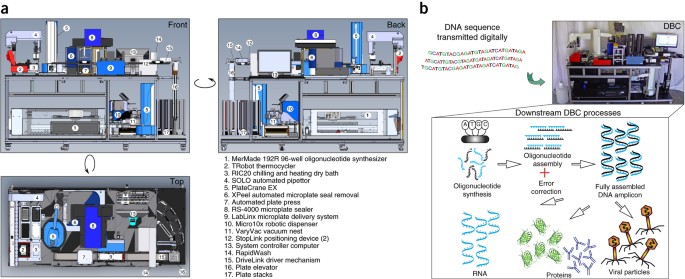
Components that constitute the DBC are marked as indicated in the following equipment list.
Schematic of the sequence of processes that are automated to occur on the DBC subsequent to recognizing the oligonucleotide synthesis file and the name of the instruments used in these
processes are shown.
(a) Genome of the phage ΦX174, was synthesized in vitro from oligonucleotides while including or not including an error-correction step. Error-corrected genome produced many more functional
viral particles (as demonstrated by plaque formation on susceptible E. coli strain HF4704, Supplementary Methods), than the genome that was not subjected to an error-correction step during
synthesis. (b) In vitro coupled transcription-translation reaction (PURExpress, New England Biolabs) was used to produce green fluorescent protein (GFP) from DNA templates that were
error-corrected or not subjected to the error-correction reaction. A no template control was included. Relative fluorescence units (RFU) (Ex: 480nm; Em: 510nm) was measured every 20min after
the first 60min of incubation at 37˚C. A consistent lag in the GFP yield was observed when a non-error corrected template was used (green curve vs orange curve).
T7- or tac- promoter driven- green fluorescent protein (GFP) encoding template with or without the T7 terminator was transcribed and translated using PURExpress in vitro Protein Synthesis
Kit (New England Biolabs) or E. coli S30 Extract System for Linear Templates (Promega). Transcription-translation reactions were incubated for two hours at 37˚C before measuring relative
fluorescence units (RFU) (Ex: 480nm; Em: 510nm). (a) To avoid DNA purification on the DBC, unpurified DNA amplicon directly from a PCR was tested for the production of protein, when
incubated with in vitro coupled transcription-translation systems. 3-6 μl of unpurified T7-GFP template was used in 25 μl of PURExpress and the RFUs were recorded. (b) Two in vitro
transcription-translation systems were compared, PURExpress in vitro Protein Synthesis Kit (New England Biolabs) or E. coli S30 Extract System for Linear Templates (Promega), for the
production of GFP using none or 3 μl of unpurified PCR product carrying T7 or the tac promoter. (c) Robustness of the PURExpress system was tested by pre-incubating the reaction mix under
sub-optimal conditions such as -20˚C or 4˚C for 16 hours prior to the coupled transcription-translation reaction for the ease-of-use in the DBC. Temperature conditions were tested with
T7-promoter driven GFP templates with or without the T7-terminator to assess the importance of the T7-terminator regulatory element.
DNA template carrying encoding GFP with T7 -promoter and -terminator as regulatory elements was transcribed and translated using the PURExpress system. A negative control for the in vitro
coupled transcription-translation reaction was included with no DNA template added to the reaction. These two reactions were tested for protein production on the Typhoon Imager (Amersham
Biosciences) using the Green laser setting along with a GFP standard (10 μg) purchased from Vector Laboratories. GFP fluorescence from the DBC sample is indicative of the production of
properly-folded and functional GFP.
Lucentis (a) and Herceptin (b) were synthesized by assembling a tac-promoter driven bi-cistronic light and heavy chain ORFs and translating this construct using the E. coli S30 Extract
System for Linear Templates (Promega). Translation was done in the presence of FluoroTect™ GreenLys in vitro Translation Labeling System (Promega), which enabled fluorescence-based
visualization of the protein. Lucentis (a) is a combination of two polypeptides of very similar molecular weight (24 kDa and 25 kDa), which could not be successfully resolved with our
system. In both (a) and (b), the control and the antibody lanes were contrasted more than the protein marker lane to visualize the proteins produces.
Hemagglutinin antigen H7 was assembled from oligo nucleotides in the DBC and ligated into a self-amplifying RNA replicon based on the genome of the Venezuelan equine encephalitis virus
before transcription. To verify the presence of H7 coding region within the RNA replicon mRNA, an oligo dT primer was used in RT-PCR (NEB ProtoScript, with or without RT, +/-) to convert the
RNA to cDNA. cDNA, thus generated, was subsequently used as a PCR template for PCR with primers flanking the H7 coding region (1790bp).
Cells were fixed and immuno-stained with an anti-H7 primary antibody and Alexa Flour 488 secondary antibody. (a) Bright-field image of H7 Replicon transfected Vero cells. (b) IFA of H7
Replicon transfected cells showing H7 specific expression. (c) Overlay of H7 and DAPI channels. (d) Overlay of H7, DAPI, and bright-field channels.
Each of the nine DNA templates (listed on Supplementary Table 4) assembled on the DBC, was subjected to GC-flux analysis using GPMiner (http://gpminer.mbc.nctu.edu.tw/) across the entire
length of the template using a 60-nucleotide window. The number of windows (X-axis) was plotted against the GC% of the windows (Y-axis).
Full-length DNA and protein gels corresponding to those in Figure 2 are shown. The expected DNA and protein products are indicated by arrows.
Anyone you share the following link with will be able to read this content: