- Select a language for the TTS:
- UK English Female
- UK English Male
- US English Female
- US English Male
- Australian Female
- Australian Male
- Language selected: (auto detect) - EN
Play all audios:
Access through your institution Buy or subscribe In rats, the thymus is a pinkish-gray, soft organ located along the ventral aspect of the trachea, dorsal to the sternum at the thoracic
inlet. It is composed of two distinct encapsulated lobes. In contrast to most mammals, in rats, the thymus does not completely involute and atrophy with age, although thymic weight does
decrease by a factor of 7 between 3 months and 18 months of age1. The thymus is a primary lymphoid organ that serves the immune system by providing an optimal microenvironment for developing
T cells. The microenvironment of the organ is peculiar among lymphoid organs, as the supporting stroma consists of reticular epithelial cells2. Lymphocyte precursors from the bone marrow
travel to the thymus and develop into thymocytes (immature lymphocytes) and, subsequently, T lymphocytes (or T cells). Mature T lymphocytes exit the thymus and comprise the peripheral T-cell
population responsible for directing many components of the adaptive immune system. Congenital absence of a thymus (as in nude mice and rats) or surgical removal at an early age results in
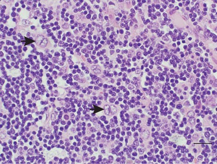
severe immunodeficiency and a high susceptibility to infection3. Thymomas are composed of a mixture of lymphocytes and epithelial cells. The epithelial cells are considered the tumor cell
component in thymomas, whereas lymphocytes are the neoplastic component in thymic lymphomas. In thymomas, the relative composition ranges from almost entirely epithelial cells to almost
entirely lymphocytes with various proportions between4,5. A definitive diagnosis may be challenging in lymphocyte-rich thymomas because the histological sections may resemble a lymphoma more
than a thymoma. The presence of small groups of epithelial cells in enlarged thymuses and the absence of involvement of other organs are generally considered diagnostic features of
thymomas6,7. This is a preview of subscription content, access via your institution ACCESS OPTIONS Access through your institution ADDITIONAL ACCESS OPTIONS: * Log in * Learn about
institutional subscriptions * Read our FAQs * Contact customer support REFERENCES * Plecas-Solarovic, B. et al. Morphometrical characteristics of age-associated changes in the thymus of old
male Wistar rats. _Anat. Histol. Embryol._ 35, 380–386 (2006). Article CAS Google Scholar * Henk-Jan Schuurman, C.F.K. & Kendall, M.D. Thymic microenvironment at the light microscopic
level. _Microscopy Res. Technique_ 38, 216–226 (1997). Article Google Scholar * Miller, J.F. The discovery of thymus function and of thymus-derived lymphocytes. _Immunol. Rev._ 185, 7–14
(2002). Article CAS Google Scholar * Kuper, C.F., Beems, R.B. & Hollanders, V.M. Spontaneous pathology of the thymus in aging Wistar (Cpb:WU) rats. _Vet. Pathol._ 23, 270–277 (1986).
Article CAS Google Scholar * Murray, A.B. et al. Incidence, morphology, and ultrastructure of spontaneous thymoma--the most common neoplasm in W/Nhg rats. _J. Natl. Cancer Inst._ 75,
369–379 (1985). CAS PubMed Google Scholar * Kaspareit-Rittinghausen, J., Deerberg, F. & Sommer, R. Atypical epithelial thymomas in rats. _Lab. Anim._ 23, 337–339 (1989). Article CAS
Google Scholar * Kuper, C.F. & Beems, R.B. Thymoma, epithelial, rat. in _Hemopoietic System_ (eds. Jones, T.C. et al.) 280–286 (Springer, Berlin, 1990). Chapter Google Scholar *
Altman, N.H. & Goodman, D.G. Neoplastic diseases. in _The Laboratory Rat, Volume I: Biology and Diseases_ (eds. Baker, H.J., Lindsey, J.R. & Weisbroth, S.H.) 353–355 (Academic, New
York, 1979). Google Scholar * Hinsull, S.M. & Bellamy, D. Spontaneous thymoma in an inbred strain of rat. _J. Natl. Cancer Inst._ 58, 1609–1614 (1977). Article CAS Google Scholar *
Naylor, D.C., Krinke, G.J. & Ruefenacht, H.J. Primary tumours of the thymus in the rat. _J. Comp. Pathol._ 99, 187–203 (1988). Article CAS Google Scholar * Yamada, S. et al.
Spontaneous thyoma in Buffalo rats. _Gann._ 64, 287–291 (1973). CAS PubMed Google Scholar * Kato, F. & Watanabe, M. Motor dysfunction in spontaneous thymoma rats, Buffalo/Mna. _J.
Pharmacobiodyn._ 5, 727–733 (1982). Article CAS Google Scholar Download references ACKNOWLEDGEMENTS We thank Matthew Davis (Georgia State University) for veterinary technical support and
for taking the gross photographs. RIGHTS AND PERMISSIONS Reprints and permissions ABOUT THIS ARTICLE CITE THIS ARTICLE Diagnosis | Thymoma. _Lab Anim_ 38, 116–117 (2009).
https://doi.org/10.1038/laban0409-116 Download citation * Issue Date: April 2009 * DOI: https://doi.org/10.1038/laban0409-116 SHARE THIS ARTICLE Anyone you share the following link with will
be able to read this content: Get shareable link Sorry, a shareable link is not currently available for this article. Copy to clipboard Provided by the Springer Nature SharedIt
content-sharing initiative