- Select a language for the TTS:
- UK English Female
- UK English Male
- US English Female
- US English Male
- Australian Female
- Australian Male
- Language selected: (auto detect) - EN
Play all audios:
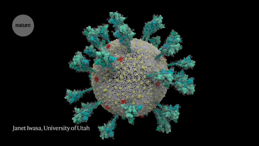
The coronavirus sports a luxurious sugar coat. “It’s striking,” thought Rommie Amaro, staring at her computer simulation of one of the trademark spike proteins of SARS-CoV-2, which stick out
from the virus’s surface. It was swathed in sugar molecules, known as glycans. “When you see it with all the glycans, it’s almost unrecognizable,” says Amaro, a computational biophysical
chemist at the University of California, San Diego. Many viruses have glycans covering their outer proteins, camouflaging them from the human immune system like a wolf in sheep’s clothing.
But last year, Amaro’s laboratory group and collaborators created the most detailed visualization yet of this coat, based on structural and genetic data and rendered atom-by-atom by a
supercomputer. On 22 March 2020, she posted the simulation to Twitter. Within an hour, one researcher asked in a comment: what was the naked, uncoated loop sticking out of the top of the
protein? Amaro had no idea. But ten minutes later, structural biologist Jason McLellan at the University of Texas at Austin chimed in: the uncoated loop was a receptor binding domain (RBD),
one of three sections of the spike that bind to receptors on human cells (see ‘A hidden spike’). In Amaro’s simulation, when the RBD lifted up above the glycan cloud, two glycans swooped in
to lock it into place, like a kickstand on a bicycle. When Amaro mutated the glycans in the computer model, the RBD collapsed. McLellan’s team built a way to try the same experiment in the
lab, and by June 2020, the collaborators had reported that mutating the two glycans reduced the ability of the spike protein to bind to a human cell receptor1 — a role that no one has
previously recognized in coronaviruses, McLellan says. It’s possible that snipping out those two sugars could reduce the virus’s infectivity, says Amaro, although researchers don’t yet have
a way to do this. Since the start of the COVID-19 pandemic, scientists have been developing a detailed understanding of how SARS-CoV-2 infects cells. By picking apart the infection process,
they hope to find better ways to interrupt it through improved treatments and vaccines, and learn why the latest strains, such as the Delta variant, are more transmissible. What has emerged
from 19 months of work, backed by decades of coronavirus research, is a blow-by-blow account of how SARS-CoV-2 invades human cells (see ‘Life cycle of the pandemic coronavirus’). Scientists
have discovered key adaptations that help the virus to grab on to human cells with surprising strength and then hide itself once inside. Later, as it leaves cells, SARS-CoV-2 executes a
crucial processing step to prepare its particles for infecting even more human cells. These are some of the tools that have enabled the virus to spread so quickly and claim millions of
lives. “That’s why it’s so difficult to control,” says Wendy Barclay, a virologist at Imperial College London. BARBED AND READY It starts with the spikes. Each SARS-CoV-2 virion (virus
particle) has an outer surface peppered with 24–40 haphazardly arranged spike proteins that are its key to fusing with human cells2. For other types of virus, such as influenza, external
fusion proteins are relatively rigid. SARS-CoV-2 spikes, however, are wildly flexible and hinge at three points, according to work published in August 2020 by biochemist Martin Beck at the
Max Planck Institute of Biophysics in Frankfurt, Germany, and his colleagues3. That allows the spikes to flop around, sway and rotate, which could make it easier for them to scan the cell
surface and for multiple spikes to bind to a human cell. There are no similar experimental data for other coronaviruses, but because spike-protein sequences are highly evolutionarily
conserved, it is fair to assume the trait is shared, says Beck. Early in the pandemic, researchers confirmed that the RBDs of SARS-CoV-2 spike proteins attach to a familiar protein called
the ACE2 receptor, which adorns the outside of most human throat and lung cells. This receptor is also the docking point for SARS-CoV, the virus that causes severe acute respiratory syndrome
(SARS). But compared with SARS-CoV, SARS-CoV-2 binds to ACE2 an estimated 2–4 times more strongly4, because several changes in the RBD stabilize its virus-binding hotspots5. Worrying
variants of SARS-CoV-2 tend to have mutations in the S1 subunit of the spike protein, which hosts the RBDs and is responsible for binding to the ACE2 receptor. (A second spike subunit, S2,
prompts viral fusion with the host cell’s membrane.) The Alpha variant, for example, includes ten changes in the spike-protein sequence, which result in RBDs being more likely to stay in the
‘up’ position6. “It is helping the virus along by making it easier to enter into cells,” says Priyamvada Acharya, a structural biologist at the Duke Human Vaccine Institute in Durham, North
Carolina, who is studying the spike mutations. The Delta variant, which is now spreading around the world, hosts multiple mutations in the S1 subunit, including three in the RBD that seem
to improve the RBD’s ability to bind to ACE2 and evade the immune system7. RESTRICTED ENTRY Once the viral spikes bind to ACE2, other proteins on the host cell’s surface initiate a process
that leads to the merging of viral and cell membranes (see ‘Viral entry up close’). The virus that causes SARS, SARS-CoV, uses either of two host protease enzymes to break in: TMPRSS2
(pronounced ‘tempress two’) or cathepsin L. TMPRSS2 is the faster route in, but SARS-CoV often enters instead through an endosome — a lipid-surrounded bubble — which relies on cathepsin L.
When virions enter cells by this route, however, antiviral proteins can trap them. SARS-CoV-2 differs from SARS-CoV because it efficiently uses TMPRSS2, an enzyme found in high amounts on
the outside of respiratory cells. First, TMPRSS2 cuts a site on the spike’s S2 subunit8. That cut exposes a run of hydrophobic amino acids that rapidly buries itself in the closest membrane
— that of the host cell. Next, the extended spike folds back onto itself, like a zipper, forcing the viral and cell membranes to fuse. The virus then ejects its genome directly into the
cell. By invading in this spring-loaded manner, SARS-CoV-2 infects faster than SARS-CoV and avoids being trapped in endosomes, according to work published in April by Barclay and her
colleagues at Imperial College London9. The virus’s speedy entry using TMPRSS2 explains why the malaria drug chloroquine didn’t work in clinical trials as a COVID-19 treatment, despite early
promising studies in the lab10. Those turned out to have used cells that rely exclusively on cathepsins for endosomal entry. “When the virus transmits and replicates in the human airway, it
doesn’t use endosomes, so chloroquine, which is an endosomal disrupting drug, is not effective in real life,” says Barclay. The discovery also points to protease inhibitors as a promising
therapeutic option to prevent a virus from using TMPRSS2, cathepsin L or other proteases to enter host cells. One TMPRSS2 inhibitor, camostat mesylate, which is approved in Japan to treat
pancreatitis, blocked viral entry into lung cells8, but the drug did not improve patients’ outcomes in an initial clinical trial11. “From my perspective, we should have such protease
inhibitors as broad antivirals available to fight new disease outbreaks and prevent future pandemics at the very beginning,” says Stefan Pöhlmann, director of the Infection Biology Unit at
the German Primate Center in Göttingen, who has led research on ACE2 binding and the TMPRSS2 pathway. DEADLY COMPETITION The next steps of infection are murkier. “There are a lot more black
boxes once you are inside the cell,” says chemist Janet Iwasa at the University of Utah in Salt Lake City, who is developing an annotated animation of the viral life cycle. “There’s more
uncertainty, and competing hypotheses.” After the virus shoots its RNA genome into the cell, ribosomes in the cytoplasm translate two sections of viral RNA into long strings of amino acids,
which are then snipped into 16 proteins, including many involved in RNA synthesis. Later, more RNAs are generated that code for a total of 26 known viral proteins, including structural ones
used to make new virus particles, such as the spike, and other accessory proteins. In this way, the virus begins churning out copies of its own messenger RNA. But it needs the cell’s
machinery to translate those mRNAs into proteins. How a rampant coronavirus variant blunts our immune defences Coronaviruses take over that machinery in many ways. Virologist Noam
Stern-Ginossar and her team at the Weizmann Institute of Science in Rehovot, Israel, zoomed in on three mechanisms by which SARS-CoV-2 suppresses the translation of host mRNA in favour of
its own. None are exclusive to this virus, but the combination, speed and magnitude of the effects seem unique, says Stern-Ginossar. First, the virus eliminates the competition: viral
protein Nsp1, one of the first proteins translated when the virus arrives, recruits host proteins to systematically chop up all cellular mRNAs that don’t have a viral tag. When
Stern-Ginossar’s team put that same tag on the end of a host mRNA, the mRNA was not chopped up12. Second, infection reduces overall protein translation in the cell by 70%. Nsp1 is again the
main culprit, this time physically blocking the entry channel of ribosomes so mRNA can’t get inside, according to work from two research teams13,14. The little translation capacity that
remains is dedicated to viral RNAs, says Stern-Ginossar. Finally, the virus shuts down the cell’s alarm system. This happens in numerous ways, but Stern-Ginossar’s team identified one clear
mechanism for SARS-CoV-2: the virus prevents cellular mRNA from getting out of the nucleus, including instructions for proteins meant to alert the immune system to infection. A second team
confirmed this finding, and again pointed to Nsp1: the protein seems to jam up exit channels in the nucleus so nothing can escape15. Because gene transcripts can’t get out of the nucleus,
the infected cells don’t release many interferons — these are signalling proteins that alert the immune system to the presence of a virus. SARS-Cov-2 is particularly efficient at shutting
down this alarm system: compared with other respiratory viruses, including SARS-CoV and respiratory syncytial virus, SARS-CoV-2 infection induces significantly lower levels of interferons16.
And this June, researchers reported mutations in the Alpha variant that seem to enable it to subdue interferon production even more efficiently17. “It’s clear that SARS-CoV-2 is a very fast
virus that has a unique ability to prevent our immune system from recognizing and combating infection in the first stages,” says Stern-Ginossar. By the time the immune system does realize
there is a virus, there is so much of it that immune-response proteins sometimes flood the bloodstream at a faster rate than normal — which can cause damage. Doctors saw early in the
pandemic that some people with COVID-19 who become very ill are harmed by an overactive immune response to SARS-CoV-2, as well as by the virus itself. Some proven treatments work by
dampening down this immune response. RENOVATION STATION Once the virus has taken over host translation, it starts a home makeover, extensively remodelling the interior and exterior of the
cell to its needs. First, some of the newly made viral spike proteins travel to the surface of the cell and poke out of the host-cell membrane. There, they activate a host calcium-ion
channel, which expels a fatty coating onto the outside of the cell — the same coating found on cells that naturally fuse together, such as muscle cells. At this point, the infected cell
fuses to neighbouring cells expressing ACE2, developing into massive individual respiratory cells filled with up to 20 nuclei. These fused structures, called syncytia, are induced by viral
infections such as HIV and herpes simplex virus, but not by the SARS virus, says molecular biologist Mauro Giacca at King’s College London, who led the team that published the finding in
April18. He hypothesizes that forming syncytia allows infected cells to thrive for long periods of time, churning out more and more virions. “This is not a hit-and-run virus,” he says. “It
persists.” A second team, led by researcher Qiang Sun at the Chinese Academy of Medical Sciences in Beijing, found that some COVID-19-infected cells even form syncytia with lymphocytes — one
of the body’s own immune cells19. This is a known mechanism of immune evasion by tumour cells, but not by viruses. It suggests that infected cells avoid immune detection by simply grabbing
on to and merging with nearby immune scouts. On the inside of the cell, even more change is occurring. Like other coronaviruses, SARS-CoV-2 transforms the long, thin endoplasmic reticulum
(ER), a network of flat membranes involved in protein synthesis and transport, into double-membrane spheres, as if the ER were blowing bubbles. These double-membrane vesicles (DMVs) might
provide a safe place for viral RNA to be replicated and translated, shielding it from innate immune sensors in the cell, but that hypothesis is still being investigated. Proteins involved in
making DMVs could be good drug targets, because they seem to be necessary for viral replication. For instance, a host protein, TMEM41B, is needed to mobilize cholesterol and other lipids to
expand the ER membranes so that all the virus parts will fit inside20. “When you take TMEM41B out, it has a major impact on infection,” says Vineet Menachery, a coronavirus researcher at
the University of Texas Medical Branch in Galveston, who was involved in the research. The coronavirus transmembrane protein Nsp3 could also be a target: it creates a crown-like pore in the
walls of the DMVs to shuttle out newly made viral RNA21. Most viruses that have an outer wrapping, known as an envelope, form this feature by assembly directly at the edge of the cell,
co-opting some of the cell’s own plasma membrane on their way out. But newly made coronavirus proteins take a different path. ENJOYING OUR LATEST CONTENT? LOGIN OR CREATE AN ACCOUNT TO
CONTINUE * Access the most recent journalism from Nature's award-winning team * Explore the latest features & opinion covering groundbreaking research Access through your
institution or Sign in or create an account Continue with Google Continue with ORCiD


:max_bytes(150000):strip_icc():focal(999x0:1001x2)/plane-crash-2-2000-ca65e24ec06447c89741c8f4766e3888.jpg)

:max_bytes(150000):strip_icc():focal(319x0:321x2)/people_social_image-60e0c8af9eb14624a5b55f2c29dbe25b.png)