- Select a language for the TTS:
- UK English Female
- UK English Male
- US English Female
- US English Male
- Australian Female
- Australian Male
- Language selected: (auto detect) - EN
Play all audios:
ABSTRACT PURPOSE To determine the prevalence and features of the different types of involvement of the optic nerve in ocular toxoplasmosis. METHODS Retrospective cross-sectional study. All
patients with active ocular toxoplasmosis, consulting in the Uveitis Section of the Ophthalmology Department were selected. The involvement of the optic nerve was classified in the following
categories: juxtapapillary retinochoroiditis, pure papillitis, neuroretinitis, distant lesion, and mixed lesion. RESULTS The prevalence of involvement of the optic nerve found was 5.3%. The
optic nerve involvement with the presence of a concurrent active distant lesion, occurred in 22 eyes (43.1%). A juxtapapillary lesion was found in 18 eyes (35.3%). Eight eyes (15.7%)
presented lesions characterised as mixed. Isolated papillitis occurred in 3 eyes (5.9%). Forty-seven lesions (95.9%) were unilateral and two (4.1%) were bilateral. Twenty-eight eyes (54.9%)
had pre-existing lesions and 23 (45%) were primary lesions. Visual acuity improved in 35 eyes (71.4%) and remained unchanged in 14 eyes (28.5%). CONCLUSION The involvement of the optic nerve
most frequently found in ocular toxoplasmosis was optic nerve oedema with a concurrent distant active lesion. The second type of lesion most often found was juxtapapillary
retinochoroiditis. Involvement was monocular in most cases and the visual prognosis was favourable. SIMILAR CONTENT BEING VIEWED BY OTHERS FREQUENCY AND VISUAL OUTCOMES OF OCULAR
TOXOPLASMOSIS IN AN ADULT BRAZILIAN POPULATION Article Open access 09 February 2021 CLINICAL MANIFESTATIONS AND VISUAL OUTCOMES ASSOCIATED WITH OCULAR TOXOPLASMOSIS IN A BRAZILIAN POPULATION
Article Open access 04 February 2021 COMPARISON BETWEEN THE AREAS OF SCARRED AND ACTIVE TOXOPLASMIC RETINOCHOROIDITIS Article Open access 24 November 2020 INTRODUCTION Uveitis is a
worldwide prevalent disease, affecting people at productive age and it is a major cause of blindness.1 Ocular toxoplasmosis, in turn, is one of the most frequent aetiologies of posterior
uveitis.2 In the State of Rio Grande do Sul, Brazil, it assumes considerable proportions, becoming a public health problem of extreme relevance.3 Ocular involvement in toxoplasmosis may
result from acquired infection after birth or from the congenital form of the disease.4, 5, 6, 7, 8 The diagnosis is basically clinical, complemented by serologic tests in order to obtain
evidence of previous exposure to the parasite.9 The involvement of the optic nerve in ocular toxoplasmosis was already described by several authors at the beginning of last century, and more
recently new studies on this subject have appeared. _Toxoplasma gondii_ may cause a lesion in the optic disc because of contiguousness;10, 11 by direct involvement11, 12, 13, 14, 15, 16, 17
or even become involved when a retinochoroiditis lesion is located far from the optic nerve.8, 9 The contiguous form was described by Jensen many years ago18 as a specific entity in four
cases of juxtapapillary choroiditis, later considered as probably due to tuberculous aetiology. In 1952, Wilder recognised _T. gondii_ histopathologically in necrotic retinochoroidal lesion
in adult patients.19, 20, 21 Lesions had granulomatous character and marked necrosis in many instances led to a pathologic diagnosis of tuberculosis. So, currently it is accepted that most
cases of Jensen's choroiditis are of toxoplasmic aetiology. This type of lesion consists of a typical area of retinochoroiditis in contact with a swollen optic disc and accompanied by a
typical sectorial deficit in the visual field. The direct involvement of the optic nerve by _T. gondii_ was demonstrated histopathologically by the presence of different forms of the
parasite inside it.22 At ophthalmoscopic observation, this involvement may be subdivided into pure anterior neuritis or papillitis and neuroretinitis.11 In pure papillitis the parasite
affects the optic disc directly, causing a swollen papilla with sheathing of the peripapillary veins and there may be no concurrent active retinochoroiditis lesion.8 However, in the majority
of these cases a peripheral scar lesion and vitreitis is always present over the optic disc.13, 17 Neuroretinitis is characterised usually by a swollen optic nerve accompanied of
papillo-macular or serous macular detachment of the retina associated to hard exudates in the macula distributed in a star shape.14, 16, 23, 24 We have observed another form of involvement
of the optic disc due to toxoplasmosis that would be caused secondarily by an active distant retinal lesion.9 In these cases there is an active focal necrotising retinochoroiditis lesion
located at variable distances from the optic disc that presents changes that resembles papillitis.8 There are no detailed studies about this type of optic nerve involvement in the
literature. The objective of this study was to determine the total prevalence and the different types of involvement of the optic nerve in ocular toxoplasmosis, as well as to study several
accompanying clinical parameters. MATERIALS AND METHODS The present retrospective cross-sectional study was performed in the Uveitis Section of the Ophthalmology Department at Hospital de
Clínicas de Porto Alegre. All patients attended in our Uveitis Section (926 records) who presented active ocular toxoplasmosis in the 1987–2001 period were reviewed. Patients who were
immunocompromised and those who had a previous optic nerve lesion by causes other than toxoplasmosis were excluded. Patients with atrophic optic nerve caused by toxoplasmosis were also
excluded because this alteration is considered a sequelae and in this study we wanted to evaluate the evolution of active lesions. To be included in the study, the patient had to present
alterations of the optic nerve in the form of swollen disc or papillitis concomitantly with the presence of an active ocular lesion caused by _T. gondii_. The diagnosis of ocular
toxoplasmosis was based on the presence of typical retinochoroiditis lesions and IgM- and/or IgG-specific antibodies for toxoplasmosis. The methods used for serology were indirect
immunofluorescence reactions (IFI), ELFA (enzyme-linked fluorescent assay) and MEIA (microparticle enzyme immunoassay) for IgM and IgG. Serological tests were also performed to detect other
infectious diseases such as syphilis, rubella, herpes, cytomegalovirus, and tuberculosis. Fundoscopy was performed under mydriasis, with direct and indirect ophthalmoscope, and fundoscopic
biomicroscopy with 78 diopter lens. Angiography and retinography were also performed in all patients for documentation. All patients were treated with the usual therapeutic scheme used in
our Department.25 (Sulphadiazine 500 mg 2 cp four time/day, pyrimethamine 25 mg 1 cp two time/day, Folinic acid 7.5 mg every day and oral corticosteropids – prednisone 1 mg/kg/day). The
files were analysed using a protocol containing the following data: patient identification, date of initial exam, eye affected, laterality, type of optic nerve lesion according to
classification described elsewhere and initial and final visual acuity. The presence of pre-existing retinochoroiditis scars in the affected eye was also studied. The visual acuity was
measured with Snellen chart at 6 m (better optical correction). The final visual acuity was measured when the lesion was considered cured (1–3 months). The optic nerve involvement was
classified as follows: * 1) _Juxtapapillary retinochoroiditis:_ retinochoroidal lesion contiguous to the swollen optic disc (Figure 1 ). * 2) _Pure Papillitis_: swollen optic disc and
sheathing of the peripapillary veins in the presence of a healed toxoplasmic retinochoroiditis lesion (Figure 2 ). * 3) _Neuroretinitis_: swollen optic disc, papillo-macular or serous
macular detachment of the retina with hard exudates at the macula. * 4) _Distant lesion:_ swollen optic disc in the presence of a distant active lesion (Figure 3). * 5) _Mixed lesion:_
presence of more than one type of involvement previously described concurrently (Figure 4 ). Statistical analysis of the data was performed by SPSS program version 10. The _χ_2 Pearson
statistical test and _t_-Student's test were also applied. The level of significance considered was 0.05. The present study was approved by the Ethics Comitee of the Hospital de
Clínicas de Porto Alegre. RESULTS Of the 926 patients, 49 patients (51 eyes) had involvement of the optic nerve with a total prevalence of 5.03%. The mean age of the patients with changes in
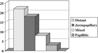
the optic disc was 27 years and 4 months (with a variation of 6–65 years) (Table 1). The optic nerve involvement concomitantly with a distant lesion occurred in 22 eyes (43.1%). The
involvement of the optic disc by a juxtapapillary lesion occurred in 18 eyes (35.3%). Eight eyes (15.7%) presented more than one type of lesion concurrently, being characterised as mixed. Of
these eight eyes, six eyes (75%) presented a distant lesion associated with neuroretinitis and two (25%) presented with juxtapapillary lesion and neurorretinitis. The pure papillitis
occurred in three eyes (5.9%) (Graph 1). No cases of isolated neuroretinitis were found. Forty-seven had (95.9%) unilateral lesions and two (4.1%) had bilateral (Table 1). Twenty-eight eyes
(54.9%) had pre-existing lesions and 23 (45%) were primary lesions (Table 1). After treatment the visual acuity improved in 35 eyes (71.4%), and remained unchanged in 14 eyes (28.5%) (Graph
2). Patients with mixed lesions had the best visual prognosis because 75% of them improved visual acuity. The percentage of improval visual acuity was 66.6, 63.63, and 61.1% in papillitis,
distant lesions, and juxtapapillary lesions, respectively. DISCUSSION Ocular toxoplasmosis is very frequent in Rio Grande do Sul, and it is the main cause of uveitis in our Department.3
After many years of observations, we could verify a wide variety of clinical presentations of this disease.26 Concomitantly, we have had opportunity to observe and treat a considerable
number of patients presenting with optic nerve alterations. Although the prevalence found in our study seems not to be very high (5.3%) this type of involvement represents concern for the
clinician because of severity of the disease and the diagnostic difficulties with other types of neuritis. The optic nerve involvement in the presence of an active distant retinochoroiditis
was the most frequent type of lesion found in our study. Some authors27, 28 do not consider optic disc swelling caused by distant lesion as true papillitis but as a reactive oedema of the
optic nerve due to a hypersensitivity reaction, and then the alteration is then called secondary papillitis.27 This study shows that they evolve favourably with treatment and usually do not
leave any permanent change in the optic nerve (63.63% of patient had improve visual acuity after resolution of inflammation). The second most numerous group was represented by a swollen
optic disc with a juxtapapillary lesion. Although the clinical appearance is usually very serious, with a large amount of exudates, hemorrhage, optic nerve oedema and low visual acuity, this
type of change responds rapidly to medication, reabsorbing the oedema and leaving a typical retinal pigmented lesion. A scar may not be apparent at the beginning of the disease and became
evident later.17 In some cases, a glial reaction remains on the optic disc. Eight eyes presented mixed lesions and all these cases neuroretinitis was present. In 1977, Gass29 postulated the
pathophysiological mechanisms of this entity. It is possible that papillitis and the macular star found, together with a distant toxoplasmic retinochoroiditis, occurs as a hypersensitivity
reaction and not due to direct damage caused by the parasite to the optic nerve as described in isolated neuroretinitis.29 Toxoplasmosis is an infrequent cause of pure papillitis. In our
study, three eyes presented this manifestation (5.9%). The findings that suggest the direct injury of the optic nerve by the parasite are the presence of optic nerve inflammation, vitreitis,
a healed toxoplasmic lesion, and the presence of toxoplasmic antibodies. Other causes of swollen optic disc, such as demyelination diseases, vasculitis of the optic disc, ischaemic optic
neuropathy, increased intracranial pressure, sarcoidosis, systemic lupus erythematosus, toxocariasis, optic nerve tumor, and other infectious causes of retinitis must always be excluded.13
In the present study, 28 eyes already had a toxoplasmic lesion before the optic nerve become involved, suggesting recurrences of an old disease and in 23 eyes the lesion of the optic nerve
was the first ocular manifestation of ocular infection by the parasite. There was a marked dominance of unilateral lesions in our sample. Only two patients presented a change in the optic
nerve in both eyes. One of these patients had proven congenital toxoplasmosis and the other had probable of congenital infection, but we did not confirm the diagnosis. Some patients of this
study performed visual field testing, that was recorded using the Humphrey 24/2 FastPac Strategy but we did not evaluate this records because few patients was submitted to this examination.
Stanford _et al_30 suggest in their study that toxoplasmic retinochoroidal scars close to the optic disc are associated with absolute defects. In the other hand, lesions that are far from
the disc are more likely to produce relative defects. The involvement of the optic nerve in ocular toxoplasmosis may lead an inexperienced clinician to think that visual acuity may be
severely and permanently affected. However, in most of the cases we initially observe a low visual acuity that after treatment and with the healing of the lesion, improves significantly.
Concerning this point our findings are coincident with those in the literature.13, 14, 16, 17, 24, 31 The final visual acuity depends basically on the location of the retinal lesion, and not
on the involvement of the optic disc. REFERENCES * Michel SS, Foster CS . Definition, classification, etiology and epidemiology. In: Foster CS, Vitale AT (eds). _Diagnosis and Treatment of
Uveitis_. WB. Saunders Company: Philadelphia, 2002, pp 17–26. Google Scholar * Oréfice F, Bonfioli AA . Toxoplasmose. In: Oréfice F (ed). _Uveíte: Clinica & Cirurgia: Atlas &
Texto_. Cultura Médica: Rio de Janeiro, 2000, pp 619–680. Google Scholar * Melamed J, Güntzel I, Lindenmeyer R . Uveitis in Southern Brazil. _Uveitis in Third Millennium_. Elsevier Science:
Amsterdam, 2000, pp 247–250. Google Scholar * Perkins ES . Ocular Toxoplasmosis. _Brit J Ophthal_ 1973; 57: 1–17. Article CAS Google Scholar * Melamed J . Peculiaridades da toxoplasmose
ocular no Rio Grande do Sul. _Arq Bras Oftal_ 1988; 51(5): 197–200. Google Scholar * Silveira C, Belfort Jr R, Burnier Jr M, Nussenblatt R . Acquired toxoplasmic infection as the cause of
toxoplasmic retinochoroiditis in families. _Am J Ophtalmol_ 1988; 106: 362–364. Article CAS Google Scholar * Nussenblatt RB, Belfort Jr R . Ocular toxoplasmosis – an old disease
revisited. _JAMA_ 1994; 271: 304–307. Article CAS Google Scholar * Gonçalves EC, Oréfice F, Mendes AG, Pedroso EP . Toxoplasmose ocular adquirida tardia. Relato de três casos simultâneos
da mesma família. _Revista Brasileira de Oftalmologia_ 1995; 54(5): 57–60. Google Scholar * Melamed J . Toxoplasmose ocular. In: Lavinsky J (ed). _Doenças Prevalentes da Retina e Vítreo_.
Cultura Médica: Rio de Janeiro, 2002, pp 597–620. Google Scholar * Uchida Y, Kakehashi Y, Kameyama K . Juxtapapillary retinochoroiditis with psychiatric disorder possibly caused by
toxoplasma. _Am J Ophhalmol_ 1978; 86: 791–793. Article CAS Google Scholar * Banta JT, Davis JL, Lam BL . Presumed toxoplasmic anterior optic neuropathy. _Ocul Immunol Inflamm_ 2002;
10(3): 201–211. Article Google Scholar * Berengo A, Frezzoti R . Active neuro-ophtalmic toxoplasmosis. A clinical study on 19 patients. _Adv Ophthalmol_ 1962; 12: 265. Google Scholar *
Folk JC, Lobes L . Presumed toxoplasmic papillitis. _Ophthalmol_ 1984; 91: 64–67. Article CAS Google Scholar * Moreno RJ, Weisman J, Waller S . Neuroretinitis: an unusual presentation of
ocular toxoplasmosis. _Ann Ophthalmol_ 1992; 24: 68–70. CAS PubMed Google Scholar * Williams N, Miller NR . Neuroretinitis. In: Pepose JS, Holland GN, Wilhelmus KR (eds). _Ocular
Infection & Immunity_, 1st ed. Mosby: St Louis, 1996, pp 601–608. Google Scholar * Fish RH, Hoskins JC, Kline CLB . Toxoplasmosis neuroretinitis. _Ophthalmol_ 1993; 100: 1177–1182.
Article CAS Google Scholar * Song A, Scott IU, Davis JL, Lam BL . Atypical anterior optic neuropathy caused by toxoplasmosis. _Am J Ophthalmol_ 2002; 133: 162–164. Article Google Scholar
* Jensen E . Retino-choroiditis juxtapapillaris. _Arch F Ophthal_ 1908; 69: 41. Google Scholar * Wilder HC . Toxoplasma chorioretinitis in adults; a preliminary study of forty-one cases
diagnosed by microscopic examination. _Arch Ophthalmol_ 1952; 47(4): 425. Article CAS Google Scholar * Wilder HC . Toxoplasma chorioretinitis in adults. _Arch Ophthalmol_ 1952; 48(4):
127–136. Article CAS Google Scholar * Holland GN, Lewis KG, O'Connor GR . Ocular toxoplasmosis: a 50th anniversary tribute to the contribuitions of Heleonor Campbell Wilder Foester.
_Arch Ophthalmol_ 2002; 120(8): 1081–1084. Article Google Scholar * Manshot WA, Daamen CBF . Connatal ocular toxoplasmosis. _Arch Ophtalmol_ 1965; 74: 48. Article Google Scholar *
Maitland CG, Miller NR . Neuroretinitis. _Arch Ophthalmol_ 1984; 102: 1146–1150. Article CAS Google Scholar * Küçükerdönmez C, Akova Y, Yilmaz G . Ocular toxoplasmosis presenting as
neuroretinitis: report of two cases. _Ocul Immunol Inflamm_ 2002; 10(3): 229–233. Article Google Scholar * Melamed J . Tratamento da toxoplasmose ocular. _Rev Bras Oftal_ 1998; 57(2):
159–163. Google Scholar * Melamed J, Eckert GU, Cagliari PZ . Atypical lesions in ocular toxoplasmosis. _Uveitis in Third Milleniun_. Elsevier Science: Buenos Aires, 2000, pp 265–268.
Google Scholar * Schlaegel Jr TF . Toxoplasmosis. In: Duane TD (ed). _Clin Ophthalmol_. Hagerstown: Harper & Row, 1982, Vol. 4, Chap 51. Google Scholar * Bosch-Driessen LE, Berendschot
TT, Ongkosuwito JV, Rothova A . Ocular toxoplasmosis: clinical features and prognosis of 154 patients. _Ophthalomology_ 2003; 109: 869–878. Article Google Scholar * Gass JDM . Optic nerve
diseases that may masquerade as macular disease. In: Gass JDM (ed). _Stereoscopic Atlas of Macular Diseases Diagnosis and Treatment_. Mosby: USA, 1997, pp 996–999. Google Scholar *
Stanford MR, Tomlin EA, Comyn O, Holland K, Pavesio C . The visual field in toxoplasmic retinochoroiditis. _Brit Journ Ophthalmol_ 2005; 89: 812–814. Article CAS Google Scholar * Smith
JR, Cunninggham ET . Atypical presentations of ocular toxoplasmosis. _Curr Opin Ophthalmol_ 2002; 13(6): 387–392. Article Google Scholar Download references AUTHOR INFORMATION AUTHORS AND
AFFILIATIONS * Department of Ophthalmology, Medical School, Federal University of Rio Grande do Sul, Porto Alegre, Brazil G U Eckert, J Melamed & B Menegaz Authors * G U Eckert View
author publications You can also search for this author inPubMed Google Scholar * J Melamed View author publications You can also search for this author inPubMed Google Scholar * B Menegaz
View author publications You can also search for this author inPubMed Google Scholar CORRESPONDING AUTHOR Correspondence to J Melamed. RIGHTS AND PERMISSIONS Reprints and permissions ABOUT
THIS ARTICLE CITE THIS ARTICLE Eckert, G., Melamed, J. & Menegaz, B. Optic nerve changes in ocular toxoplasmosis. _Eye_ 21, 746–751 (2007). https://doi.org/10.1038/sj.eye.6702319
Download citation * Received: 02 September 2005 * Accepted: 29 January 2006 * Published: 31 March 2006 * Issue Date: 01 June 2007 * DOI: https://doi.org/10.1038/sj.eye.6702319 SHARE THIS
ARTICLE Anyone you share the following link with will be able to read this content: Get shareable link Sorry, a shareable link is not currently available for this article. Copy to clipboard
Provided by the Springer Nature SharedIt content-sharing initiative KEYWORDS * ocular toxoplasmosis * papillitis * neuroretinitis * juxtapapillary retinochoroiditis