- Select a language for the TTS:
- UK English Female
- UK English Male
- US English Female
- US English Male
- Australian Female
- Australian Male
- Language selected: (auto detect) - EN
Play all audios:
Sir, Acute macular neuroretinopathy (AMNR) is a rare condition characterized by a sudden onset of mild visual impairment in one or both eyes of young patients, predominantly women.
Typically, the neuroretinopathy consists of reddish-brown wedge-shaped macular lesions, pointing towards the centre of the fovea.1 The diagnosis is clinical, aided by the exact
correspondence of the lesion with the scotoma identified on the Amsler grid. The precise location of the lesions is uncertain.1 We evaluated a case of AMNR with optical coherence tomography
(OCT) and fundus fluorescein angiography (FFA). CASE REPORT A 30-year-old man presented with blurring of vision in the left eye for 6 months. His best-corrected visual acuity was 6/6 in the
right eye and 6/9 in the left. There was no history of headache, flu-like illness, trauma, or administration of intravenous epinephrine/contrast agents. Anterior segment examination was
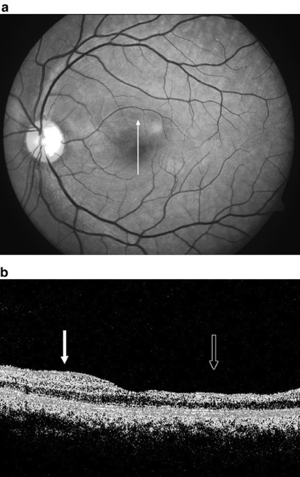
normal in both eyes. Slit-lamp biomicroscopy of the left fundus showed a subtle red wedge-shaped lesion above the fovea pointing towards its centre (Figure 1(a)). The right fundus was
normal. Amsler grid examination of the left eye showed a scotoma corresponding exactly to the macular lesion. FFA revealed no hypofluorescence of the affected area. Optical coherence
tomography of the left eye revealed distinct retinal thinning in the area of the lesion, more prominent in the inner retinal layers (Figure 1(b)). COMMENT The dark-red appearance of the
lesions in AMNR is attributed to a focal atrophic thinning of retina.1 Opinion is divided on the location of the lesions. Biomicroscopic interpretation is variable.1, 2 The striking
correspondence of the lesions to the paracentral scotomata on Amsler grid and documentation of an abnormality of the early receptor potential point to the involvement of the outer retinal
layers.1, 3 The retinal haemorrhages, acute oedematous appearance of the macula, suspected retinal capillary dilation, and an association with epinephrine administration indicate inner
retinal involvement.1, 2, 4 To the best of our knowledge, there is only one earlier report of OCT findings in AMNR: Feigl and Hass5 observed a band of high reflectivity over the retinal
pigment epithelium, and implicated an inflammatory process in the outer retina. They found no retinal thinning in their case. Our OCT findings were different from those of their study. The
differences could partly be explained by the long duration of the lesions in our patient. In late AMNR, the focal retinal thinning is almost impossible to appreciate by biomicroscopy or
stereo photography.4 We found a definite evidence of focal retinal thinning in our patient in the area affected by AMNR, more evident in the inner retina, although it may not be possible to
pinpoint the exact retinal layers involved. REFERENCES * Turbeville SD, Cowan LD, Gass JDM . Acute macular neuroretinopathy: a review of the literature. _Surv Ophthalmol_ 2003; 48: 1–11.
Article Google Scholar * Bos PJ, Deutman AF . Acute macular neuroretinopathy. _Am J Ophthalmol_ 1975; 80: 573–584. Article CAS Google Scholar * Sieving PA, Fishman GA, Salzano T, Rabb
MF . Acute macular neuroretinopathy: early receptor potential change suggests photoreceptor pathology. _Br J Ophthalmol_ 1984; 68: 229–234. Article CAS Google Scholar * O'Brien DM,
Farmer SG, Kalina RE Leon JA . Acute macular neuroretinopathy following intravenous sympathomimetics. _Retina_ 1989; 9: 281–286. Article CAS Google Scholar * Feigl B, Haas A . Optical
coherence tomography (OCT) in acute macular neuroretinopathy. _Acta Opthalmol Scand_ 2000; 78: 714–716. Article CAS Google Scholar Download references ACKNOWLEDGEMENTS We acknowledge the
support of Aravind Eye Research Foundation for this project. AUTHOR INFORMATION AUTHORS AND AFFILIATIONS * Retina-Vitreous Service, Aravind Eye Hospital & Postgraduate Institute of
Ophthalmology, Madurai, Tamil Nadu, India D Shukla, A Arora, S Ambatkar, K Ramasamy & N Perumalsamy Authors * D Shukla View author publications You can also search for this author
inPubMed Google Scholar * A Arora View author publications You can also search for this author inPubMed Google Scholar * S Ambatkar View author publications You can also search for this
author inPubMed Google Scholar * K Ramasamy View author publications You can also search for this author inPubMed Google Scholar * N Perumalsamy View author publications You can also search
for this author inPubMed Google Scholar CORRESPONDING AUTHOR Correspondence to D Shukla. RIGHTS AND PERMISSIONS Reprints and permissions ABOUT THIS ARTICLE CITE THIS ARTICLE Shukla, D.,
Arora, A., Ambatkar, S. _et al._ Optical coherence tomography findings in acute macular neuroretinopathy. _Eye_ 19, 107–108 (2005). https://doi.org/10.1038/sj.eye.6701414 Download citation *
Published: 16 April 2004 * Issue Date: 01 January 2005 * DOI: https://doi.org/10.1038/sj.eye.6701414 SHARE THIS ARTICLE Anyone you share the following link with will be able to read this
content: Get shareable link Sorry, a shareable link is not currently available for this article. Copy to clipboard Provided by the Springer Nature SharedIt content-sharing initiative

:max_bytes(150000):strip_icc():focal(216x0:218x2)/benedict-cumberbatch-1-435-4-20cc736017b24435a3498a49d7c22b0e.jpg)