- Select a language for the TTS:
- UK English Female
- UK English Male
- US English Female
- US English Male
- Australian Female
- Australian Male
- Language selected: (auto detect) - EN
Play all audios:
Sir, Ocular involvement is rare in multiple myeloma and is mainly due to the presence of paraproteins.1, 2, 3 Direct infiltration of plasma cells into the eye is exceedingly rare. Here, we
report a case of abnormal plasma cell infiltration in the eye of a patient with myeloma, which presented as a pseudohypopyon. CASE REPORT A 75-year-old female, who had been diagnosed with
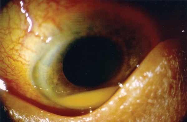
myeloma 4 months ago, was referred for an ophthalmic evaluation. She had complained of a 2-week history of visual deterioration with mild discomfort in the right eye. Ocular examination
revealed conjunctival hyperemia, posterior synechiae, fine keratic precipitates, a hypopyon (Figure 1), and a mild vitritis in the right eye with a visual acuity of 6/18. The left eye was
normal. Clinically there was no evidence of systemic infection. Vitreous and aqueous taps obtained did not reveal any microbial growth, but demonstrated the presence of small and mature
multinucleated plasma cells on microscopy (Figure 2a Giemsa staining). Flow cytometric immunophenotyping of cells both from the blood and from the aqueous sample (Figure 2b–d) demonstrated
the presence of the myeloma phenotype characterised by CD19−/CD20−/CD45−/CD38+/CD138+/CD56+.4, 5, 6 The patient, already on thalidomide at this point, was commenced on a course of mild
topical steroids (0.5% prednisolone) four times daily as well as 0.5% cyclopentolate bd. Although there was symptomatic improvement, a small pseudohypopyon remained after 8 weeks of therapy
with no change in visual acuity. Sadly, this patient succumbed to the disease 3 months later. COMMENT Direct ocular involvement in myeloma has been reported,7, 8 but only one report has
demonstrated definitively, by the use of immunohistochemistry, the identity of the malignant cells within the anterior chamber. 8 Myeloma plasma cells are phenotypically abnormal, and have
been shown to be distinguishable from normal plasma cells with flow cytometric analysis on the basis of their low expression of CD19 and CD45 and higher levels of expression of CD138 and
CD38 with or without increased expression CD56.4, 5, 6 Our study has demonstrated that flow cytometric analysis for the detection of malignant phenotypes is possible in small aqueous
samples, and may be useful in resolving the cause of hypoyon in these rare cases. REFERENCES * Bronstein M . Ocular involvement in multiple myeloma. _Arch Ophthalmol_ 1955; 55: 188–192.
Article Google Scholar * Orellana J, Friedman AH . Ocular manifestations of multiple myeloma, Waldenstrom's macroglobulinemia, and benign monoclonal gammopathy. _Surv Ophthalmol_
1981; 26: 157–169. Article CAS Google Scholar * Knapp AJ, Gartner S, Henkind P . Multiple myeloma and its ocular manifestations. _Surv Ophthalmol_ 1987; 31: 343–351. Article CAS Google
Scholar * Witzig TE, Kimlinger TK, Greipp PR . Detection of peripheral blood myeloma cells by three-color flow cytometry. _Curr Top Microbiol Immunol_ 1995; 194: 3–8. CAS PubMed Google
Scholar * Pagnucco G, Vanelli L, Gervasi F . Multidimensional flow cytometry immunophenotyping of hematologic malignancy. _Ann N Y Acad Sci_ 2002; 963: 313–321. Article Google Scholar *
Leo R, Boeker M, Peest D, Hein R, Bartl R, Gessner JE _et al_. Multiparameter analyses of normal and malignant human plasma cells: CD38++, CD56+, CD54+, cIg+ is the common phenotype of
myeloma cells. _Ann Hematol_ 1992; 64: 132–139. Article CAS Google Scholar * Shakin EP, Augsburger JJ, Eagle Jr RC, Ehya H, Shields JA, Fischer D _et al_. Multiple myeloma involving the
iris. _Arch Ophthalmol_ 1988; 106: 524–526. Article CAS Google Scholar * Tranos PG, Andreou PS, Wickremasinghe SS, Brazier JD . Pseudohypopyon as a feature of multiple myeloma. _Arch
Ophthalmol_ 2002; 120: 87–88. PubMed Google Scholar Download references AUTHOR INFORMATION AUTHORS AND AFFILIATIONS * Department of Ophthalmology and Visual Sciences, Eye and ENT Centre,
Queens Medical Centre, University of Nottingham, Clifton Boulevard, Nottingham, NG7 2UH, UK J H Chan & H S Dua * Department of Ophthalmology, Royal Free Hospital, Pond Street, London,
NW3 2 QG, UK P G Tranos & J D Jagger * Department of Haematology, St Mary's Hospital, Praed Street, London, W2 1NY, UK F L Lim Authors * J H Chan View author publications You can
also search for this author inPubMed Google Scholar * H S Dua View author publications You can also search for this author inPubMed Google Scholar * P G Tranos View author publications You
can also search for this author inPubMed Google Scholar * J D Jagger View author publications You can also search for this author inPubMed Google Scholar * F L Lim View author publications
You can also search for this author inPubMed Google Scholar CORRESPONDING AUTHOR Correspondence to J H Chan. RIGHTS AND PERMISSIONS Reprints and permissions ABOUT THIS ARTICLE CITE THIS
ARTICLE Chan, J., Dua, H., Tranos, P. _et al._ Pseudohypopyon due to malignant infiltration of the anterior chamber in multiple myeloma. _Eye_ 19, 112–113 (2005).
https://doi.org/10.1038/sj.eye.6701410 Download citation * Published: 23 April 2004 * Issue Date: 01 January 2005 * DOI: https://doi.org/10.1038/sj.eye.6701410 SHARE THIS ARTICLE Anyone you
share the following link with will be able to read this content: Get shareable link Sorry, a shareable link is not currently available for this article. Copy to clipboard Provided by the
Springer Nature SharedIt content-sharing initiative