- Select a language for the TTS:
- UK English Female
- UK English Male
- US English Female
- US English Male
- Australian Female
- Australian Male
- Language selected: (auto detect) - EN
Play all audios:
ABSTRACT Although most colorectal cancer develops based on the adenoma–adenocarcinoma sequence, morphologically, colorectal cancer is not a homogeneous disease entity. Generally, there are
two distinct morphological types: polypoid and ulcerative colorectal tumours. Previous studies have demonstrated that K-_ras_ codon 12 mutations are preferentially associated with polypoid
growth of colorectal cancer; however, little is known about the molecular mechanism that determines ulcerative growth of colorectal cancer. β-catenin complex plays a critical role both in
tumorigenesis and morphogenesis. We examined the differential expression of β-catenin and its related factors among different types of colorectal cancer in order to determine any
relationship with gross tumour morphology. Immunohistochemical staining of β-catenin, E-cadherin and MMP-7 was performed on 51 tumours, including 26 polypoid tumours and 25 ulcerative
tumours. Protein truncation tests and single-strand conformational polymorphism for mutation of the adenomatous polyposis coli tumour suppressor gene, as well as single-strand conformational
polymorphism for the mutation of β-catenin exon 3 were also done. Nuclear expression of β-catenin was observed in 18 out of 25 (72%) cases of ulcerative colorectal cancer and seven out of
26 (26.9%) cases of polypoid colorectal cancer. A significant relationship of nuclear β-catenin expression with ulcerative colorectal cancer was found (_P_<0.001). However, this finding
was independent of adenomatous polyposis coli tumour suppressor gene mutation and E-cadherin expression. Together with previous data, we propose that different combinations of genetic
alterations may underlie different morphological types of colorectal cancer. These findings should be taken into consideration whenever developing a new genetic diagnosis or therapy for
colorectal cancer. SIMILAR CONTENT BEING VIEWED BY OTHERS MOLECULAR CHARACTERIZATION OF ULCERATIVE COLITIS-ASSOCIATED COLORECTAL CARCINOMAS Article 14 December 2020 LOSS OF HES1 EXPRESSION
IS ASSOCIATED WITH EXTRACELLULAR MATRIX REMODELING AND TUMOR IMMUNE SUPPRESSION IN _KRAS_ MUTANT COLON ADENOCARCINOMAS Article Open access 25 September 2023 SMOC2, AN INTESTINAL STEM CELL
MARKER, IS AN INDEPENDENT PROGNOSTIC MARKER ASSOCIATED WITH BETTER SURVIVAL IN COLORECTAL CANCERS Article Open access 03 September 2020 MAIN Colorectal carcinogenesis involves a multistep
progression of genetic mutations. Based on the adenoma–carcinoma sequence, much research has focused on mutation detection; sequential genetic alterations have been illustrated as a linear
process (Fearon and Vogelstein, 1990; Kinzler and Vogelstein, 1996). Although this approach represents a well-known paradigm for the sequential development of cancer driven by the
accumulation of genetic defects, more and more cases of carcinogenesis have been reported in contrast to the linear and clonal development of cancer (Sedivy et al, 2000). Recently, a
non-linear, chaodynamic model of carcinogenesis has been suggested by others. In this model, genetic instability among cells produces a tremendous and chaotic diversity that may lead to
cancer (Coffey, 1998). Furthermore, from a holistic point of view, not only genetic events may drive the onset of cancer. In biology, deterministic and non-deterministic phenomena co-exist.
Clinically, in terms of gross morphology, colorectal cancer (CRC) is not a homogeneous disease entity (Kudo, 1993; Hamilton, 1995; Kato et al, 1997). In general, there are two common but
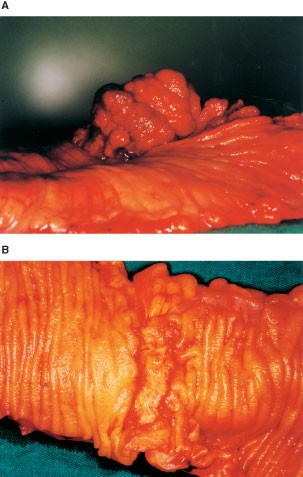
distinct morphological types, namely polypoid and ulcerative colorectal tumours (Hamilton, 1995). These are distinguished by different gross appearances: either an exophytic or endophytic
growth pattern (Figure 1). Polypoid tumours always show distinguished protruding or exophytic growth towards the bowel lumen (Figure 1A). Ulcerative tumours have an ulcer that usually
exhibits endophytic growth toward the bowel wall itself; thus the tumour's floor may be below the surface of the surrounding mucosa (Figure 1B). These divergent morphological features
lead us to question whether different genetic or different combinations of genetic alterations are what are involved in colorectal carcinogenesis. In the model proposed by Vogelstein et al
(1988), adenomatous _polyposis coli_ (APC) tumour suppressor gene mutation is found in the earliest stage, before proceeding to K-_ras_ mutation as well as to other tumour suppressor genes,
such as p53 and DCC. Paralleling increases in adenoma size and grade of dysplasia ensue during colorectal tumour progression. However, nothing has been described on whether different genetic
alterations occurred between these two distinct types of colorectal cancer during multistep carcinogenesis. Although data from synchronous adenoma and carcinoma analyses emphasised that the
accumulation of genetic alterations is more important than their order (Fearon and Vogelstein, 1990; Kinzler and Vogelstein, 1996), some researchers are still interested in establishing an
ideal linear process. Nonetheless, there are reports indicating that the significantly low frequency of K-_ras_ mutation is associated with superficial or non-polypoid type colorectal
adenoma or carcinoma (Yamagata et al, 1994, 1995). Others and we have further demonstrated significant correlation between polypoid growth of CRC and K-_ras_ codon 12 mutation (Chiang et al,
1998). However, with regard to the ulcerative type of colorectal cancer, this remains poorly understood. We investigated the expression of β-catenin, which plays an important role in the
morphogenesis and carcinogenesis of colorectal cancer (Gumbiner, 1996). Apart from its involvement in cell adhesion and the Wingless/Wnt signaling pathway, β-catenin may play a direct role
in colorectal carcinogenesis because it binds the products of the APC tumour suppressor gene. When APC is mutated, which occurs in up to 80% of colorectal cancer, β-catenin accumulates and
translocates to the nucleus, where it binds the transcription factors of the TCF/LEF gene family and activates the expression of target genes (Korinek et al, 1997). We attempted to detect if
any differential expression of β-catenin was related to different morphological growth patterns in colorectal carcinomas. Factors related to altered β-catenin expression including
E-cadherin expression, mutations of the APC tumour suppressor gene or the β-catenin gene itself were also investigated. Materialising matrix metalloproteinase-7 (MMP-7) expression, one of
the downstream targets of β-catenin, was also analysed in relation to β-catenin expression. MATERALS AND METHODS SAMPLE COLLECTION We collected 51 primary colorectal carcinoma tissue samples
from sporadic colorectal cancer patients who underwent colectomies at Chang Gung Memorial Hospital (CGMH). All samples were collected immediately after resection and stored in a −80°C
freezer. Normal mucosa samples were removed at the same time from sites about 10 cm from each tumour. Whole tumour specimens were then prepared for routine histopathological examination.
Formalin-fixed, paraffin-embedded colorectal carcinoma tissue samples were preserved in the tissue archives of the Pathology Department at CGMH. Detailed morphological descriptions,
histopathological data and clinical data were obtained for each case from the Cancer Registry of the Department of Colorectal Surgery at CGMH. Tumours with exophytic cauliflower-like
appearances with or without a very shallow ulcer only, and with a height exceeding half their diameter, were classified as polypoid (Figure 1A). Tumours within depressed ulcers with or
without very low elevated edges, and showing endophytic growth, were classified as ulcerative (Figure 1B). IMMUNOHISTOCHEMISTRY Standard immunohistochemical detection with minor
modifications was performed on sections from the archival, paraffin-embedded tissue to detect E-cadherin, β-catenin and matrilysin proteins. Five-micron sections mounted onto slides were
deparaffinised and rehydrated in graded alcohols and distilled water. Endogenous peroxidase activity was inhibited by incubation with 3% hydrogen peroxide in methanol for 20 min. Antigen
retrieval was done by microwaving at high power for two cycles of 5 min each, with a 10-min break between cycles in citrate buffer at pH 6.0. Non-specific binding of secondary antibodies was
blocked by incubation in 10% normal rabbit serum. Incubations with primary antibodies were done at 37°C for 120 min for anti E-cadherin (Transduction Laboratories, Lexington, KY, USA) at
100 × dilution, 60 min at 37°C for anti-β-catenin (Transduction Laboratories) at 200 × dilution and 1 : 300 for MMP-7 (Chemicon, Hofhem, Germany). After three washes with phosphate buffered
saline (PBS), the slides were incubated with biotinylated antimouse immunoglobulin and stained using the Ultra Tech Detection System Kit (Immunotech, Cedex, France). 3,3′ Diaminobenzidine
(DAB) was used as the chromogen. The slides were counterstained with hematoxylin and dehydrated in graded alcohols, air-dried and mounted using a resin-based mounting medium (Immunotech)
under coverslips. STAIN INTERPRETATION Slides were independently examined by two experienced observers (JM Chiang and KF Ng) who were blind to the clinicopathological data of the tumour and
to the initial results of the other observer. In areas of well-preserved tissue, the staining intensity of the cell membrane or cytoplasm was evaluated using the staining of adjacent
non-involved normal mucosa as the internal control for each section. Membrane expression of E-cadherin or β-catenin was considered preserved when staining of cancer cells was as strong as
that of normal glands, and the proportion of positive cancer cells in each section was more than 90%. If there was identifiable positive staining in less than 10% of the cancer cells, the
tumours were recorded as having a loss of E-cadherin or β-catenin membrane expression. Nuclear staining of positive cells was defined as an intense brown colour in the nucleus (Figure 2).
The pattern of nuclear staining was defined as follows: negative group, no less than 5% scattered positive cells without any clusters; focal group, positive cells clustered in focal areas,
when >5%, but <50% of the nuclei were stained; diffuse, over-expressed group, positive cells distributed diffusely, homogeneously or heterogeneously, when ⩾50% of the nuclei were
stained. APC AND Β-CATENIN GENE MUTATIONAL ANALYSIS Genomic DNA from each tumour sample and corresponding normal mucosa was extracted. SSCP analysis of β-catenin exon 3 was performed using
the following primer pair: exon 3, 5′-GATTTGATGGAGTTGGACATGG-3′ and 5′-TGTTCTTGAGTGAAGGACTGAG-3′. Samples were amplified through 35 cycles on a thermocycler (Perkin-Elmer) at 95°C
denaturation for 50 s, 57°C annealing for 30 s and 72°C extension for 10 min. Polymerase chain reaction (PCR) was performed in a volume of 25 μl with 20 ng of genomic DNA in a PCR buffer
containing 1.5 mM MgCl2, 200 μM each deoxyribonucleoside triphosphate, 5 pmol of each primer and 0.5 units of Taq polymerase (Perkin-Elmer, Branchburg, NJ, USA). For the investigation of the
APC gene, exon 15 mutations were performed using the protein truncation test (PTT) on genomic DNA as described previously (Van der Luijt, 1994). Exons 1–14 were screened by SSCP using
published oligonucleotides and PCR conditions (Van der Luijt, 1994; Li et al, 1999). After PCR amplification, products were loaded onto 12.5% polyacrylamide gels from a GeneGel Excel
12.5/2.4 kit (Pharmacia Biotech, AB Uppsala, Sweden) and underwent electrophoresis at 20°C. The single and double strands of the PCR products were visualised by silver staining as described
previously. STATISTICS Comparative data were analysed using the Mann–Whitney _U_-test, the Pearson's correlation coefficient and the chi-square test. A two-sided _P_<0.05 was
determined as statistically significant. RESULTS We collected 51 colorectal carcinoma samples, including 25 ulcerative tumours and 26 polypoid tumours. The clinicopathological parameters,
including age, gender, tumour stage, tumour differentiation and tumour size were comparable between the two groups, except for right colon predominance in the polypoid CRC group and
significantly deeper invasion depth in the ulcerative CRC group (Table 1). The difference in depth of invasion between these two groups were mainly reflected in the smaller-sized (⩽4 cm)
tumour group, while no difference in invasion depth was found for the larger-sized (>4 cm) tumour group (Table 2). Regarding β-catenin and E-cadherin expression by immunohistochemical
staining, loss of membrane staining was observed in 23 out of the 51 (45%) and 22 out of the 51 (43%) cases, respectively. Forty-nine per cent (25 out of 51) of cases showed nuclear
β-catenin expression and always showed a reciprocal loss of membrane staining except for two preserved cases (Table 3). There was no significantly different expression of E-cadherin found
between polypoid and ulcerative groups. However, a significant difference in membrane β-catenin staining was found between ulcerative (17 out of 25, 68%) and polypoid (six out of 26, 23%)
groups. A significant difference in nuclear expression of β-catenin (_P_=0.001) was also observed between the ulcerative (18 out of 25, 72%) and polypoid (seven out of 26) groups of CRC
(Table 3) when we defined more than 25% of cells with nuclear β-catenin expression as positive, and negative was defined as expression in less than 25% of the cells. Nonetheless, nuclear
β-catenin expression was typically heterogenous; we, therefore, analysed the extent of nuclear β-catenin expression with relation to these two distinct morphological types of CRC. We found a
significant difference between polypoid and ulcerative types of CRC. However, we did not find a significant difference related to the depth of invasion or the location of the tumour (Table
4). The frequency of APC mutations was 39 out of 51 (76%) cases. Among the 39 mutations found, 34 were detected in exon 15 truncated proteins (Figure 3), two mutations were found in exon 12,
one in exon 10 and one in exon 6. Nineteen out of 25 (76%) APC mutations were found in polypoid tumours and 20 out of 26 (77%) were found in ulcerative tumours (Table 3). No β-catenin exon
3 mutation was detected in this study. Nuclear β-catenin expression was analysed in relation to its related factors. There were no correlations found among nuclear β-catenin expression and
APC gene mutations (Table 5). In addition, loss of E-cadherin expression was not significantly related to nuclear β-catenin expression. Twenty-two per cent (11 out of 51) of the cases
demonstrated positive matrilysin (MMP-7) staining, and were observed in all eight mucinous colorectal carcinomas, one in poorly differentiated CRC and the other two in well differentiated
CRC. Nonetheless, of these 11 cases, only three demonstrated nuclear β-catenin expression. DISCUSSION We clearly demonstrated that nuclear β-catenin expression was significantly related to
ulcerative growth of CRC. This result is comparable to results in a recent report (Aust et al, 2001) describing altered distribution of β-catenin in ulcerative colitis-related colorectal
cancer. Morphologically, the gross appearance of advanced sporadic CRC is quite variable. It is usually difficult to categorise all tumours into either the polypoid or ulcerative groups
because intermediate or mixed types showing variable amounts of both components can be found. Other less frequently encountered types are flat or plateau tumours or the rare pipe-like shaped
(linitis plastica) tumours. This striking diversity in the gross appearance of CRC may reflect the underlying chaotic genetic instability. In our study, we investigated purely polypoid and
purely ulcerative tumours. The implications of our findings are further discussed below. Contradictory to previous studies showing that K-_ras_ codon 12 mutation is selectively related to
polypoidal growth of CRC (Yamagata et al, 1994; Chiang et al, 1998), our study indicated that there were different combinations of genetic alterations occurring in morphologically different
tumours during colorectal carcinogenesis. Therefore, reports trying to define the sequence or the specific sites of genetic alterations during multi-step colorectal carcinogenesis should be
very carefully considered because the same combinations of genetic alterations may not always be accumulated among the morphological types of CRC. Our findings highlight that future
development of responsible genes for gene therapy or genetic diagnosis for CRC may need to be individualised. Although many _in vitro_ studies using cell lines have proven that mutations in
the APC tumour suppressor gene occurs in most colorectal cancer and leads to the activation of β-catenin (Munemitsu et al, 1995; Morin et al, 1997), this probably is not always the case _in
vivo_. In our study, we showed that nuclear β-catenin expression was independent of APC tumour suppressor gene mutation, as was reported previously (Kobayashi et al, 2000). Furthermore, we
did not find any β-catenin mutation itself. The question follows, therefore, what regulates or forces the nuclear translocation of β-catenin? Other genetic or epigenetic events may be
present for modulating nuclear translocation. Tyrosine phosphorylation of β-catenin by a biochemical molecule such as intestinal trefoil factor has been reported (Liu et al, 1997). While,
retinoic acid (RA) has been shown to decrease the activity of the β-catenin- lymphoid enhancer binding factor/T-cell factor signalling pathway. RA activity was also independent of APC tumour
suppression and ubiquitination-dependent degradation of cytoplasmic β-catenin (Easwaran et al, 1999). Although a significant relationship exists between ulcerative CRC and nuclear β-catenin
expression, we did not find a significant increase in the extent of nuclear β-catenin expression related to the depth of invasion (Table 4). These findings indicate that although nuclear
β-catenin expression may determine the ulcerative growth pattern of CRC, the depth of invasion may be determined by several other factors, and might be the result of a more complex process
between tumour and stroma interaction. This finding also implies that higher amounts of nuclear β-catenin expression are probably necessary for the ulcerative growth from the early stage of
tumour progression, while lower amounts of nuclear β-catenin expression may be sufficient to induce polypoid tumour growth (Brabletz et al, 2000). This finding supports a previous
observation showing that β-catenin occurred in the highest concentrations in the invading line of endophytic growth of tumour cells. (Brabletz et al, 1998). Although we found a significant
correlation between nuclear beta-catenin expression and ulcerative growth and also observed that most of the ulcerative tumours were rectal carcinomas (Table 1), we did not observe a
significant relationship between tumour localisation and nuclear on tumour localisation. Furthermore, the small number of cases in this study limited further analysis of whether there are
different carcinogenesis pathways in the colon and rectum. Precisely how nuclear β-catenin expression confers ulcerative or endophytic growth to CRC remains poorly understood. Further
analyses of several downstream factors of the nuclear β-catenin/TCF complex, including c-MYC (He et al, 1998), cyclin D1 (Tetsu and McCormick, 1999), gastrin (Koh et al, 2000), PPARS (He et
al, 1999) MMP-7 (Brabletz et al, 1999; Crawford et al, 1999) are warranted. Some factors such as c-_myc_, cyclin D and gastrin reportedly relate to tumour proliferation, while others are
related to tumour invasion. MMP-7 is regulated by β-catenin expression (Brabletz et al, 1999) and is a proteolytic enzyme related to tumour invasion. It may well be reasonable to relate
proteolysis to ulcerative growth. However, in our study, we did not observe parallel expression between β-catenin and MMP-7, which is the case in cell culture studies. These negative
findings may be explained similarly to other down-regulators such as tumour growth factor (TGF)-β, which is involved in a more complex process _in vivo_ than _in vitro_ (Gaire et al, 1994).
Finally, the finding that nuclear β-catenin expression is closely related to ulcerative growth of CRC further supports a previous study that showed analogies between embryonic gastrulation
and β-catenin expression (Kirchner and Brabletz, 2000). Strong, diffuse β-catenin nuclear expression was observed as necessary for mesenchymal transition of tumour cells that expressed
invasion behaviour, while weak nuclear β-catenin expression was only enough for epithelial transitions responsible for cell proliferation (Kirchner and Brabletz, 2000). The connection of our
data and the data from embryonic development, thus, supports the concept that tumorigenesis has the properties of a complex developmental disorder (Dean, 1998). In summary, we observed that
the different combinations of genetic alterations may selectively underlie different types of CRC. Nuclear β-catenin expression is related to the ulcerative growth patterns of CRC. Although
the precise mechanism remains poorly understood, its expression is independent of APC mutation. These observations further highlight the heterogenous nature of CRC, which should be kept in
mind when developing a new gene therapy or a new genetic diagnosis for CRC. CHANGE HISTORY * _ 16 NOVEMBER 2011 This paper was modified 12 months after initial publication to switch to
Creative Commons licence terms, as noted at publication _ REFERENCES * Aust DE, Terdiman JP, Willenbucher RF, Chew K, Ferrell L, Florendo C, Molinaro-Clark A, Baretton GB, Lohrs U, Waldman
FM (2001) Altered distribution of beta-catenin, and its binding proteins E-cadherin and APC, in ulcerative colitis-related colorectal cancers. _Mod Pathol_ 14: 29–39 Article CAS Google
Scholar * Brabletz T, Herrmann K, Jung A, Faller G, Kirchner T (2000) Expression of nuclear β-catenin and C-myc is correlated to tumor size but not to proliferative activity of colorectal
adenomas. _Am J Pathol_ 156: 865–870 Article CAS Google Scholar * Brabletz T, Jung A, Dag S, Hlubek F, Kirchner T (1999) β-catenin regulates the expression of matrix metalloproteinase-7
in human colorectal cancer. _Am J Pathol_ 155: 1033–1038 Article CAS Google Scholar * Brabletz T, Jung A, Herrmann K, Gunther K, Hohenberger W, Kirchner T (1998) Nuclear overexpression of
the oncoprotein β-catenin in colorectal cancer is localized predominantly at the invasion front. _Pathol Res Pract_ 194: 701–704 Article CAS Google Scholar * Chiang JM, Chou WY, Chou TB
(1998) K-ras codon 12 mutation determines the polypoid growth of colorectal cancer. _Cancer Res_ 58: 3289–3293 CAS PubMed Google Scholar * Coffey DS (1998) Self-organization, complexity
and chaos: the new biology for medicine. _Nat Med_ 4: 882–885 Article CAS Google Scholar * Crawford HC, Fingleton BM, Rudolph-Owen LA, Heppner Goss KJ, Rubinfeld B, Polakis P, Matrisian
LM (1999) The metalloproteinase matrilysin is a target of β-catenin transactivation in intestinal tumors. _Oncogene_ 18: 2883–2891 Article CAS Google Scholar * Dean M (1998) Cancer as a
complex developmental disorder. _Cancer Res_ 58: 5633–5636 CAS PubMed Google Scholar * Easwaran V, Pishvaian M, Salimuddin BS (1999) Cross-regulation of β-catenin- LEF/TCF and retinoid
signaling pathways. _Curr Biol_ 9: 1415–1418 Article CAS Google Scholar * Fearon ER, Vogelstein B (1990) A genetic model for colorectal tumorigenesis. _Cell_ 61: 759–767 Article CAS
Google Scholar * Gaire M, Magbanua Z, McDonnell S, McNeil L, Lovett DH, Matrisian LM (1994) Structure and expression of the human gene for the matrix metalloproteinase matrilysin. _J Biol
Chem_ 269: 2032–2040 CAS PubMed Google Scholar * Gumbiner BM (1996) Cell adhesion: the molecular basis of tissue architecture and morphogenesis. _Cell_ 84: 345–357 Article CAS Google
Scholar * Hamilton SR (1995) Pathologic features of colorectal cancer. In: _Cancer of the Colon, Rectum, and Anus_ Cohen AM, Winawer ST, Friedman MA, Gunderson LL (eds) pp 189–191, New
York: McGraw-Hill Google Scholar * He TC, Chan TA, Vogelstein B, Kinzler KW (1999) PPARS is an APC-regulated target of nonsteroidal anti-inflammatory drugs. _Cell_ 99: 335–345 Article CAS
Google Scholar * He TC, Spark AB, Rago C, Hermeking H, Zawel L, da Costa LT, Morin PJ, Vogelstein B, Kinzler KW (1998) Identification of c-MYC as a target of the APC pathway. _Science_
281: 1509–1512 Article CAS Google Scholar * Kato S, Fujii T, Oda Y (1997) Differences of genetic alterations between polypoid, flat and depressed growth-type colorectal cancers.
_Gastroenterology_ 112: A589 (abstract) * Kinzler KW, Vogelstein B (1996) Lessons from hereditary colorectal cancer. _Cell_ 87: 159–170 Article CAS Google Scholar * Kirchner T, Brabletz T
(2000) Patterning and nuclear β-catenin expression in the colonic adenoma-carcinoma sequence-analogies with embryonic gastrulation. _Am J Pathol_ 157: 1113–1121 Article CAS Google Scholar
* Kobayashi M, Honma T, Matsuda Y, Suzuki Y, Narisawa R, Ajioka Y, Asakura H (2000) Nuclear translocation of beta-catenin in colorectal cancer. _Br J Cancer_ 82: 1689–1693 Article CAS
Google Scholar * Koh TJ, Bulitta CJ, Fleming JV, Dockray GJ, Varro A, Wang TC (2000) Gastrin is a target of the β-catenin/TCF-4 growth-signaling pathway in a model of intestinal polyposis.
_J Clin Invest_ 106: 533–539 Article CAS Google Scholar * Korinek V, Barker N, Morin PJ (1997) Constitutive transcriptional activation by a beta-catenin-Tcf complex in APC −/− colon
carcinoma. _Science_ 275: 1784–1787 Article CAS Google Scholar * Kudo S (1993) Endoscopic mucosal resection of flat and depressed types of early colorectal cancer. _Endoscopy_ 25: 455–461
Article CAS Google Scholar * Li G, Tamura K, Yamamoto Y, Sashio H, Utsunomiya J, Yamaura T, Shimoyama T, Furayama J (1999) Molecular and clinical study of familial adenomatous polyposis
for genetic testing and management. _J Exp Clin Cancer Res_ 18: 519–529 CAS PubMed Google Scholar * Liu D, el-Harriry I, Karayiannakis AM, Wilding J, Chinery R, Kmiot W, McCrea PD,
Gullick WJ, Pignatelli M (1997) Phosphorylation of β-catenin and epidermal growth factor receptors by intestinal trefoil factor. _Lab Invest_ 77: 557–563 CAS PubMed Google Scholar * Morin
PJ, Sparks AB, Korinek V, Barker N, Clevers H, Vogelstein B, Kinzler KW (1997) Activation of β-catenin – Tcf signaling in colon cancer by mutations in β-catenin or APC. _Science (Washington
DC)_ 275: 1787–1790 Article CAS Google Scholar * Munemitsu S, Albert I, Souza B, Rubinfeld B, Polakis P (1995) Regulation of intracellular β-catenin levels by the adenomatous polyposis
coli (APC) tumor suppressor protein. _Proc Natl Acad Sci USA_ 92: 3046–3050 Article CAS Google Scholar * Sedivy R, Wolf B, Kalipciyan M, Steger GG, Karner-Hanusch J, Mader RM (2000)
Genetic analysis of multiple synchronous lesions of the colon adenoma-carcinoma sequence. _Br J Cancer_ 82: 1276–1282 Article CAS Google Scholar * Tetsu O, McCormick F (1999) Beta-catenin
regulates expression of cyclin D1 in colon carcinoma cells. _Nature_ 398: 422–426 Article CAS Google Scholar * van der Luijt R, Khan PM, Vasen H, van Leeuwen C, Tops C, Roest P, den
Dunnen J, Fodde R (1994) Rapid detection of translation-terminating mutations at the adenomatous polyposis coli (APC) gene by direct protein truncation test. _Genomics_ 20: 1–4 Article CAS
Google Scholar * Vogelstein B, Fearon ER, Hamilton SR, Kern SE, Preisinger AC, Leppert M, Nakamura Y, White R, Smits AM, Bos JL (1988) Genetic alterations during colorectal tumor
development. _N Engl J Med_ 319: 525–532 Article CAS Google Scholar * Yamagata S, Muto T, Uchida Y, Masaki T, Higuchi Y, Sawada T, Hirroka T (1995) Polypoid growth and K-ras codon 12
mutation in colorectal cancer. _Cancer (Phila)_ 75: 953–957 Article CAS Google Scholar * Yamagata S, Muto T, Uchida Y, Masaki T, Sawada T, Tsuno N, Hirooka T (1994) Lower incidence of
k-ras codon 12 mutation in flat colorectal adenomas than in polypoid adenomas. _Jap J Cancer Res_ 85: 147–151 Article CAS Google Scholar Download references AUTHOR INFORMATION AUTHORS AND
AFFILIATIONS * Division of Colon and Rectal Surgery, Human Molecular Genetics Laboratory, Chang Gung Memorial Hospital, 199 Tung Hwa North Road, Taipei, 333, Taiwan J M Chiang, Y H Wu Chou,
T C Chen, K F Ng & J L Lin * Department of Pathology, Chang Gung Memorial Hospital, 199 Tung Hwa North Road, Taipei, 333, Taiwan J M Chiang, Y H Wu Chou, T C Chen, K F Ng & J L Lin
Authors * J M Chiang View author publications You can also search for this author inPubMed Google Scholar * Y H Wu Chou View author publications You can also search for this author inPubMed
Google Scholar * T C Chen View author publications You can also search for this author inPubMed Google Scholar * K F Ng View author publications You can also search for this author inPubMed
Google Scholar * J L Lin View author publications You can also search for this author inPubMed Google Scholar CORRESPONDING AUTHOR Correspondence to J M Chiang. RIGHTS AND PERMISSIONS From
twelve months after its original publication, this work is licensed under the Creative Commons Attribution-NonCommercial-Share Alike 3.0 Unported License. To view a copy of this license,
visit http://creativecommons.org/licenses/by-nc-sa/3.0/ Reprints and permissions ABOUT THIS ARTICLE CITE THIS ARTICLE Chiang, J., Chou, Y., Chen, T. _et al._ Nuclear β-catenin expression is
closely related to ulcerative growth of colorectal carcinoma. _Br J Cancer_ 86, 1124–1129 (2002). https://doi.org/10.1038/sj.bjc.6600214 Download citation * Received: 30 July 2001 * Revised:
31 January 2002 * Accepted: 31 January 2002 * Published: 02 April 2002 * Issue Date: 08 April 2002 * DOI: https://doi.org/10.1038/sj.bjc.6600214 SHARE THIS ARTICLE Anyone you share the
following link with will be able to read this content: Get shareable link Sorry, a shareable link is not currently available for this article. Copy to clipboard Provided by the Springer
Nature SharedIt content-sharing initiative KEYWORDS * β-catenin * colorectal cancer * polypoid tumour * ulcerative tumour * carcinogenesis